Ectopic ACTH syndrome in a dog with a mesenteric
neuroendocrine tumour: a case report
V.A. Castillo
1, P.P. Pessina
2, J.D. Garcia
1, P. Hall
1, M.F. Gallelli
1,
D.D. Miceli
1, M.F. Cabrera Blatter
11Faculty of Veterinary Sciences, University of Buenos Aires, Ciudad Autonoma de Buenos Aires, Buenos Aires, Argentina
2Faculty of Veterinary Medicine, University of the Republic, Montevideo, Rep. Oriental del Uruguay
ABSTRACT: Ectopic ACTH secretion is provoked by extra-pituitary tumours that secrete ACTH, constituting an infrequent type of Cushing Syndrome in the dog. Neuroendocrine tumours (NET) are characterised by the synthesis of peptides with hormone activity. A dog with clinical diagnosis of Cushing’s syndrome and presenting an abdominal tumour located in the area of the left adrenal gland was sent to the hospital. Cortisol was not inhib-ited at four and eight hours after the application of low-dose dexamethasone and the cortisol/creatinine ratio was elevated (93 × 10-6, referencevalues < 10 × 10–6). Plasma ACTH measurements were high (28.6 pmol/l, reference
values 5.5–14.3 pmol/l). On computed tomography, the tumour was found in the meso-epigastrium, with both adrenal glands hyperplasic and no alteration of the pituitary image. The tumour was located between the two layers of the meso-colon and was removed using laparoscopy. After surgery, ACTH concentrations became normal and clinical signs remitted. The histopathological diagnosis was NET, with positive ACTH immunostaining.
Keywords: neuroendocrine tumour; apudoma; ectopic ACTH
Supported by the University of Buenos Aires, Argentina (Grant No. UBACyT 20020100100246).
Cushing’s syndrome (CS) is a frequent endo-crine pathology in the dog. Its main cause is exces-sive production of adrenocorticotropic hormone (ACTH) by a corticotroph-cell adenoma. It can also arise from tumours in one of the adrenal glands, though recently the presence of illegitimate re-ceptors in these glands is also considered a source (Kooistra and Galac 2012). In humans ectopic ACTH secretion can be associated with various solid tumours, mostly of neuroendocrine origin and has been called Ectopic ACTH syndrome (EAS) (Alexandrakis and Grossman 2010; Newell-Price et al. 1998; Isidori and Lenzi 2007; Pivonello et al. 2008). There are few registered cases in dogs, one of which was reported by Churcher (1999) and an-other more recent case by Galac et al. (2005), with no other cases reported to the best of the authors’ knowledge.
Neuroendocrine tumours (NET) or apudomas are characterised by cells that capture amine precur-sors (Amine Precursor Uptake Decarboxylase cells,
APUD) and by decarboxylation convert the amines into peptides with different functions. These tumours are most frequently located in the endocrine pan-creas (insulinomas and glucagonomas), the adrenal medulla (pheochromocytomas), intestines and lungs. Other locations have been described for neuroen-docrine tumours in dogs, although extremely rarely (Patnaik et al. 1981; Harkema et al. 1992; Sako et al. 2005; Vergine et al. 2005; Kuwata et al. 2010) and are not associated with ACTH secretion or with clinical symptoms compatible with hypercortisolism.
This report concerns a dog diagnosed with CS in-voked by an extra-adrenal, extra-pituitary tumour that we discovered to be an ACTH-secreting NET located in the mesentery.
Case description
that was referred to our hospital from Uruguay with a clinical diagnosis of Cushing’s syndrome. The owner described an increase in weight and abdomen size over the last four months, and had noted polyuria-polydipsia and polyphagia in the last 30 days before consulting. Routine laboratory results (Table 1) showed alteration of the following param-eters: hepatic enzymes, electrolytes (Na and K) and glycaemia, all within the reference range of values of the acting laboratory. The urine cortisol/creatinine ratio (UCCR) was determined in order to screen for CS (Rijnberk et al.1988; Kooistra and Galac 2012), and gave a value of 93 × 10–6 (reference value < 10 × 10–6). Once the diagnosis of CS was suspected, inhi-bition with low dose dexamethasone intravenously (i.v.) was conducted (Feldman et al.1996) as a di-agnostic and differential test, showing no inhibi-tion at 4 h and 8 h (basal cortisol: 185 nmol/l; 4 h: 183.4 nmo/l; 8 h: 217.4 nmol/l). Based on these results, ultrasonographic evaluation of the abdo-men was indicated which revealed the presence of a hypoechogenic formation of 5.2 cm with irregular borders in the left hemi-abdomen, associated with the area of the left adrenal gland. Owing to the suspicion of neoplasia in the left adrenal gland, the owner was advised to take the case to a hospital in Buenos Aires, Argentina for adrenalectomy and post-surgical follow-up.
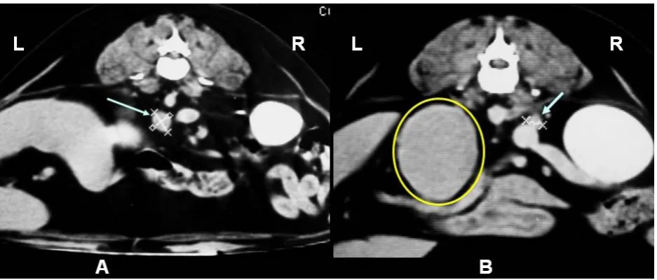
Computed tomography. A computed tomog-raphy (CT) of the sellar area (3 mm serial coronal sections every 9 s of scanning), of the abdomen in general and the adrenal glands in particular
(3 mm serial transversal sections of 5 s scanning) with the dog in sternal recumbence was indicated prior to surgery. The study was carried out with and without i.v. iodine contrast medium (Telebrix® Coronario, Temis Lostalo, Argentina).
The results of the study indicated that both adre-nal glands preserved their shape and were hyper-plastic (right adrenal gland: 8 × 7 mm; left adrenal gland: 12 × 14 mm). In the left epi-mesogastric region the presence of a 5 × 6 cm high and 8 cm long tumour was reported (Figure 1). It was unrelated to any adjacent organ, showing smooth slightly irregular borders. The images of the rest of the abdominal structures did not reveal any alteration except for hepatomegaly, typical of CS. Exploration of the sellar region showed the pituitary and the rest of the encephalic parenchyma to be without any morphological alterations. Additionally, due to the discovery of the extra-adrenal tumour, explora-tion of the thorax was carried out. All structures therein were of normal morphology.
Endocrine biochemistry. Based on the CT re-port, it was suspected that the CS might be caused by ectopic ACTH secretion. Therefore, measure-ments of this hormone and of cortisol, to be carried out in the same plasma sample, were requested. ACTH and cortisol were measured before surgery and this was repeated at the time of discharge from the hospital (four days after surgery) before return-ing to the country of origin. For both hormones, the blood sample was collected into refrigerated plastic tubes with anticoagulant (EDTA) and
[image:2.595.67.536.520.720.2]tinin and immediately centrifuged. The serum was then stored at –30 °C until the assays were carried out. Hormone analyses were carried out in the Biochemistry laboratory of the Veterinary Medicine Hospital of the Faculty of Veterinary Sciences of the University of Buenos Aires using enzyme immunoassay commercial kits from Alpco Diagnostics (Windham, NH, USA). For ACTH, the Adrenocorticotropic hormone Elisa (ACTH Elisa) was used, with an intra- and inter-assay coefficient of variation of 6.7 and 7.1%, respectively, and with a sensitivity of 0.46 pg/ml. For cortisol, a Cortisol ELISA was used, with an intra- and inter-assay co-efficient of variation of 5.0% and 8.1%, respectively, and a sensitivity of 0.4 µg/dl.
The CRH test which discriminates between pi-tuitary-dependent and ectopic ACTH (Nieman et al. 1993; Isidori et al. 2003) could not be carried out owing to the lack of necessary reagents in our country.
Laparotomy. The patient was subjected to mid-line abdominal surgery and a 5 × 7cm high-width and 9 cm long tumour, located in the epigastrium-meso-gastrium region between the two layers of the meso-colon, was removed. The tumour was situated close to the left adrenal gland, was very vascularised and had distinct irregular borders. The pancreas, kidneys, duodenum, colon, stomach, ab-dominal lymph nodes and liver were all carefully explored and neither metastasis nor other evident alterations were found, except for hepatomegaly which had already been observed in the ultrasound and CT evaluations. Therefore, the mass was con-sidered a primary tumour.
The patient was released 72 h post surgery and returned to Uruguay 48 h later (5th day after sur-gery) where clinical and biochemical follow-up was carried out.
Histopathology and immunohistochemistry. The tumour was examined using histopathology (haematoxylin-eosin staining) and immunohisto-chemistry (IHC) in 10% buffered formalin, with an indication for ACTH staining due to the sus-picion of ectopic ACTH secretion. Sections of 3 µm were stained with haematoxylin-eosin (HE). Immunohistochemical staining was performed us-ing the avidin-biotin complex (ABC) and the im-munoperoxidase detection system (Millipore IHC Select) technique, using a mouse monoclonal an-tibody (1 : 400 in PBS) against the one to 24 amino acid region of ACTH (Santa Cruz Biotechnology, SPM333). The chromogen 3,3'-diaminobenzidine
(DAB) was used for revealing. A DC180 digital camera and a tri-ocular DMLS Leica microscope were used to capture the images.
Ethical approval. The Ethics Committee of the Faculty of Veterinary Science (CICUAL) ap-proved the present study, according to the laws on experimentation in animals in Argentina and World Health Organization recommendations. The signed consent of the dog’s owner was obtained for conducting the requested studies, surgery and publication of the data.
RESULTS
Endocrine biochemistry
Prior to surgery, plasma ACTH concentration was 28.6 pmol/l (reference value in our laborato-ry: 5.5–14.3 pmol/l) and cortisol was 220.7 nmol/l (reference value: 22.07–110.4 nmol/l). Plasma con-centrations of ACTH decreased to 11 pmol/l and cortisol to 88.3 nmol/l in the samples obtained four days after surgery.
Histopathology and immunohistochemistry
The histopathological report identified polygonal-cuboidal cells that were grouped forming rosettes and a pseudo alveolar structure, with a vascularised fibrous stroma of carcinoid aspect, nuclei largely hyperchromatic and atypical and with eosinophilic cytoplasm, all compatible with a neuroendocrine tumour (Ashley 1990). Immunohistochemistry was positive for ACTH, showing strong and diffuse nu-clear and cytoplasmic immunoreactivity of the tu-mour cells to the anti-ACTH antibody (Figure 2).
Clinical evolution after surgery
ratio and possible risk of adrenal insufficiency, hy-drocortisone was indicated: 0.5mg/kg every 12 h for three days, followed by the same dose once a day for three more days, then 0.25 mg/kg/day for three days and finally, this same dose every 48 h (last three doses). The day after suspending the hydrocortisone administration, electrolyte analysis was repeated, showing normal values for Na, K and the Na/K ratio (148 mmol/l, 4.2 mmol/l and 35.2/1, respectively). Sixty days after surgery, evaluation back in the owner’s own country revealed a reduc-tion in abdomen and weight (8.700 kg) and no clini-cal symptoms compatible with CS. Biochemistry results at this time showed a decrease in hepatic enzymes, an electrolyte analysis with no alterations and normal glycaemia (Table 1). A year after
sur-gery (evaluated in Uruguay) the patient remained stable, without clinical signs of a recurrence of the pathology and with a normal biochemical analysis.
DISCUSSION AND CONCLUSIONS
In this report, we describe a CS due to ectopic ACTH secretion by a NET found in the abdomen located in the meso-colon, confirmed by IHC.
Similar cases have been reported by Churcher (1999) and Galac et al. (2005), both of which are similar to the present case in certain aspects and different in others. With regard to the location of the NET, in the case reported by Churcher (1999), the hypercortisolism was associated with the pres-ence of a hepatic carcinoid, while the case reported by Galac et al. (2005) was related to a NET located in the pancreas. In our study, the tumour did not belong to any organ in particular, and was located in the mesentery. Other authors have also reported cases of NET associated with ectopic ACTH se-cretion with infrequent or unusual presentations (Johnson 1982; Otsuka et al. 2005; Thomas et al. 2013). The most frequent locations for the ACTH-secreting carcinoid are the lungs, pancreas, thy-roid and adrenal medulla (Clark and Carney 1987; Terzolo et al. 2001; Otzuka et al. 2005).
[image:4.595.66.532.80.220.2]With respect to similarities, none of the three cases showed cortisol suppression after dexametha-sone administration because the axis was already inhibited (Churcher 1999; Galac et al. 2005). The lack of dexamethasone inhibition is to be expected in cases of adrenal neoplasias and of pituitary mac-roadenomas (Kooistra et al. 1997; Newel-Price et
Table 1. Biochemical data from the patient with ectopic ACTH syndrome
Biochemistry Pre-surgery diagnosis After surgery 60 days 1 year
AP (UI/l) 1250 450 200
GPT/ALT (UI/l) 120 60 36
GOT/AST (UI/l) 55 32 45
Glucose (mmol/l) 5.9 3.8 4.8
Na+ (mEq/l) 147 157 152
K+ (mEq/l) 5.0 4.8 5.2
[image:4.595.64.291.559.675.2]AP = alkaline phosphatase (normal value: up to 300 UI/l); GPT/ALT = pyruvic glutamatetransaminase/alanine ami-notransferase (normal value: up to 50 UI/l); GOT/AST = oxa-lacetic glutamate transaminase/aspartate aminotransferase (normal value: up to 50 UI/l); glucose (normal value: 3.4– 6.2 mmol/l); Na = sodium (normal value: 140–155 mEq/l), K = potassium (normal value: 3.7–5.5 mEq/l)
Figure 2. Histopathology (a) and IHC (b, c). (a) The disposition of the neoplastic cells, forming groups some like cords and others like rosettes, with very pycnotic nuclei, can be seen by H-E (40×). (b) Negative control of ICH (stained with hematoxylin), (c) brown positive staining for ACTH in both nucleus and cytoplasm. Polyclonal anti ACTH anti-body, ABC method, hematoxylin counterstain (40×)
al. 1998; Kooistra and Galac 2012), both scenarios having been ruled out after the image studies were carried out. Nevertheless, in human medicine, this lack of suppression of the axis together with nega-tive images for the pituitary and adrenal glands make it necessary to rule out the presence of car-cinoids that can produce EAS (Newell-Price 1998; Newell-Price 2006).
In the CT study, the pituitary image was normal, with both adrenal glands hyperplasic, similarly to what has been reported in dogs. The finding of an intra-abdominal tumour mass in addition to clinical and biochemical features of CS led to the suspicion of ectopic ACTH secretion, which was clinically confirmed by the high plasma levels of ACTH and cortisol. In the cases of EAS report-ed in human mreport-edicine, plasma concentrations of both hormones are characteristically higher than in pituitary-dependent Cushing patients (Howlett et al. 1986; Newel-Price et al 2006; Alexandrakis and Grossamn 2010; Ma et al. 2010). High concen-trations of ACTH without evidence of a pituitary tumour are suggestive of EAS according to previ-ous reports. In human medicine, the CRH test is indicated to differentiate between corticotroph ad-enoma and ectopic ACTH secretion. The majority of patients with EAS do not respond to CRH, but some do respond normally (Malchoff et al. 1988; Neiman et al. 1993; Isidori et al. 2003). In veterinary medicine, only in the case reported by Galac et al. (2005) was this test performed. Unfortunately, in our country the CRH is unavailable commercially and the test could not be performed. Thus, the plasma ACTH and cortisol concentrations were taken into account as suggestive of EAS. Our findings of plasma ACTH and cortisol without the imaging of a pitui-tary tumour and our discovery of an extra-adrenal tumour in the abdomen are in agreement both with reports in human medicine and the results of Galac et al. (2005). In the case of Churcher (1999), ACTH concentrations were within the reference values given by the author, this being an unexpected pat-tern for a case of ectopic ACTH secretion.
Potassium levels were normal in our case; in con-trast, according to human reports and those of Galac et al. (2005) and Churcher (1999), hypokalaemia would be expected (Howlet et al. 1986; Ulick et al. 1992; Isidori and Lenzi 2005). It is possible that the relatively short period of time between the appear-ance of clinical symptoms, diagnosis and treatment did not allow time for the proposed mechanisms leading to hypokalaemia to develop. The altered
electrolyte levels compatible with adrenal insuffi-ciency, which were observed later and which neces-sitated hydrocortisone supplementation for a short period, can be explained by the rapid decrease in ACTH after the tumour was removed. The ACTH stimulation test could not be performed in order to confirm the adrenal insufficiency owing to con-siderations of time: the owner’s dog remained in Buenos Aires in order to establish hydrocortisone therapy based on the results of the ionogram, which were suggestive of adrenal insufficiency. The fact that this episode was not repeated after suspending hydrocortisone administration and that electrolyte analysis remained normal indicates that pituitary secretion of ACTH (once excess cortisol from the ectopic ACTH was inhibited) returned to normal, as did the pituitary axis.
The substantial difference with the other two reports in dogs is that the IHC for ACTH was positive, indicating that the tumour was producing the hormone through a subpopulation of ACTH-secreting tumour cells, thus representing a clear case of ectopic ACTH syndrome. In the other two reports mentioned above, the IHC failed to stain positive for ACTH, which is possible in these situa-tions and has also been reported in humans (Coates et al. 1986; Alexandrakis and Grossman 2010).
The fact that after the removal of the tumour the clinical symptoms produced by the hypercor-tisolism remitted totally and that both hepatic enzymes and ACTH and cortisol concentrations re-turned to normal without the need for adrenal cor-tex inhibition therapy confirms that the cause of the CS was the abdominal tumour, with no metastasis nor other adrenal or pituitary tumours present at the time of the surgery. This is an important point because in the case reported by Galac et al. (2005), there was metastasis of the primary tumour and it was necessary to administer trilostane to control the hypercortisolism and in the case reported by Churcher (1999), the dog died after the surgery.
or scintigraphy with octreotide) to detect the pos-sibility of carcinoids should be considered in these cases. Lastly, ectopic ACTH secretion as an under-lying cause of CS in the dog should be considered as a potential aetiology for this disease.
REFERENCES
Alexandraki KI, Grossman AB (2010): The ectopic ACTH syndrome. Review Endocrinology Metabolism Disorder 11, 117–126.
Ashley DJB (1990): Carcinoid (amine-secreting) tumour. In: Evans’ Histological Appearances of Tumours, 4th ed. Churchill Livingstone, London. 341–353.
Churcher RK (1999): Hepatic carcinoid, hypercorti-solism and hypokalaemia in a dog. Austraian Veteri-nary Journal 77, 641–645.
Clark ES, Carney JA (1987): Pancreatic islet cell tumor associated with Cushing’s syndrome. American Jour-nal Surgery Pathology 8, 917–924.
Coates PJ, Doniach I, Howlet TA, Rees LH, Besser GM (1986): Immunocytochemical study of 18 tumours causing ectopic Cushing’s syndrome. Journal of Clin-ical Pathology 39, 955–960.
Galac S, Kooistra HS, Voorhout G, van den Ingh TSGAM, Mol JA, van den Berg G, Meij BP (2005): Hypercorticism in a dog due to ectopic secretion of adreno-corticotropic hormone. Domestic Animal Endocrinology 28, 338–348.
Feldman EC, Nelson RW, Feldman MS (1996): Use of low- and high-dose dexamethasone tests for distin-guishing pituitary-dependent from adrenal tumor hyperadrenocorticism in dogs. Journal American Vet-erinary Medical Association 209, 772–775.
Harkema JR, Jones SE, Naydan DK, Wilson DW (1992): An atypical neuroendocrine tumor in the lung of a Beagle dog. Veterinary Pathology 29, 175–179. Howlett TA, Drury PL, Perry L, Doniach I, Rees LH,
Besser GM (1986): Diagnosis and management of ACTH-dependent Cushing’s syndrome: comparison of the features in ectopic and pituitary ACTH produc-tion. Clinical Endocrinology 24, 699–713.
Isidori AM, Kaltsas GA, Mohammed S, Morris DG, Jenkins P, Chew SL, Monson JP, Besser GM, Grossman AB (2003): Discriminatory value of the low-dose dexamethasone suppression test in establishing the diagnosis and dif-ferential diagnosis of Cushing’s syndrome. Journal Clin-ical Endocrinology Metabolism 88, 5299–5306.
Isidori AM, Lenzi A (2007): Ectopic ACTH syndrome. Arquivos Brasileiros Endocrinologia and Metabologia 51, 1217–1225.
Johnson FD (1982): Ectopic ACTH syndrome in APUD tumors. Oncology 39, 358–361.
Kooistra HS, Voorhout G, Mol JA, Rijnberk, A (1997): Correlation between impairment of glucocorticoid feedback and the size of the pituitary gland in dogs with pituitary dependent hyperadrenocorticism. Jour-nal Endocrinology152, 387–394.
Kooistra HS, Galac S (2012): Recent Advances in the Diagnosis of Cushing’s Syndrome in Dogs. Topics in Companion Animal Medicine 27, 21–24.
Kuwata K, Shibutani M, Kemmochi Y, Taniai E, Morita R, Ogawa B, Mitsumori K (2010): A neuroendocrine carcinoma of undetermined origin in a dog. Journal of Toxicology Pathology 23, 151–155.
Ma Y, Aitelli C, Dobson RW, Konduri K (2010): Ectopic adrenocorticotropic hormone syndrome: a diagnostic challenge and review of the literature. Proceedings (Baylor University Medical Center) 23, 426–428. Malchoff CD, Orth DN, Abboud C, Carney JA, Pairolero
PC, Carey PM (1988): ectopic ACTH syndrome caused by a bronchial carcinoid tumor responsive to dexa-methasone, metyrapone and corticotrophin-realising factor. American Journal Medicine 84, 760–764. Newell-Price J, Trainer P, Besser M, Grossman A (1998):
The diagnosis and differential diagnosis of Cushing’s syndrome and pseudo-Cushing’s states. Endocrine Reviews 19, 647–672.
Newell-Price J, Bertagna X, Grossman AB, Nieman LK (2006): Cushing’s syndrome. Lancet 367, 1605–1617. Nieman LK, Oldfield EH, Wesley R, Chrousos GP, Lo-riaux DL, Cutler GB Jr (1993): A simplified morning ovine corticotrophin releasing hormone stimulation test for the differential diagnosis of adrenocorticotro-pin-dependent Cushing’s syndrome. Journal Clinical Endocrinology Metabolism77, 1308–1312.
Otsuka F, Miyoshi T, Murakami K, Inagaki K, Takeda M, Ujike K, Ogura T, Omori M, Doihara H, Tanaka Y, Hashi-moto K, Makino H (2005): An extra-adrenal abdominal pheochromocytoma causing ectopic ACTH syndrome. American Journal Hypertension 18, 1364–1368. Patnaik AK, Lieberman PH, Hurvitz AI, Johnson GF
(1981): Canine hepatic carcinoids. Veterinary Pathol-ogy 18, 445–453.
Pivonello R, De Martino MC, De Leo M, Lombardi G, Colao A (2008): Cushing’s syndrome. Endocrinology Metabolism Clinics North America 37, 135–149. Rijnberk A, van Wees A, Mol JA (1988): Assessment of
two tests for the diagnosis of canine hyperadrenocor-ticism. Veterinary Record 122, 178–180.
Corresponding Author:
Victor A. Castillo, University of Buenos Aires, Faculty of Veterinary Sciences, Ciudad Autonoma de Buenos Aires, Argentina E-mail: vcastill@fvet.uba.ar
carcinoma in the nasal cavity of ten dogs. Journal Comparative Pathology 133, 155–163.
Terzolo M, Reimondo G, Ali A, Bovio S, Daffara F, Pac-cotti P, Angeli, A (2001): Ectopic ACTH syndrome: molecular bases and clinical heterogeneity. Annals Oncology 12, S83–S87.
Thomas T, Zender S, Terkamp C, Jaeckel E, Manns MP (2013): Hypercortisolaemia due to ectopic adrenocor-ticotropic hormone secretion by a nasal paragangli-oma: a case report and review of the literature. BMC Research Notes 6, 331–635.
Ulick S, Wang JZ, Blumenfeld JD, Pickering TG (1992): Cortisol inactivation overload: a mechanism of min-eralocorticoid hypertension in the ectopic adrenocor-ticotropin syndrome. Journal Clinical Endocrinology and Metabolism 74, 963–967.
Vergine M, Pozzo S, Pogliani E, Rondena M, Roccabianca P, Bertazzolo W (2005): Common bile duct obstruction due to a duodenal gastrinoma in a dog. The Veterinary Journal 170, 141–143.