3-Methylbenzene-1,2-diamine
Xiao-Li Yang,* Zhi-Qiang Feng and Ling-Yun Hao
School of Material Engineering, Jinling Institute of Technology, Nanjing 211169, People’s Republic of China
Correspondence e-mail: [email protected]
Received 27 October 2012; accepted 8 November 2012
Key indicators: single-crystal X-ray study;T= 291 K; mean(C–C) = 0.002 A˚;
Rfactor = 0.045;wRfactor = 0.124; data-to-parameter ratio = 11.8.
The title compound, C7H10N2, was synthesized from 2-methyl-6-nitroaniline by a reduction reaction. In the crystal, molecules are linkedviaN—H N hydrogen bonds, forming two-dimensional networks lying parallel to (100). These networks are stabilized by C—H and N—H inter-actions.
Related literature
The title compound is an important organic synthesis inter-mediate. For background to its applications, see: Wen et al. (2009). For the synthetic procedure, see: Liet al.(2011). For bond-length data, see: Allenet al.(1987).
Experimental
Crystal data
C7H10N2
Mr= 122.17
Monoclinic,P21=c a= 11.836 (2) A˚
b= 7.7160 (15) A˚
c= 7.7430 (15) A˚ = 90.72 (3)
V= 707.1 (2) A˚3
Z= 4
MoKradiation = 0.07 mm1
T= 291 K 0.30.20.1 mm
Enraf–Nonius CAD-4 diffractometer
Absorption correction: scan (Northet al., 1968)
Tmin= 0.979,Tmax= 0.993
2693 measured reflections
1300 independent reflections 962 reflections withI> 2(I)
Rint= 0.060
3 standard reflections every 200 reflections
intensity decay: 1%
Refinement
R[F2> 2(F2)] = 0.045
wR(F2) = 0.124
S= 1.00 1300 reflections 110 parameters
H atoms treated by a mixture of independent and constrained refinement
max= 0.13 e A˚ 3
min=0.12 e A˚ 3
Table 1
Hydrogen-bond geometry (A˚ ,). Cg1 is the centroid of C1–C6 ring.
D—H A D—H H A D A D—H A
N1—H1A N2i
0.91 (2) 2.36 (2) 3.237 (2) 163.5 (16) N2—H2A N1ii 0.84 (2) 2.48 (2) 3.248 (2) 152.2 (19) C2—H2 Cg1iii
0.93 2.85 3.6713 (18) 148 N2—H2B Cg1iv
0.90 (2) 2.776 (18) 3.5192 (17) 141.2 (16)
Symmetry codes: (i) xþ1;yþ1 2;zþ
1
2; (ii) xþ1;yþ1;zþ1; (iii)
x;yþ1 2;z
3
2; (iv)x;y 1 2;z
1 2.
Data collection: CAD-4 Software (Enraf–Nonius, 1985); cell refinement: CAD-4 Software; data reduction: XCAD4 (Harms & Wocadlo,1995); program(s) used to solve structure: SHELXS97
(Sheldrick, 2008); program(s) used to refine structure:SHELXL97
(Sheldrick, 2008); molecular graphics:SHELXTL(Sheldrick, 2008); software used to prepare material for publication:SHELXTL.
This work was supported by the Doctoral Fund of Jinling Institute of Technology (jit-b-201227). The authors thank the Center of Test and Analysis, Nanjing University, for the data collection.
Supplementary data and figures for this paper are available from the IUCr electronic archives (Reference: BQ2378).
References
Allen, F. H., Kennard, O., Watson, D. G., Brammer, L., Orpen, A. G. & Taylor, R. (1987).J. Chem. Soc. Perkin Trans. 2, pp. S1–19.
Enraf–Nonius (1985).CAD-4 Software. Enraf–Nonius, Delft, The Nether-lands.
Harms, K. & Wocadlo, S. (1995).XCAD4. University of Marburg, Germany. Li, Y., Zhang, H. B., Liu, Y. Y., Li, F. S. & Liu, X. N. (2011).J. Mol. Struct.997,
110–116.
North, A. C. T., Phillips, D. C. & Mathews, F. S. (1968).Acta Cryst.A24, 351– 359.
Sheldrick, G. M. (2008).Acta Cryst.A64, 112–122.
Wen, H. L., Yao, K. S., Zhang, Y. D., Zhou, Z. M. & Kirschning, A. (2009).
Catal. Commun.10, 1207–1211.
Structure Reports Online
supporting information
Acta Cryst. (2012). E68, o3346 [doi:10.1107/S1600536812046120]
3-Methylbenzene-1,2-diamine
Xiao-Li Yang, Zhi-Qiang Feng and Ling-Yun Hao
S1. Comment
Aromatic amines are important organic intermediates and widely used in chemical, pharmaceutical, agricultural and
photographic chemicals. Aromatic amines are mainly synthesized by the reduction of aromatic nitro compounds.
Compared with other reduction methods, catalytic transfer hydrogenation (CTH) is environmentally friendly, high yield
and good selectivity (Wen et al., 2009), in which hydrogen gas is replaced by a hydrogen donor such as hydrazine
hydrate. Here, the title compound, (I), was synthesized by the CTH of 2-methyl-6-nitroaniline, and we report the crystal
structure of (I).
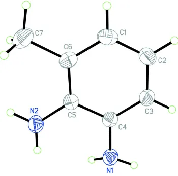
In the molecule of (I), (Fig.1), the bond lengths (Allen et al., 1987) and angles are within normal ranges. In the crystal,
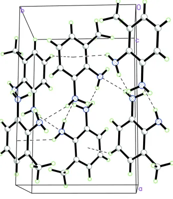
there are two C—H···Cg1 and N—H···Cg1 interactions (Cg1 is the centroid of C1-C6 ring). The molecules are linked
each other by the two intermolecular N—H···N hydrogen bonds to form a three-dimensional network, which seem to be
very effective in the stabilization of the crystal structure (Fig. 2.).
S2. Experimental
The title compound, (I) was prepared by the literature method (Li et al., 2011). Crystals suitable for X-ray analysis were
obtained by dissolving (I) (0.5 g) in ethyl acetate (20 ml) and evaporating the solvent slowly at room temperature for
about 7 d.
S3. Refinement
H atoms were positioned geometrically, with C—H = 0.93 Å for aromatic H, and constrained to ride on their parent
Figure 1
The molecular structure of (I), with the atom-numbering scheme. Displacement ellipsoids are drawn at the 50%
Figure 2
A packing diagram of (I). Hydrogen bonds are shown by dashed lines.
3-Methylbenzene-1,2-diamine
Crystal data
C7H10N2 Mr = 122.17
Monoclinic, P21/c Hall symbol: -P 2ybc a = 11.836 (2) Å b = 7.7160 (15) Å c = 7.7430 (15) Å β = 90.72 (3)° V = 707.1 (2) Å3 Z = 4
F(000) = 264 Dx = 1.148 Mg m−3
Mo Kα radiation, λ = 0.71073 Å Cell parameters from 25 reflections θ = 9–13°
Enraf–Nonius CAD-4 diffractometer
Radiation source: fine-focus sealed tube Graphite monochromator
ω/2θ scans
Absorption correction: ψ scan (North et al., 1968)
Tmin = 0.979, Tmax = 0.993 2693 measured reflections
1300 independent reflections 962 reflections with I > 2σ(I) Rint = 0.060
θmax = 25.4°, θmin = 1.7° h = −14→14
k = −9→9 l = 0→9
3 standard reflections every 200 reflections intensity decay: 1%
Refinement
Refinement on F2 Least-squares matrix: full R[F2 > 2σ(F2)] = 0.045 wR(F2) = 0.124 S = 1.00 1300 reflections 110 parameters 0 restraints
Primary atom site location: structure-invariant direct methods
Secondary atom site location: difference Fourier map
Hydrogen site location: inferred from neighbouring sites
H atoms treated by a mixture of independent and constrained refinement
w = 1/[σ2(F
o2) + (0.073P)2] where P = (Fo2 + 2Fc2)/3 (Δ/σ)max < 0.001
Δρmax = 0.13 e Å−3 Δρmin = −0.12 e Å−3
Special details
Geometry. All e.s.d.'s (except the e.s.d. in the dihedral angle between two l.s. planes) are estimated using the full covariance matrix. The cell e.s.d.'s are taken into account individually in the estimation of e.s.d.'s in distances, angles and torsion angles; correlations between e.s.d.'s in cell parameters are only used when they are defined by crystal symmetry. An approximate (isotropic) treatment of cell e.s.d.'s is used for estimating e.s.d.'s involving l.s. planes.
Refinement. Refinement of F2 against ALL reflections. The weighted R-factor wR and goodness of fit S are based on F2, conventional R-factors R are based on F, with F set to zero for negative F2. The threshold expression of F2 > σ(F2) is used only for calculating R-factors(gt) etc. and is not relevant to the choice of reflections for refinement. R-factors based on F2 are statistically about twice as large as those based on F, and R- factors based on ALL data will be even larger.
Fractional atomic coordinates and isotropic or equivalent isotropic displacement parameters (Å2)
x y z Uiso*/Ueq
C1 0.15863 (14) 0.55588 (19) 0.1128 (2) 0.0551 (5)
H1 0.0829 0.5612 0.0799 0.066*
C2 0.23534 (15) 0.6592 (2) 0.0300 (2) 0.0578 (5)
H2 0.2116 0.7340 −0.0573 0.069*
C3 0.34751 (13) 0.65096 (18) 0.07727 (19) 0.0493 (4)
H3 0.3996 0.7206 0.0211 0.059*
C4 0.38402 (12) 0.54018 (16) 0.20768 (17) 0.0408 (4)
C5 0.30472 (12) 0.43631 (17) 0.29360 (17) 0.0399 (4)
C6 0.19124 (12) 0.44367 (18) 0.2446 (2) 0.0478 (4)
C7 0.10562 (18) 0.3339 (3) 0.3346 (3) 0.0760 (6)
N1 0.49730 (11) 0.5349 (2) 0.26271 (18) 0.0508 (4)
H2B 0.2929 (18) 0.257 (2) 0.474 (3) 0.074 (6)*
H7A 0.031 (2) 0.350 (3) 0.279 (3) 0.097 (7)*
H7B 0.127 (2) 0.211 (3) 0.323 (3) 0.111 (8)*
H7C 0.1046 (18) 0.375 (3) 0.458 (3) 0.091 (7)*
Atomic displacement parameters (Å2)
U11 U22 U33 U12 U13 U23
C1 0.0485 (9) 0.0534 (9) 0.0630 (10) 0.0031 (7) −0.0144 (8) −0.0054 (8)
C2 0.0675 (11) 0.0475 (9) 0.0578 (10) −0.0018 (8) −0.0169 (8) 0.0077 (8)
C3 0.0583 (10) 0.0435 (8) 0.0461 (9) −0.0086 (7) −0.0023 (7) 0.0014 (7)
C4 0.0458 (8) 0.0359 (7) 0.0408 (8) −0.0009 (6) 0.0020 (6) −0.0065 (6)
C5 0.0445 (8) 0.0354 (7) 0.0398 (7) 0.0032 (6) 0.0025 (6) −0.0045 (6)
C6 0.0424 (8) 0.0444 (8) 0.0566 (9) −0.0021 (6) 0.0008 (7) −0.0062 (7)
C7 0.0493 (11) 0.0862 (16) 0.0926 (17) −0.0115 (10) 0.0066 (11) 0.0150 (13)
N1 0.0437 (8) 0.0531 (9) 0.0558 (8) −0.0016 (6) 0.0033 (6) 0.0063 (7)
N2 0.0482 (8) 0.0534 (8) 0.0557 (8) 0.0009 (7) 0.0021 (7) 0.0128 (7)
Geometric parameters (Å, º)
C1—C2 1.373 (2) C5—N2 1.4092 (19)
C1—C6 1.389 (2) C6—C7 1.499 (2)
C1—H1 0.9300 C7—H7A 0.98 (2)
C2—C3 1.374 (2) C7—H7B 0.99 (2)
C2—H2 0.9300 C7—H7C 1.01 (2)
C3—C4 1.388 (2) N1—H1A 0.91 (2)
C3—H3 0.9300 N1—H1B 0.89 (2)
C4—N1 1.4023 (19) N2—H2A 0.84 (2)
C4—C5 1.408 (2) N2—H2B 0.90 (2)
C5—C6 1.392 (2)
C2—C1—C6 121.70 (15) C1—C6—C5 118.94 (14)
C2—C1—H1 119.2 C1—C6—C7 120.69 (16)
C6—C1—H1 119.2 C5—C6—C7 120.37 (15)
C1—C2—C3 119.44 (15) C6—C7—H7A 109.4 (13)
C1—C2—H2 120.3 C6—C7—H7B 108.7 (16)
C3—C2—H2 120.3 H7A—C7—H7B 108.3 (19)
C2—C3—C4 120.86 (15) C6—C7—H7C 106.2 (12)
C2—C3—H3 119.6 H7A—C7—H7C 110.6 (19)
C4—C3—H3 119.6 H7B—C7—H7C 113 (2)
C3—C4—N1 121.78 (14) C4—N1—H1A 116.5 (12)
C3—C4—C5 119.42 (14) C4—N1—H1B 115.0 (11)
N1—C4—C5 118.72 (13) H1A—N1—H1B 112.1 (17)
C6—C5—C4 119.64 (13) C5—N2—H2A 118.1 (14)
C6—C5—N2 122.45 (14) C5—N2—H2B 115.9 (12)
C4—C5—N2 117.83 (14) H2A—N2—H2B 118.8 (19)
C2—C3—C4—N1 −177.35 (13) C2—C1—C6—C7 179.18 (17)
C2—C3—C4—C5 −0.6 (2) C4—C5—C6—C1 −0.9 (2)
C3—C4—C5—C6 1.19 (19) N2—C5—C6—C1 −177.77 (13)
N1—C4—C5—C6 178.00 (12) C4—C5—C6—C7 179.99 (16)
C3—C4—C5—N2 178.19 (12) N2—C5—C6—C7 3.1 (2)
Hydrogen-bond geometry (Å, º)
Cg1 is the centroid of C1–C6 ring.
D—H···A D—H H···A D···A D—H···A
N1—H1A···N2i 0.91 (2) 2.36 (2) 3.237 (2) 163.5 (16)
N2—H2A···N1ii 0.84 (2) 2.48 (2) 3.248 (2) 152.2 (19)
C2—H2···Cg1iii 0.93 2.85 3.6713 (18) 148
N2—H2B···Cg1iv 0.90 (2) 2.776 (18) 3.5192 (17) 141.2 (16)