Crystal structure of (7-chloro-2-oxo-2
H
-chromen-4-yl)methyl
N
,
N
-dimethyl-carbamodithioate
H. D. Kavitha,aM. Vinduvahini,b
N. M. Mahabhaleshwaraiah,cO. Kotreshdand H. C. Devarajegowdae*
a
Department of Physics, Govt. Science College, Hassan 573 201, Karnataka, India, b
Department of Physics, Sri D Devaraja Urs Govt. First Grade College, Hunsur-571105, Mysore District, Karnataka, India,cDepartment of Chemistry, Karnatak University’s Karnatak Science College, Dharwad, Karnataka 580001, India, dDepartment of Chemistry, Karnatak Science College, Karnatak University, Dharwad, Karnataka 580001, India, andeDepartment of Physics, Yuvaraja’s College (Constituent College), University of Mysore, Mysore 570 005, Karnataka, India. *Correspondence e-mail: hcdevarajegowda@ycm.uni-mysore.ac.in
Received 5 March 2015; accepted 19 March 2015
Edited by R. F. Baggio, Comisio´n Nacional de Energı´a Ato´mica, Argentina
In the title compound, C13H12Cl N O2S2, the 2H-chromene
ring system is almost planar, with a maximum deviation of 0.005 (2) A˚ . The packing features C—H S hydrogen bonds and – interactions between fused benzene rings of chromene [shortest centroid–centroid distances = 3.6553 (13) and 3.5551 (13) A˚ ].
Keywords:crystal structure; 2H-chromene; C—H S hydrogen bonds;– interactions.
CCDC reference:1055112
1. Related literature
For biological applications of coumarins and dithio-carbamates, see: Boas et al. (2004); D’hooghe & De Kimpe (2006); Ferna´ndezet al.(1995); Raoet al.(1981); Trkovniket al. (1983). For a related structure and the synthesis, see: Mahabaleshwaraiahet al.(2012).
2. Experimental
2.1. Crystal data
C13H12ClNO2S2 Mr= 313.81
Monoclinic,P21=c a= 9.7244 (4) A˚ b= 7.1157 (3) A˚ c= 20.0896 (9) A˚
= 94.404 (3)
V= 1386.01 (10) A˚3 Z= 4
MoKradiation
= 0.57 mm1 T= 296 K
0.240.200.12 mm
2.2. Data collection
Bruker SMART CCD area-detector diffractometer
Absorption correction: scan (SADABS; Sheldrick, 2007) Tmin= 0.770,Tmax= 1.000
16144 measured reflections 4752 independent reflections 2805 reflections withI> 2(I) Rint= 0.029
2.3. Refinement
R[F2> 2(F2)] = 0.053 wR(F2) = 0.147 S= 1.03 4752 reflections
174 parameters
H-atom parameters constrained max= 0.32 e A˚
3 min=0.35 e A˚ 3
Table 1
Hydrogen-bond geometry (A˚ ,).
D—H A D—H H A D A D—H A
C16—H16A S3i 0.97 2.84 3.707 (2) 150
Symmetry code: (i)x;yþ1 2;zþ
1 2.
Data collection: SMART (Bruker, 2001); cell refinement: SAINT (Bruker, 2001); data reduction:SAINT; program(s) used to solve structure:SHELXS97(Sheldrick, 2008); program(s) used to refine structure: SHELXL2014 (Sheldrick, 2015); molecular graphics: ORTEP-3 for Windows(Farrugia, 2012); software used to prepare material for publication:SHELXL2014.
Acknowledgements
The authors acknowledge the Universities Sophisticated Instrumental Centre, Karnatak University, Dharwad for the data collection and the UGC, India, for financial assistance.
data reports
Supporting information for this paper is available from the IUCr electronic archives (Reference: BG2550).
References
Boas, U., Gertz, H., Christensen, J. B. & Heegaard, P. M. H. (2004). Tetrahedron Lett.45, 269–272.
Bruker (2001).SMARTandSAINT.Bruker AXS Inc., Madison, Wisconsin, USA.
D’hooghe, M. & De Kimpe, N. (2006).Tetrahedron,62, 513–535.
Farrugia, L. J. (2012).J. Appl. Cryst.45, 849–854.
Ferna´ndez, J. M. G., Mellet, C. O., Blanco, J. L. J., Mota, J. F., Gadelle, A., Coste-Sarguet, A. & Defaye, J. (1995).Carbohydr. Res.268, 57–71. Mahabaleshwaraiah, N. M., Ravi, H. R., Vinduvahini, M., Sreepad, H. R. &
Kotresh, O. (2012).Acta Cryst.E68, o3001.
Rao, A. K., Raju, M. S. & Raju, K. M. (1981).J. Indian Chem. Soc.58, 1021– 1023.
Sheldrick, G. M. (2007).SADABS. Bruker AXS Inc., Madison, Wisconsin, USA.
Sheldrick, G. M. (2008).Acta Cryst.A64, 112–122. Sheldrick, G. M. (2015).Acta Cryst.C71, 3–8.
supporting information
sup-1
Acta Cryst. (2015). E71, o263–o264
supporting information
Acta Cryst. (2015). E71, o263–o264 [doi:10.1107/S2056989015005678]
Crystal structure of (7-chloro-2-oxo-2
H
-chromen-4-yl)methyl
N
,
N
-dimethyl-carbamodithioate
H. D. Kavitha, M. Vinduvahini, N. M. Mahabhaleshwaraiah, O. Kotresh and H. C.
Devarajegowda
S1. Comment
Synthetic coumarins are widely used as aroma chemicals because of their odour strength, tenacity, stability to alkali and
relatively cheap price; applications include their use as a sweetener and fixative (in perfume); fragrance enhancers (for
natural essential oils); blenders (in soaps and detergents); aroma enhancers (in tobacco); and for imparting pleasant
odours to industrial products. The coumarins have been the subject of extensive studies because of their interesting
biological activities and have, in fact, been used as therapeutic agents for the treatment of various diseases. Coumarins
show quite diverse biological activities, including anticoagulant, anti-allergic, anthelmintic, diuretic, insecticidal and
antibiotic properties (Trkovnik et al., 1983); Rao et al., 1981). The great electrophilicity of the nitrogen atom as
compared to that of sulfur makes the latter more acidic and an active centre in the nucleophilic attack. The fact is that the
sulfur anion formed is more stabilized by negative charge distribution. Furthermore, functionalized carbamates are an
important class of compounds and their medicinal and biological properties warrant study (D'hooghe et al., 2006).
Organic dithiocarbamates are valuable synthetic intermediates (Boas et al., 2004), which are ubiquitously found in a
variety of biologically active compounds. Functionalization of the carbamate moiety offers an attractive method for the
generation of derivatives, which may constitute interesting medicinal and biological properties (Fernández et al., 1995).
The compound herein reported, (7-chloro-2-oxo-2H-chromen-4-yl)methyl dimethylcarbamodithioate (Fig. 1) presents a
planar 2H-chromene ring system [maximum deviation: 0.005 (2) Å for atom C13]. The crystal structure shows
intermolecular C—H···S bonds·(C16-H16A···S3, H···S= 2.84 Å; C-H···S: 150°) and π–π interactions between fused
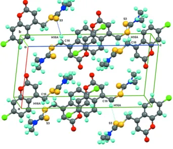
benzene rings of chromene [shortest centroid–centroid distances = 3.6553 (13) Å and 3.5551 (13) Å]. A packing view is
shown in Fig. 2.
S2. Experimental
All the chemicals used were of analytical reagent grade and were used directly without further purification. The title
compound was synthesized according to the reported method (Mahabaleshwaraiah et al., 2012). The compound is
recrystallized by ethanol- chloroform mixture. Colourless needles of the title compound were grown from a mixed
solution of Ethanol/Chloroform (V/V = 2/1) by slow evaporation at room temperature. Yield =72%; m.p.:405–407 K.
S3. Refinement
All H atoms were positioned geometrically, with C—H = 0.93 Å for aromatic H, C—H = 0.97 Å for methylene H and C
—H = 0.96 Å for methyl H, and refined using a riding model with Uiso(H) = 1.5Ueq(C) for methyl H and Uiso(H) =
Figure 1
The molecular structure of the title compound. Displacement ellipsoids are drawn at the 50% probability level.
Figure 2
[image:4.610.135.477.349.644.2]supporting information
sup-3
Acta Cryst. (2015). E71, o263–o264
(7-Chloro-2-oxo-2H-chromen-4-yl)methyl N,N-dimethylcarbamodithioate
Crystal data
C13H12ClNO2S2
Mr = 313.81 Monoclinic, P21/c
a = 9.7244 (4) Å b = 7.1157 (3) Å c = 20.0896 (9) Å β = 94.404 (3)° V = 1386.01 (10) Å3
Z = 4 F(000) = 648
Dx = 1.504 Mg m−3
Melting point: 407 K
Mo Kα radiation, λ = 0.71073 Å Cell parameters from 2446 reflections θ = 2.0–25.0°
µ = 0.57 mm−1
T = 296 K Plate, colourless 0.24 × 0.20 × 0.12 mm
Data collection
Bruker SMART CCD area-detector diffractometer
Radiation source: fine-focus sealed tube Graphite monochromator
ω and φ scans
Absorption correction: ψ scan (SADABS; Sheldrick, 2007) Tmin = 0.770, Tmax = 1.000
16144 measured reflections 4752 independent reflections 2805 reflections with I > 2σ(I) Rint = 0.029
θmax = 32.2°, θmin = 2.0°
h = −14→14 k = −10→10 l = −27→30
Refinement
Refinement on F2
Least-squares matrix: full R[F2 > 2σ(F2)] = 0.053
wR(F2) = 0.147
S = 1.03 4752 reflections 174 parameters 0 restraints
Primary atom site location: structure-invariant direct methods
Secondary atom site location: difference Fourier map
Hydrogen site location: inferred from neighbouring sites
H-atom parameters constrained w = 1/[σ2(F
o2) + (0.0636P)2 + 0.4178P]
where P = (Fo2 + 2Fc2)/3
(Δ/σ)max = 0.043
Δρmax = 0.32 e Å−3
Δρmin = −0.35 e Å−3
Special details
Experimental. IR (KBr, cm-1): 1722 (C=O), 1381 (C=S), 894(C—N). GCMS: m/e: 313; 1H NMR (400 MHz, CDCl 3, \?,
p.p.m): 3.38 (s, 3H, N—CH3), 3.47 (s, 3H, N—CH3), 4.80 (s, 2H, C4—CH2), 6.56 (s, 1H, C3—H), 7.45–7.92 (m, 3H, Ar
—H). Mol. Formula: C13H12Cl N O2S22; Elemental analysis: C, 49.75; H, 3.85; N, 4.46 (calculated); C, 49.67; H, 3.76; N,
4.39 (found).
Geometry. All e.s.d.'s (except the e.s.d. in the dihedral angle between two l.s. planes) are estimated using the full covariance matrix. The cell e.s.d.'s are taken into account individually in the estimation of e.s.d.'s in distances, angles and torsion angles; correlations between e.s.d.'s in cell parameters are only used when they are defined by crystal symmetry. An approximate (isotropic) treatment of cell e.s.d.'s is used for estimating e.s.d.'s involving l.s. planes.
Refinement. Refinement of F2 against ALL reflections. The weighted R-factor wR and goodness of fit S are based on F2,
conventional R-factors R are based on F, with F set to zero for negative F2. The threshold expression of F2 > 2σ(F2) is
used only for calculating R-factors(gt) etc. and is not relevant to the choice of reflections for refinement. R-factors based on F2 are statistically about twice as large as those based on F, and R- factors based on ALL data will be even larger.
Fractional atomic coordinates and isotropic or equivalent isotropic displacement parameters (Å2)
x y z Uiso*/Ueq
S2 0.19845 (6) 0.93877 (9) 0.24696 (3) 0.05245 (18) S3 0.25794 (7) 0.52236 (9) 0.23740 (3) 0.05581 (19) O4 0.27371 (14) 0.7395 (2) 0.50559 (7) 0.0489 (4) O5 0.48199 (17) 0.7569 (3) 0.47037 (9) 0.0673 (5) N6 0.37617 (18) 0.7922 (3) 0.17126 (9) 0.0522 (5) C7 −0.0832 (2) 0.7267 (3) 0.53965 (11) 0.0416 (5) C8 −0.1507 (2) 0.7599 (3) 0.47806 (12) 0.0442 (5) H8 −0.2465 0.7639 0.4730 0.053* C9 −0.0740 (2) 0.7870 (3) 0.42398 (11) 0.0413 (5) H9 −0.1192 0.8099 0.3823 0.050* C10 0.07040 (19) 0.7808 (3) 0.43027 (10) 0.0349 (4) C11 0.1331 (2) 0.7472 (3) 0.49375 (10) 0.0369 (4) C12 0.0577 (2) 0.7200 (3) 0.54875 (11) 0.0442 (5) H12 0.1017 0.6977 0.5908 0.053* C13 0.15923 (19) 0.8083 (3) 0.37605 (10) 0.0368 (4) C14 0.2966 (2) 0.8017 (3) 0.38965 (11) 0.0419 (5) H14 0.3529 0.8210 0.3549 0.050* C15 0.3606 (2) 0.7662 (3) 0.45538 (11) 0.0455 (5) C16 0.0936 (2) 0.8373 (4) 0.30683 (11) 0.0501 (6) H16A 0.0133 0.9169 0.3099 0.060* H16B 0.0609 0.7164 0.2898 0.060* C17 0.2867 (2) 0.7429 (3) 0.21486 (10) 0.0414 (5) C18 0.4622 (3) 0.6497 (5) 0.14199 (14) 0.0774 (9) H18A 0.5282 0.6018 0.1758 0.116* H18B 0.5098 0.7053 0.1068 0.116* H18C 0.4050 0.5488 0.1243 0.116* C19 0.4015 (3) 0.9852 (5) 0.15168 (14) 0.0722 (8) H19A 0.3167 1.0548 0.1500 0.108* H19B 0.4372 0.9864 0.1084 0.108* H19C 0.4673 1.0418 0.1837 0.108*
Atomic displacement parameters (Å2)
U11 U22 U33 U12 U13 U23
supporting information
sup-5
Acta Cryst. (2015). E71, o263–o264
C15 0.0340 (11) 0.0521 (13) 0.0503 (12) 0.0019 (9) 0.0017 (9) −0.0057 (10) C16 0.0333 (11) 0.0751 (17) 0.0423 (12) 0.0060 (10) 0.0052 (9) 0.0067 (11) C17 0.0291 (10) 0.0612 (14) 0.0328 (10) −0.0022 (9) −0.0047 (7) 0.0045 (9) C18 0.0583 (17) 0.120 (3) 0.0564 (16) 0.0281 (17) 0.0192 (13) 0.0130 (16) C19 0.0536 (16) 0.098 (2) 0.0651 (17) −0.0159 (15) 0.0067 (13) 0.0305 (16)
Geometric parameters (Å, º)
Cl1—C7 1.737 (2) C10—C11 1.392 (3) S2—C17 1.783 (2) C10—C13 1.454 (3) S2—C16 1.788 (2) C11—C12 1.386 (3) S3—C17 1.663 (2) C12—H12 0.9300 O4—C11 1.371 (2) C13—C14 1.343 (3) O4—C15 1.378 (3) C13—C16 1.499 (3) O5—C15 1.198 (3) C14—C15 1.438 (3) N6—C17 1.329 (3) C14—H14 0.9300 N6—C19 1.455 (4) C16—H16A 0.9700 N6—C18 1.466 (3) C16—H16B 0.9700 C7—C12 1.369 (3) C18—H18A 0.9600 C7—C8 1.376 (3) C18—H18B 0.9600 C8—C9 1.378 (3) C18—H18C 0.9600 C8—H8 0.9300 C19—H19A 0.9600 C9—C10 1.401 (3) C19—H19B 0.9600 C9—H9 0.9300 C19—H19C 0.9600
C14—C13—C10 118.78 (18) N6—C19—H19C 109.5 C14—C13—C16 122.65 (18) H19A—C19—H19C 109.5 C10—C13—C16 118.55 (17) H19B—C19—H19C 109.5 C13—C14—C15 123.11 (19)
C12—C7—C8—C9 0.0 (3) C11—C10—C13—C16 177.7 (2) Cl1—C7—C8—C9 −179.30 (16) C9—C10—C13—C16 −2.9 (3) C7—C8—C9—C10 0.3 (3) C10—C13—C14—C15 0.9 (3) C8—C9—C10—C11 −0.4 (3) C16—C13—C14—C15 −177.3 (2) C8—C9—C10—C13 −179.84 (19) C11—O4—C15—O5 −180.0 (2) C15—O4—C11—C12 −179.28 (19) C11—O4—C15—C14 −0.3 (3) C15—O4—C11—C10 0.5 (3) C13—C14—C15—O5 179.2 (2) C9—C10—C11—O4 −179.57 (18) C13—C14—C15—O4 −0.4 (3) C13—C10—C11—O4 −0.1 (3) C14—C13—C16—S2 −20.1 (3) C9—C10—C11—C12 0.2 (3) C10—C13—C16—S2 161.75 (16) C13—C10—C11—C12 179.74 (18) C17—S2—C16—C13 85.7 (2) C8—C7—C12—C11 −0.1 (3) C19—N6—C17—S3 −179.84 (18) Cl1—C7—C12—C11 179.17 (15) C18—N6—C17—S3 −3.0 (3) O4—C11—C12—C7 179.83 (18) C19—N6—C17—S2 0.5 (3) C10—C11—C12—C7 0.0 (3) C18—N6—C17—S2 177.35 (18) C11—C10—C13—C14 −0.6 (3) C16—S2—C17—N6 −176.86 (15) C9—C10—C13—C14 178.8 (2) C16—S2—C17—S3 3.46 (16)
Hydrogen-bond geometry (Å, º)
D—H···A D—H H···A D···A D—H···A
C16—H16A···S3i 0.97 2.84 3.707 (2) 150