Received 10 June 2016 Accepted 14 July 2016
Edited by M. Weil, Vienna University of Technology, Austria
Keywords:crystal structure; powder diffraction; density functional theory; citrate; potassium salt.
CCDC references:1493594; 1481346; 1493593
Supporting information:this article has supporting information at journals.iucr.org/e
Alagappa Rammohanaand James A. Kadukb*
a
Atlantic International University, Honolulu, HI, USA, andbIllinois Institute of Technology, Chicago, IL, USA. *Correspondence e-mail: [email protected]
The crystal structure of anhydrous tripotassium citrate, [K3(C6H5O7)]n, has been
solved and refined using laboratory X-ray powder diffraction data, and optimized using density functional techniques. The three unique potassium cations are 6-, 8-, and 6-coordinate (all irregular). The [KOn] coordination
polyhedra share edges and corners to form a three-dimensional framework, with channels running parallel to the c axis. The only hydrogen bond is an intramolecular one involving the hydroxy group and the central carboxylate group, with graph-set motifS(5).
1. Chemical context
In the course of a systematic study of the crystal structures of group 1 (alkali metal) citrate salts to understand the anion’s conformational flexibility, ionization, coordination tendencies, and hydrogen bonding, we have determined several new crystal structures. Most of the new structures were solved using powder X-ray diffraction data (laboratory and/or synchrotron), but single crystals were used where available. The general trends and conclusions about the 16 new compounds and 12 previously characterized structures are being reported separately (Rammohan & Kaduk, 2016a). Five of the new structures, viz. NaKHC6H5O7, NaK2C6H5O7,
Na3C6H5O7, NaH2C6H5O7, and Na2HC6H5O7, have been
published recently (Rammohan & Kaduk, 2016b,c,d,e; Rammohan et al., 2016), and two additional structures, viz. KH2C6H5O7 and KH2C6H5O7(H2O)2, have been
2. Structural commentary
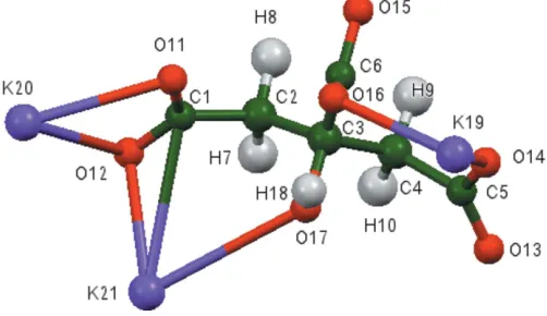
The asymmetric unit of the title compound is shown in Fig. 1. The r.m.s. deviation of the non-hydrogen atoms between the Rietveld-refined and the DFT-optimized structures is 0.117 A˚ (Fig. 2). The maximum deviation is 0.260 A˚ , at O14. The good agreement between the two structures is strong evidence that the experimental structure is correct (van de Streek & Neumann, 2014). This discussion uses the DFT-optimized structure. Most of the bond lengths, bond angles, and torsion angles fall within the normal ranges indicated by a Mercury MogulGeometry Check (Macraeet al., 2008). Only the C4— C5 bond length [refined = 1.511 (5), optimized = 1.536,Mogul
average = 1.498 (12) A˚ ,Z-score = 3.1], and the C3—C2—C1 [refined = 115 (2), optimized = 115.0, Mogul average = 103 (2)] and O17—C3—C2 angles [refined = 107 (2),
opti-mized = 109.6, Mogul average = 106 (2)] are flagged as
unusual. The citrate anion occurs in the trans,trans -confor-mation, which is one of the two low-energy conformations of an isolated citrate. The central carboxylate group and the hydroxy group occur in the normal planar arrangement. Both
terminal carboxylate groups O11/O12 and O13/O14 chelate to a single potassium cation (K20 for each). The terminal carboxylate oxygen atom O12 and the hydroxy O17 atom chelate to K21, and the terminal carboxylate oxygen atoms O13 and O17 chelate to K19. The terminal/central pairs O11/ O16, O14/O16, O11/O15, and O14/O15 chelate to K21, K19, K19, and K21, respectively. The three potassium cations K19, K20, and K21 are 6-, 8-, and 6-coordinate, respectively (all irregular, using a K—O cut-off distance of 3.24 A˚ ). Their bond-valence sums are 1.12, 1.03, and 1.12 valence units. The metal-oxygen bonding is ionic, based on the cation charges and the Mulliken overlap populations.
Although the lattice parameters of anhydrous tripotassium citrate are in general similar to those of the monohydrate (Carrellet al., 1987; CSD code ZZZHVI01), consistent with the difference in water content, the powder patterns differ considerably. Visual examination of the structures shows that the arrangements of the citrate anions are very different. A mechanism for the transformation of one phase into the other is not obvious.
The Bravais–Friedel–Donnay–Harker (Bravais, 1866; Friedel, 1907; Donnay & Harker, 1937) morphology suggests that we might expect blocky morphology for anhydrous tripotassium citrate, with {011} as the principal faces. A second-order spherical harmonic texture model was included in the refinement. The texture index was only 1.001, indicating that preferred orientation was not significant for this rotated flat plate specimen.
1160
Rammohan and Kaduk [K3(C6H5O7)] Acta Cryst.(2016). E72, 1159–1162research communications
Figure 1
[image:2.610.45.296.74.219.2]The asymmetric unit of the title compound, showing the atom numbering. Atoms are represented by 50% probability spheroids.
Figure 2
[image:2.610.313.567.85.124.2]Comparison of the refined and optimized structures of anhydrous tripotassium citrate. The refined structure is in red, and the DFT-optimized structure is in blue.
Table 1
Hydrogen-bond geometry (A˚ ,).
D—H A D—H H A D A D—H A
[image:2.610.310.564.508.699.2]O17—H18 O16 0.983 1.814 2.552 129.1
Figure 3
[image:2.610.46.294.547.709.2]3. Supramolecular features
The [KOn] coordination polyhedra share edges and corners to
form a three-dimensional framework (Fig. 3), with channels running down the c axis. The only hydrogen bond is an intramolecular one (Table 1) involving the hydroxy group and the central carboxylate group, with graph-set motifS(5). The Mulliken overlap population in the hydrogen-acceptor bond is 0.076 e. By the correlation in Rammohan & Kaduk (2016a), this hydrogen bond accounts for 15.1 kcal per mole of crystal energy.
4. Database survey
Details of the comprehensive literature search for citrate structures are presented in Rammohan & Kaduk (2016a). A reduced-cell search of the cell of anhydrous tripotassium citrate in the Cambridge Structural Database (Groom et al., 2016) (increasing the default tolerance from 1.5 to 2.0%) yielded 208 hits, but limiting the chemistry to C, H, K, and O only resulted in no hits. The powder pattern is now contained in the the Powder Diffraction File (ICDD, 2015) as entry 00-064-1370.
5. Synthesis and crystallization
Potassium citrate monohydrate was synthesized by dissolving 2.0796 g (10.0 mmole) H3C6H5O7(H2O) in 20 ml deionized
water. 2.0731g K2CO3(15.0 mmole, Sigma-Aldrich) was added
to the citric acid solution slowly with stirring. The resulting clear colourless solution was evaporated to dryness in a 333 K oven. The powder pattern matched PDF entry 02-064-1651, confirming the structure as potassium citrate monohydrate (Carrell et al., 1987). The monohydrate was heated at 15 K min 1 to 498 K, and held there for two minutes (the white solid started to discolour). The white solid was removed from the oven, and immediately placed in a sealed glass jar to cool.
6. Refinement details
Crystal data, data collection and structure refinement details are summarized in Table 2. The white solid was ground and blended with NIST SRM 640b Si internal standard in a mortar and pestle. The specimen was protected from the atmosphere by an 8 micron Kapton film attached to the sample holder with petroleum jelly. (The sample hydrates slowly on contact with ambient atmosphere.)
The pattern (Fig. 4) was indexed on a primitive ortho-rhombic unit cell using ITO (Visser, 1969). Manual examina-tion of the systematic absences suggested space groupPna21.
Pseudo-Voigt profile coefficients were as parameterized in Thompsonet al.(1987) with profile coefficients for Simpson’s rule integration of the Pseudo-Voigt function according to Howard (1982). The asymmetry correction of Finger et al.
(1994) was applied and microstrain broadening by Stephens (1999). The structure was solved withFOX(Favre-Nicolin &
Temperature (K) 300 300
a,b,c(A˚ ) 7.7062 (2), 12.4693 (3), 10.4241 (2) 5.43105, 5.43105, 5.43105 ,,(
) 90, 90, 90 90, 90, 90
V(A˚3) 1001.66 (3) 160.20
Z 4 8
Radiation type K1,K2,= 1.540629, 1.544451 A˚ K1,K2,= 1.540629, 1.544451 A˚
Specimen shape, size (mm) Flat sheet, 2424 Flat sheet, 2424
Data collection
Diffractometer Bruker D2 Phaser Bruker D2 Phaser
Specimen mounting Normal sample holder with Kapton film
Normal sample holder with Kapton film
Data collection mode Reflection Reflection
Scan method Step Step
2values () 2
min= 4.908, 2max= 69.916,
2step= 0.020
2min= 4.908, 2max= 69.916,
2step= 0.020
Refinement
Rfactors and goodness of fit Rp= 0.038,Rwp= 0.049,
Rexp= 0.034,R(F2) = 0.059,
2= 2.103
Rp= 0.038,Rwp= 0.049,
Rexp= 0.034,R(F2) = 0.059,
2= 2.103
No. of parameters 73 73
No. of restraints 29 29
The same symmetry and lattice parameters were used for the DFT calculation. Computer programs:DIFFRAC.Measurement(Bruker, 2009),FOX(Favre-Nicolin & Cˇ erny´, 2002),GSAS
[image:3.610.46.569.93.359.2]Cˇ erny´, 2002) using a citrate moiety and three potassium atoms as fragments. The structure was refined by the Rietveld method usingGSAS/EXPGUI(Larson & Von Dreele, 2004; Toby, 2001). All C—C and C—O bond lengths were restrained, as were all bond angles. The hydrogen atoms were included at fixed positions, which were recalculated during the course of the refinement using Materials Studio (Dassault Systemes, 2014). The Uisovalues of the atoms in the central
and outer portions of the citrate anion were constrained to be equal, and the Uiso values of the hydrogen atoms were
constrained to be 1.3those of the atoms to which they are attached.
The ADDSYM module of PLATON (Spek, 2009) suggested the presence of an additional centre of symmetry, and that the space group wasPnam. Refinement in this space group yielded poorer residuals, so we believe thatPna21is the
correct space group.
7. DFT calculations
After the Rietveld refinement, a density functional geometry optimization (fixed experimental unit cell) was carried out usingCRYSTAL09(Dovesiet al., 2005). The basis sets for the C, H, and O atoms were those of Gatti et al.(1994), and the basis set for K was that of Dovesiet al.(1991). The calculation used 8 k-points and the B3LYP functional, and took about
66 h on a 2.4 GHz PC. The Uiso values from the Rietveld
refinement were assigned to the optimized fractional coord-inates.
References
Bravais, A. (1866). In Etudes Cristallographiques. Paris: Gauthier Villars.
Bruker (2009).DIFFRAC.Measurement. Bruker AXS Inc., Madison, Wisconsin, USA.
Carrell, H. L., Glusker, J. P., Piercy, E. A., Stallings, W. C., Zacharias, D. E., Davis, R. L., Astbury, C. & Kennard, C. H. L. (1987).J. Am. Chem. Soc.109, 8067–8071.
Crystal Impact (2015). DIAMOND. Crystal Impact GbR, Bonn, Germany. http://www.crystalimpact.com/diamond.
Dassault Systemes (2014). Materials Studio. BIOVIA, San Diego, California, USA.
Donnay, J. D. H. & Harker, D. (1937).Am. Mineral.22, 446–467. Dovesi, R., Orlando, R., Civalleri, B., Roetti, C., Saunders, V. R. &
Zicovich-Wilson, C. M. (2005).Z. Kristallogr.220, 571–573. Dovesi, R., Roetti, C., Freyria-Fava, C., Prencipe, M. & Saunders,
V. R. (1991).Chem. Phys.156, 11–19.
Favre-Nicolin, V. & Cˇ erny´, R. (2002).J. Appl. Cryst.35, 734–743. Finger, L. W., Cox, D. E. & Jephcoat, A. P. (1994).J. Appl. Cryst.27,
892–900.
Friedel, G. (1907).Bull. Soc. Fr. Mineral.30, 326–455.
Gatti, C., Saunders, V. R. & Roetti, C. (1994).J. Chem. Phys.101, 10686–10696.
Groom, C. R., Bruno, I. J., Lightfoot, M. P. & Ward, S. C. (2016).Acta Cryst.B72, 171–179.
Howard, C. J. (1982).J. Appl. Cryst.15, 615–620.
ICDD (2015).PDF-4+ 2015 and PDF-4 Organics 2016 (Databases), edited by S. Kabekkodu. International Centre for Diffraction Data, Newtown Square, PA, USA.
Kaduk, J. A. & Stern, C. (2016a). Private communication (No. 1446457–1446458). CCDC, Cambridge, England.
Kaduk, J. A. & Stern, C. (2016b). Private communication (No. 1446460–1446461). CCDC, Cambridge, England.
Larson, A. C. & Von Dreele, R. B. (2004). General Structure Analysis System (GSAS). Report LAUR, 86–784 Los Alamos National Laboratory, New Mexico, USA.
Macrae, C. F., Bruno, I. J., Chisholm, J. A., Edgington, P. R., McCabe, P., Pidcock, E., Rodriguez-Monge, L., Taylor, R., van de Streek, J. & Wood, P. A. (2008).J. Appl. Cryst.41, 466–470.
Rammohan, A. & Kaduk, J. A. (2016a).Acta Cryst.B72. Submitted. Rammohan, A. & Kaduk, J. A. (2016b).Acta Cryst.E72, 170–173. Rammohan, A. & Kaduk, J. A. (2016c).Acta Cryst.E72, 403–406. Rammohan, A. & Kaduk, J. A. (2016d).Acta Cryst.E72, 793–796. Rammohan, A. & Kaduk, J. A. (2016e).Acta Cryst.E72, 854–857. Rammohan, A. Sarjeant, A. A. & Kaduk, J. A. (2016).Acta Cryst.
E72, 943–946.
Spek, A. L. (2009).Acta Cryst.D65, 148–155. Stephens, P. W. (1999).J. Appl. Cryst.32, 281–289.
Streek, J. van de & Neumann, M. A. (2014).Acta Cryst.B70, 1020– 1032.
Thompson, P., Cox, D. E. & Hastings, J. B. (1987).J. Appl. Cryst.20, 79–83.
Toby, B. H. (2001).J. Appl. Cryst.34, 210–213. Visser, J. W. (1969).J. Appl. Cryst.2, 89–95. Westrip, S. P. (2010).J. Appl. Cryst.43, 920–925.
1162
Rammohan and Kaduk [K3(C6H5O7)] Acta Cryst.(2016). E72, 1159–1162 [image:4.610.44.298.67.272.2]research communications
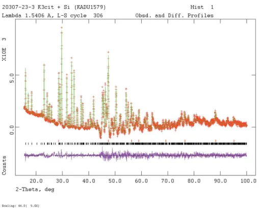
Figure 4
Rietveld plot for the refinement of K3C6H5O7. The red crosses represent the observed data points, and the green line is the calculated pattern. The magenta curve is the difference pattern, plotted at the same scale as the other patterns. The vertical scale for angles > 44.0has been multiplied by
sup-1 Acta Cryst. (2016). E72, 1159-1162
Crystal structure of anhydrous tripotassium citrate from laboratory X-ray
powder diffraction data and DFT comparison
Alagappa Rammohan and James A. Kaduk
Computing details
Data collection: DIFFRAC.Measurement (Bruker, 2009) for KADU1578_phase_1. Program(s) used to solve structure:
FOX (Favre-Nicolin & Černý, 2002) for KADU1578_phase_1. Program(s) used to refine structure: GSAS (Larson & Von
Dreele, 2004) and EXPGUI (Toby, 2001) for KADU1578_phase_1. Molecular graphics: DIAMOND (Crystal Impact,
2015) for KADU1578_phase_1. Software used to prepare material for publication: publCIF (Westrip, 2010) for
KADU1578_phase_1.
(KADU1578_phase_1) Poly[(µ-2-hydroxypropane-1,2,3-tricarboxylato0tripotassium]
Crystal data
[K3(C6H5O7)]
Mr = 306.39
Orthorhombic, Pna21
a = 7.7062 (2) Å
b = 12.4693 (3) Å
c = 10.4241 (2) Å
V = 1001.66 (3) Å3
Z = 4
T = 300 K
Fractional atomic coordinates and isotropic or equivalent isotropic displacement parameters (Å2)
x y z Uiso*/Ueq
C1 0.841 (4) 0.883 (3) 0.28611 0.023 (3)*
C2 0.926 (4) 0.915 (3) 0.411 (2) 0.021 (5)*
C3 0.814 (2) 0.8966 (12) 0.532 (3) 0.021 (5)*
C4 0.937 (4) 0.910 (4) 0.647 (3) 0.021 (5)*
C5 0.857 (4) 0.872 (3) 0.771 (3) 0.023 (3)*
C6 0.6539 (18) 0.9724 (11) 0.541 (5) 0.023 (3)*
H7 1.04032 0.90124 0.41652 0.027 (7)*
H8 0.9082 0.99396 0.3928 0.027 (7)*
H9 0.97168 0.98330 0.645913 0.027 (7)*
H10 1.01382 0.84403 0.63413 0.027 (7)*
O11 0.695 (4) 0.9217 (19) 0.252 (4) 0.023 (3)*
O12 0.936 (4) 0.816 (2) 0.226 (4) 0.023 (3)*
O13 0.935 (4) 0.821 (2) 0.859 (4) 0.023 (3)*
O14 0.697 (4) 0.894 (2) 0.787 (4) 0.023 (3)*
O15 0.6828 (17) 1.0726 (11) 0.529 (6) 0.023 (3)*
O16 0.5076 (16) 0.9267 (10) 0.533 (5) 0.023 (3)*
supporting information
sup-2 Acta Cryst. (2016). E72, 1159-1162
H18 0.6543 0.8072 0.525 0.030 (4)*
K19 0.355 (3) 0.8827 (13) 0.743 (3) 0.0442 (17)*
K20 0.1336 (10) 0.7108 (4) 0.021 (3) 0.0442 (17)*
K21 0.148 (3) 0.3753 (14) 0.797 (3) 0.0442 (17)*
Geometric parameters (Å, º)
C1—C2 1.510 (5) O13—K20viii 2.90 (3)
C1—O11 1.278 (9) O14—C5 1.270 (9)
C1—O12 1.271 (9) O14—O13 2.18 (3)
C2—C1 1.510 (5) O14—K19 2.68 (4)
C2—C3 1.544 (4) O14—K20viii 2.81 (3)
C2—H7 0.90 (3) O14—K21ii 2.90 (3)
C2—H8 1.01 (4) O15—C6 1.275 (8)
C3—C2 1.544 (4) O15—K19i 3.05 (6)
C3—C4 1.544 (5) O15—K20ix 2.987 (15)
C3—C6 1.553 (4) O15—K20x 3.051 (15)
C3—O17 1.452 (8) O15—K21ii 2.88 (5)
C4—C3 1.544 (5) O16—C6 1.266 (8)
C4—C5 1.511 (5) O16—K19 2.55 (4)
C4—H9 0.95 (4) O16—K21iii 2.81 (4)
C4—H10 1.03 (5) O17—C3 1.452 (8)
C5—C4 1.511 (5) O17—H18 0.757 (11)
C5—O13 1.268 (9) O17—K19ii 3.22 (4)
C5—O14 1.270 (9) O17—K21vi 3.23 (4)
C6—C3 1.553 (4) H18—O17 0.757 (11)
C6—O15 1.275 (8) K19—O11x 2.47 (2)
C6—O16 1.266 (8) K19—O13xi 2.88 (3)
H7—C2 0.90 (3) K19—O14 2.68 (4)
H8—C2 1.01 (4) K19—O15x 3.05 (6)
H9—C4 0.82 (4) K19—O16 2.55 (4)
H10—C4 1.03 (5) K19—O17xi 3.22 (4)
O11—C1 1.278 (9) K20—O11xi 2.96 (3)
O11—K19i 2.47 (2) K20—O12xii 2.93 (3)
O11—K20ii 2.96 (3) K20—O12xi 3.18 (3)
O11—K21iii 2.75 (4) K20—O13xiii 2.66 (3)
O12—C1 1.271 (9) K20—O13xiv 2.90 (3)
O12—K20iv 2.93 (3) K20—O14xiv 2.81 (3)
O12—K20ii 3.18 (3) K20—O15xv 2.987 (15)
O12—K21v 3.37 (4) K20—O15i 3.051 (15)
O12—K21vi 2.58 (3) K21—O11xvi 2.75 (4)
O13—C5 1.268 (9) K21—O12xvii 2.58 (3)
O13—O14 2.18 (3) K21—O14xi 2.90 (3)
O13—K19iv 3.54 (4) K21—O15xi 2.88 (5)
O13—K19ii 2.88 (3) K21—O16xvi 2.81 (4)
O13—K20vii 2.66 (3) K21—O17xvii 3.23 (4)
sup-3 Acta Cryst. (2016). E72, 1159-1162
C4—C3—C6 112 (2) O13xi—K19—O16 117.0 (10)
C4—C3—O17 111 (2) O13xi—K19—O17xi 76.7 (9)
C6—C3—O17 107.1 (12) O14—K19—O15x 85.2 (9)
C3—C4—C5 112 (2) O14—K19—O16 71.5 (11)
C4—C5—O13 126 (2) O14—K19—O17xi 114.2 (10)
C4—C5—O14 116 (2) O15x—K19—O16 147.9 (10)
O13—C5—O14 118 (2) O15x—K19—O17xi 141.9 (9)
C3—C6—O15 116.9 (13) O16—K19—O17xi 69.7 (7)
C3—C6—O16 115.3 (13) O11xi—K20—O12xii 74.9 (9)
O15—C6—O16 126.2 (16) O11xi—K20—O12xi 43.7 (5)
C1—O11—K19i 122 (3) O11xi—K20—O13xiii 153.7 (8)
C1—O11—K20ii 98.9 (17) O11xi—K20—O13xiv 105.6 (8)
C1—O11—K21iii 136 (3) O11xi—K20—O14xiv 114.7 (5)
K19i—O11—K20ii 119.7 (12) O11xi—K20—O15xv 77.6 (12)
K19i—O11—K21iii 93.6 (11) O11xi—K20—O15i 113.4 (13)
K20ii—O11—K21iii 82.3 (8) O12xii—K20—O12xi 86.3 (8)
C1—O12—K20iv 162 (3) O12xii—K20—O13xiii 86.2 (5)
C1—O12—K20ii 88.7 (17) O12xii—K20—O13xiv 154.3 (8)
C1—O12—K21vi 109 (3) O12xii—K20—O14xiv 158.6 (7)
K20iv—O12—K20ii 81.0 (7) O12xii—K20—O15xv 79.2 (12)
K20iv—O12—K21vi 85.8 (9) O12xii—K20—O15i 80.0 (10)
K20ii—O12—K21vi 84.9 (10) O12xi—K20—O13xiii 154.4 (9)
C5—O13—K19ii 92 (2) O12xi—K20—O13xiv 77.9 (4)
C5—O13—K20vii 172.2 (18) O12xi—K20—O14xiv 114.1 (8)
C5—O13—K20viii 96.3 (17) O12xi—K20—O15xv 121.3 (12)
K19ii—O13—K20vii 86.2 (9) O12xi—K20—O15i 74.6 (10)
K19ii—O13—K20viii 87.4 (10) O13xiii—K20—O13xiv 99.4 (7)
K20vii—O13—K20viii 91.3 (7) O13xiii—K20—O14xiv 77.9 (8)
C5—O14—K19 157 (3) O13xiii—K20—O15xv 81.1 (10)
C5—O14—K20viii 100.5 (18) O13xiii—K20—O15i 80.1 (10)
C5—O14—K21ii 110 (3) O13xiv—K20—O14xiv 44.9 (6)
K19—O14—K20viii 87.4 (8) O13xiv—K20—O15xv 126.3 (12)
K19—O14—K21ii 86.0 (11) O13xiv—K20—O15i 76.4 (9)
K20viii—O14—K21ii 114.1 (12) O14xiv—K20—O15xv 84.2 (14)
C6—O15—K19i 105 (4) O14xiv—K20—O15i 110.8 (11)
C6—O15—K20ix 115.2 (11) O15xv—K20—O15i 152.7 (6)
C6—O15—K20x 162.1 (14) O11xvi—K21—O12xvii 84.5 (12)
C6—O15—K21ii 96 (4) O11xvi—K21—O14xi 84.9 (8)
K19i—O15—K20ix 77.8 (11) O11xvi—K21—O15xi 82.9 (9)
K19i—O15—K20x 81.7 (12) O11xvi—K21—O16xvi 71.9 (10)
K19i—O15—K21ii 154.0 (5) O11xvi—K21—O17xvii 119.9 (10)
supporting information
sup-4 Acta Cryst. (2016). E72, 1159-1162
K20ix—O15—K21ii 79.7 (11) O12xvii—K21—O15xi 87.5 (7)
K20x—O15—K21ii 82.5 (12) O12xvii—K21—O16xvi 110.7 (10)
C6—O16—K19 117 (3) O12xvii—K21—O17xvii 72.0 (9)
C6—O16—K21iii 123 (3) O14xi—K21—O15xi 74.4 (10)
K19—O16—K21iii 120.5 (5) O14xi—K21—O16xvi 81.9 (8)
C3—O17—H18 91.9 (14) O14xi—K21—O17xvii 128.0 (11)
C3—O17—K19ii 119 (2) O15xi—K21—O16xvi 146.8 (10)
C3—O17—K21vi 123 (2) O15xi—K21—O17xvii 146.4 (9)
K19ii—O17—K21vi 92.3 (3) O16xvi—K21—O17xvii 66.8 (7)
Symmetry codes: (i) −x+1, −y+2, z−1/2; (ii) x+1/2, −y+3/2, z; (iii) −x+1/2, y+1/2, z−1/2; (iv) x+1, y, z; (v) −x+3/2, y+1/2, z−1/2; (vi) −x+1, −y+1, z−1/2; (vii) x+1, y, z+1; (viii) x+1/2, −y+3/2, z+1; (ix) −x+1/2, y+1/2, z+1/2; (x) −x+1, −y+2, z+1/2; (xi) x−1/2, −y+3/2, z; (xii) x−1, y, z; (xiii) x−1, y, z−1; (xiv)
x−1/2, −y+3/2, z−1; (xv) −x+1/2, y−1/2, z−1/2; (xvi) −x+1/2, y−1/2, z+1/2; (xvii) −x+1, −y+1, z+1/2.
(KADU1578_phase_2) Poly[(µ14-2-hydroxypropane-1,2,3-tricarboxylato0tripotassium]
Crystal data
Si
Mr = 28.09 Cubic, Fd3m a = 5.43105 Å
V = 160.20 Å3
Z = 8
T = 300 K
Fractional atomic coordinates and isotropic or equivalent isotropic displacement parameters (Å2)
x y z Uiso*/Ueq
Si1 0.125 0.125 0.125 0.01*
Geometric parameters (Å, º)
Si1—Si1i 2.3517 Si1—Si1iii 2.3517
Si1—Si1ii 2.3517 Si1—Si1iv 2.3517
Si1i—Si1—Si1ii 109.4712 Si1ii—Si1—Si1iii 109.4712
Si1i—Si1—Si1iii 109.4712 Si1ii—Si1—Si1iv 109.4712
Si1i—Si1—Si1iv 109.4712 Si1iii—Si1—Si1iv 109.4712
Symmetry codes: (i) x+1/4, y+1/4, −z; (ii) −z, x+1/4, y+1/4; (iii) y+1/4, −z, x+1/4; (iv) −x, −y, −z.
(kadu1578_DFT)
Crystal data
[K3(C6H5O7)]
Mr = 306.37
Orthorhombic, PNA21
a = 7.7074 (2) Å
b = 12.4676 (3) Å
c = 10.4222 (3) Å
V = 1001.66 Å3
Z = 4
Cu Kα radiation, λ = 1.5418 Å
T = 300 K
Data collection
Density functional calculation
h = →
k = →
sup-5 Acta Cryst. (2016). E72, 1159-1162
C4 0.91865 0.91285 0.64734 0.04090*
C5 0.83786 0.87569 0.77456 0.03110*
C6 0.64533 0.97237 0.52736 0.03110*
H7 1.04170 0.87092 0.42230 0.05310*
H8 0.94795 0.99967 0.40314 0.05310*
H9 0.94862 0.99862 0.65281 0.05310*
H10 1.04015 0.86912 0.63386 0.05310*
O11 0.69006 0.90966 0.25219 0.03110*
O12 0.93097 0.81435 0.21142 0.03110*
O13 0.92734 0.81335 0.84433 0.03110*
O14 0.68837 0.91019 0.80291 0.03110*
O15 0.67248 1.07226 0.52772 0.03110*
O16 0.49925 0.92599 0.52665 0.03110*
O17 0.74146 0.78645 0.52713 0.03110*
H18 0.61565 0.79949 0.52593 0.04040*
K19 0.35417 0.87929 0.75053 0.04550*
K20 0.13670 0.70910 0.02740 0.04550*
K21 0.14595 0.37878 0.80253 0.04550*
Bond lengths (Å)
C1—C2 1.536 C4—C5 1.536
C1—O11 1.265 C4—H9 1.096
C1—O12 1.268 C4—H10 1.093
C2—C3 1.541 C5—O13 1.268
C2—H7 1.093 C5—O14 1.265
C2—H8 1.096 C6—O15 1.263
C3—C4 1.541 C6—O16 1.266
C3—C6 1.564 O17—H18 0.983
C3—O17 1.439
Hydrogen-bond geometry (Å, º)
D—H···A D—H H···A D···A D—H···A