2,6-Dimethoxy-9,10-anthraquinone
Akira Ohta,aKazuki Hattori,aTakashi Kobayashi,b Hiroyoshi Naito,bTakeshi Kawasecand Chitoshi Kitamurac*
aDepartment of Chemistry, Faculty of Science, Shinshu University, 3-1-1 Asahi,
Matsumoto, Nagano 390-8621, Japan,bDepartment of Physics and Electronics,
Graduate School of Engineering, Osaka Prefecture University, 1-1 Gakuencho, Naka-ku, Sakai, Osaka 599-8531, Japan, andcDepartment of Materials Science and
Chemistry, Graduate School of Engineering, University of Hyogo, 2167 Shosha, Himeji, Hyogo 671-2280, Japan
Correspondence e-mail: [email protected]
Received 10 August 2012; accepted 30 August 2012
Key indicators: single-crystal X-ray study;T= 223 K; mean(C–C) = 0.004 A˚; Rfactor = 0.057;wRfactor = 0.178; data-to-parameter ratio = 15.0.
The title compound, C16H12O4, crystallizes with two half-molecules in the asymmetric unit, each of which is completed by a crystallographic inversion center. The two crystal-lographically independent molecules have almost the same geometry and are almost planar [maximum deviations = 0.018 (3) and 0.049 (3) A˚ ]. They adopt a conformation in which the Cmethyl—O bonds are directed along the molecular short axis [C—C—O—C torsion angles of 179.6 (2) and 178.0 (2)]. In the crystal, the molecular packing is
character-ized by a combination of a columnar stacking and a herringbone-like arrangement. The molecules form slipped -stacks along the b axis, in which there are two kinds of columns differing from each other in their slippage. The interplanar distances between neighboring molecules are 3.493 (3) for one column and 3.451 (2) A˚ for the other.
Related literature
For a study of the effects of alkoxy substituents on the structures and solid-state photophysics of anthraquinones, see: Ohta, Hattori, Kusumoto,et al.(2012). For the synthesis, see: Keller & Ru¨chardt (1998). For a related structure, see: Ohta, Hattori, Kawase,et al.(2012).
Crystal data
C16H12O4 Mr= 268.26 Monoclinic,P21=a a= 16.2689 (19) A˚
b= 3.9357 (4) A˚
c= 19.9510 (19) A˚
= 109.499 (3)
V= 1204.2 (2) A˚3 Z= 4
MoKradiation
= 0.11 mm 1 T= 223 K
0.580.080.06 mm
Data collection
Rigaku R-AXIS RAPID diffractometer
10392 measured reflections
2743 independent reflections 1501 reflections withI> 2(I)
Rint= 0.066
Refinement
R[F2> 2(F2)] = 0.057 wR(F2) = 0.178 S= 1.00 2743 reflections
183 parameters
H-atom parameters constrained
max= 0.26 e A˚ 3
min= 0.28 e A˚ 3
Data collection:RAPID-AUTO (Rigaku, 1999); cell refinement:
PROCESS-AUTO (Rigaku, 1998); data reduction: PROCESS-AUTO; program(s) used to solve structure: SIR2004(Burla et al., 2005); program(s) used to refine structure:SHELXL97(Sheldrick, 2008); molecular graphics:ORTEP-3 for Windows(Farrugia, 1997); software used to prepare material for publication:WinGX(Farrugia, 1999).
This work was partly supported by Grant-in-Aid for Scientific Research (C) (No. 23550161) from JSPS and Grant-in-Aid for Scientific Research on Innovative Areas (No. 23108720, "pi-Space") from MEXT.
Supplementary data and figures for this paper are available from the IUCr electronic archives (Reference: KJ2210).
References
Burla, M. C., Caliandro, R., Camalli, M., Carrozzini, B., Cascarano, G. L., De Caro, L., Giacovazzo, C., Polidori, G. & Spagna, R. (2005).J. Appl. Cryst.38, 381–388.
Farrugia, L. J. (1997).J. Appl. Cryst.30, 565. Farrugia, L. J. (1999).J. Appl. Cryst.32, 837–838.
Keller, F. & Ru¨chardt, C. (1998).J. Prakt. Chem.340, 642–648.
Ohta, A., Hattori, K., Kawase, T., Kobayashi, T., Naito, H. & Kitamura, C. (2012).Acta Cryst.E68, o2587.
Ohta, A., Hattori, K., Kusumoto, Y., Kawase, T., Kobayashi, T., Naito, H. & Kitamura, C. (2012).Chem. Lett.41, 674–676.
Rigaku (1998).PROCESS-AUTO. Rigaku Corporation, Tokyo, Japan. Rigaku (1999).RAPID-AUTO. Rigaku Corporation, Tokyo, Japan. Sheldrick, G. M. (2008).Acta Cryst.A64, 112–122.
Structure Reports
Online
supporting information
Acta Cryst. (2012). E68, o2843 [https://doi.org/10.1107/S1600536812037361]
2,6-Dimethoxy-9,10-anthraquinone
Akira Ohta, Kazuki Hattori, Takashi Kobayashi, Hiroyoshi Naito, Takeshi Kawase and Chitoshi
Kitamura
S1. Comment
9,10-Anthraquinone is an important building block of many dyes and pigments. Recently, we have investigated the
effects of alkoxy substituents on the optical properties of anthraquinones both in solution and in the solid state (Ohta,
Hattori, Kusumoto, et al., 2012). We have revealed the cystal structure of 2,3,6,7-tetramethoxy-9,10- anthraquinone
(Ohta, Hattori, Kawase, et al., 2012). In order to elucidate the substitution effects of the methoxy groups on the crystal
packing, the X-ray analysis of the title compound was performed.
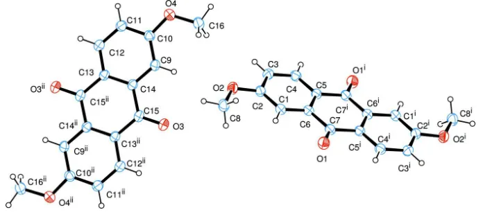
The molecular structure of the title compound is shown in Fig. 1. The title compound crystallizes with two halves of the
molecule in the asymmetric unit of the unit cell. The complete molecules are located on crystallographic inversion
centers. The molecules are almost planar with the maximum deviation of 0.018 (3) Å for C8 in one molecule and
0.049 (3) Å for C16 in another molecule. The molecules prefer the conformations in which the Cmethyl—O bonds are
directed along the short molecular axis. Thus, the torsion angles of C3—C2—O2—C8 and C11—C10—O4—C16 are
179.6 (2) and 178.0 (2)°, respectively. Theses conformations are similar to the coressponding moiety in
2,3,6,7-tetrameth-oxy-9,10-anthraquinone (Ohta, Hattori, Kawase, et al., 2012). However, there is a large difference in crystal packing
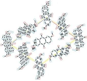
between the title compound and 2,3,6,7-tetramethoxy-9,10-anthraquinone. As shown in Fig. 2, the crystal structure is
characterized by a columnar stacking and a herrinbone-like arrangement, although 2,3,6,7-tetramethoxy-
9,10-anthra-quinone molecules took a slipped-parallel arrangement. Along the b axis, there are two columns in which molecules form
slipped π-stacks. The interplanar distances between neighboring molecules are 3.493 (3) Å for one column and 3.451 (2)
Å for another column. Furthermore, the translational shifts of neighboring molecules in the stacks are as follows: For
molecule 1 (C1–C8, O1, and O2), the slip distance between neighboring molecules is 3.94 Å, and the anthraquinone rings
in the column slipped relative to each other along the long molecular axis by 0.59 Å and along the short molecular axis
by 1.72 Å. In contrast, for molecules 2 (C9–C16, O3, and O4), the slip distance between neighboring molecules is 3.94
Å, and the anthraquinone rings in the column slipped relative to each other along the long molecular axis by 1.89 Å and
along the short molecular axis by 0.05 Å.
To examine the influence of crystal packing on the solid-state fluorescence, the fluorescence spectrum and the absolute
quantum yield of the title compound were measured with a Hamamatsu Photonics PMA11 calibrated optical
multichannel analyzer with a solid-state blue laser (λex = 377 nm) and a Labsphere IS IS-040-SF integrating sphere,
respectively. The crystals showed negligible fluorescence (Φ < 0.001). The fluorescence quenching may result from the
The title compound was prepared according to the literature procedure (Keller & Rüchardt, 1998). Single crystals suitable
for X-ray analysis were obtained by recrystallization from toluene.
S3. Refinement
All the H atoms were positioned geometrically and refined using a riding model with C—H = 0.94 Å and Uiso(H) =
1.2Ueq(C) for aromatic C—H, and C—H = 0.97 Å and Uiso(H) = 1.5Ueq(C) for CH3. The positions of methyl H atoms
[image:3.610.132.480.193.351.2]were optimized rotationally.
Figure 1
The molecular structure of the title compound, showing the atomic numbering and 50% probability displacement
Figure 2
The packing diagram of the title compound, viewed down the b axis. Hydrogen atoms are omitted for clarity.
2,6-dimethoxyanthracene-9,10-dione
Crystal data
C16H12O4
Mr = 268.26
Monoclinic, P21/a
Hall symbol: -P 2yab
a = 16.2689 (19) Å
b = 3.9357 (4) Å
c = 19.9510 (19) Å
β = 109.499 (3)°
V = 1204.2 (2) Å3
Z = 4
F(000) = 560
Dx = 1.48 Mg m−3
Mo Kα radiation, λ = 0.71073 Å
Cell parameters from 5015 reflections
θ = 3.2–27.5°
µ = 0.11 mm−1
T = 223 K
Prism, yellow
0.58 × 0.08 × 0.06 mm
Data collection
Rigaku R-AXIS RAPID diffractometer
Radiation source: fine-focus sealed x-ray tube Graphite monochromator
Detector resolution: 10 pixels mm-1
ω scans
10392 measured reflections
2743 independent reflections 1501 reflections with I > 2σ(I)
Rint = 0.066
θmax = 27.5°, θmin = 3.2°
h = −21→21
k = −4→5
Refinement on F2 Least-squares matrix: full
R[F2 > 2σ(F2)] = 0.057
wR(F2) = 0.178
S = 1.00
2743 reflections 183 parameters 0 restraints
Primary atom site location: structure-invariant direct methods
Secondary atom site location: difference Fourier map
Hydrogen site location: inferred from neighbouring sites
H-atom parameters constrained
w = 1/[σ2(F
o2) + (0.0915P)2] where P = (Fo2 + 2Fc2)/3 (Δ/σ)max < 0.001
Δρmax = 0.26 e Å−3
Δρmin = −0.28 e Å−3
Special details
Geometry. All s.u.'s (except the s.u. in the dihedral angle between two l.s. planes) are estimated using the full covariance matrix. The cell s.u.'s are taken into account individually in the estimation of s.u.'s in distances, angles and torsion angles; correlations between s.u.'s in cell parameters are only used when they are defined by crystal symmetry. An approximate (isotropic) treatment of cell s.u.'s is used for estimating s.u.'s involving l.s. planes.
Refinement. Refinement of F2 against ALL reflections. The weighted R-factor wR and goodness of fit S are based on F2,
conventional R-factors R are based on F, with F set to zero for negative F2. The threshold expression of F2 > 2σ(F2) is
used only for calculating R-factors(gt) etc. and is not relevant to the choice of reflections for refinement. R-factors based
on F2 are statistically about twice as large as those based on F, and R- factors based on ALL data will be even larger.
Fractional atomic coordinates and isotropic or equivalent isotropic displacement parameters (Å2)
x y z Uiso*/Ueq
C1 0.10535 (16) 0.2528 (6) 0.13534 (12) 0.0412 (6)
H1 0.0785 0.1497 0.1652 0.049*
C2 0.19519 (17) 0.2826 (6) 0.15742 (12) 0.0424 (6)
C3 0.23494 (17) 0.4388 (6) 0.11278 (12) 0.0452 (6)
H3 0.296 0.4582 0.1275 0.054*
C4 0.18464 (17) 0.5631 (6) 0.04772 (12) 0.0444 (6)
H4 0.2117 0.6696 0.0184 0.053*
C5 0.09401 (16) 0.5337 (6) 0.02449 (11) 0.0387 (6)
C6 0.05487 (16) 0.3753 (6) 0.06904 (11) 0.0389 (6)
C7 −0.04125 (16) 0.3291 (6) 0.04555 (11) 0.0416 (6)
C8 0.21230 (19) 0.0119 (7) 0.26849 (13) 0.0522 (7)
H8A 0.179 −0.1854 0.2455 0.078*
H8B 0.1741 0.1718 0.2805 0.078*
H8C 0.2581 −0.0583 0.3115 0.078*
O1 −0.07545 (12) 0.1767 (5) 0.08334 (8) 0.0541 (5)
O2 0.25054 (12) 0.1718 (5) 0.22088 (8) 0.0506 (5)
C9 0.47309 (16) 0.1926 (6) 0.36503 (12) 0.0401 (6)
H9 0.4168 0.1898 0.331 0.048*
C10 0.54271 (17) 0.0488 (6) 0.34963 (12) 0.0424 (6)
C11 0.62617 (17) 0.0536 (6) 0.39982 (12) 0.0444 (6)
H11 0.6731 −0.0422 0.3888 0.053*
C12 0.64007 (17) 0.1983 (6) 0.46545 (12) 0.0430 (6)
H12 0.6965 0.1982 0.4993 0.052*
C13 0.57121 (15) 0.3458 (6) 0.48250 (11) 0.0380 (6)
C15 0.41234 (15) 0.4966 (6) 0.44723 (12) 0.0409 (6)
C16 0.45117 (19) −0.1244 (8) 0.23357 (13) 0.0556 (7)
H16A 0.4122 −0.2454 0.2529 0.083*
H16B 0.4552 −0.2453 0.1924 0.083*
H16C 0.4287 0.1026 0.2196 0.083*
O3 0.33925 (12) 0.4938 (5) 0.40245 (8) 0.0542 (5)
O4 0.53622 (12) −0.1021 (5) 0.28663 (8) 0.0520 (5)
Atomic displacement parameters (Å2)
U11 U22 U33 U12 U13 U23
C1 0.0444 (15) 0.0426 (13) 0.0369 (11) −0.0015 (11) 0.0141 (10) −0.0030 (10)
C2 0.0446 (15) 0.0446 (13) 0.0363 (11) 0.0018 (11) 0.0110 (10) −0.0005 (10)
C3 0.0375 (14) 0.0536 (15) 0.0446 (13) 0.0014 (11) 0.0137 (11) 0.0008 (12)
C4 0.0445 (15) 0.0486 (14) 0.0421 (13) −0.0036 (11) 0.0172 (11) −0.0037 (11)
C5 0.0400 (14) 0.0402 (12) 0.0369 (11) −0.0008 (10) 0.0144 (10) −0.0015 (10)
C6 0.0425 (14) 0.0399 (12) 0.0361 (11) −0.0016 (10) 0.0154 (10) −0.0019 (10)
C7 0.0439 (15) 0.0441 (14) 0.0374 (12) −0.0019 (11) 0.0145 (11) −0.0030 (11)
C8 0.0523 (17) 0.0594 (16) 0.0422 (13) 0.0005 (13) 0.0122 (12) 0.0069 (12)
O1 0.0466 (11) 0.0721 (12) 0.0454 (9) −0.0061 (9) 0.0177 (8) 0.0096 (9)
O2 0.0437 (11) 0.0645 (12) 0.0410 (9) 0.0022 (8) 0.0106 (8) 0.0088 (8)
C9 0.0391 (14) 0.0429 (13) 0.0351 (11) 0.0013 (10) 0.0081 (9) 0.0056 (10)
C10 0.0459 (15) 0.0433 (13) 0.0386 (12) 0.0012 (11) 0.0149 (11) 0.0013 (11)
C11 0.0416 (15) 0.0480 (14) 0.0446 (13) 0.0034 (11) 0.0158 (11) 0.0052 (11)
C12 0.0367 (14) 0.0455 (13) 0.0448 (13) 0.0014 (10) 0.0109 (10) 0.0042 (11)
C13 0.0346 (13) 0.0396 (12) 0.0388 (12) 0.0009 (10) 0.0110 (10) 0.0053 (10)
C14 0.0354 (13) 0.0376 (12) 0.0389 (11) 0.0014 (10) 0.0108 (10) 0.0075 (10)
C15 0.0354 (14) 0.0425 (13) 0.0405 (12) 0.0011 (10) 0.0068 (10) 0.0066 (11)
C16 0.0592 (19) 0.0597 (17) 0.0421 (13) 0.0018 (13) 0.0094 (12) −0.0066 (13)
O3 0.0367 (11) 0.0701 (12) 0.0484 (10) 0.0058 (9) 0.0042 (8) −0.0043 (9)
O4 0.0524 (12) 0.0620 (11) 0.0413 (9) 0.0031 (9) 0.0153 (8) −0.0049 (8)
Geometric parameters (Å, º)
C1—C2 1.384 (4) C9—C10 1.389 (3)
C1—C6 1.390 (3) C9—C14 1.396 (3)
C1—H1 0.94 C9—H9 0.94
C2—O2 1.357 (3) C10—O4 1.362 (3)
C2—C3 1.405 (3) C10—C11 1.392 (3)
C3—C4 1.373 (3) C11—C12 1.376 (3)
C3—H3 0.94 C11—H11 0.94
C4—C5 1.395 (4) C12—C13 1.401 (3)
C4—H4 0.94 C12—H12 0.94
C5—C6 1.401 (3) C13—C14 1.401 (3)
C5—C7i 1.477 (3) C13—C15ii 1.474 (3)
C6—C7 1.486 (3) C14—C15 1.492 (3)
C7—O1 1.232 (3) C15—O3 1.226 (3)
C8—H8A 0.97 C16—H16A 0.97
C8—H8B 0.97 C16—H16B 0.97
C8—H8C 0.97 C16—H16C 0.97
C2—C1—C6 120.0 (2) C10—C9—C14 119.3 (2)
C2—C1—H1 120 C10—C9—H9 120.3
C6—C1—H1 120 C14—C9—H9 120.3
O2—C2—C1 124.8 (2) O4—C10—C9 124.3 (2)
O2—C2—C3 115.4 (2) O4—C10—C11 115.2 (2)
C1—C2—C3 119.7 (2) C9—C10—C11 120.4 (2)
C4—C3—C2 120.0 (2) C12—C11—C10 120.1 (2)
C4—C3—H3 120 C12—C11—H11 120
C2—C3—H3 120 C10—C11—H11 120
C3—C4—C5 121.0 (2) C11—C12—C13 120.8 (2)
C3—C4—H4 119.5 C11—C12—H12 119.6
C5—C4—H4 119.5 C13—C12—H12 119.6
C4—C5—C6 118.7 (2) C12—C13—C14 118.7 (2)
C4—C5—C7i 120.0 (2) C12—C13—C15ii 120.0 (2)
C6—C5—C7i 121.3 (2) C14—C13—C15ii 121.3 (2)
C1—C6—C5 120.6 (2) C9—C14—C13 120.7 (2)
C1—C6—C7 118.9 (2) C9—C14—C15 118.8 (2)
C5—C6—C7 120.5 (2) C13—C14—C15 120.6 (2)
O1—C7—C5i 121.2 (2) O3—C15—C13ii 121.4 (2)
O1—C7—C6 120.6 (2) O3—C15—C14 120.5 (2)
C5i—C7—C6 118.2 (2) C13ii—C15—C14 118.1 (2)
O2—C8—H8A 109.5 O4—C16—H16A 109.5
O2—C8—H8B 109.5 O4—C16—H16B 109.5
H8A—C8—H8B 109.5 H16A—C16—H16B 109.5
O2—C8—H8C 109.5 O4—C16—H16C 109.5
H8A—C8—H8C 109.5 H16A—C16—H16C 109.5
H8B—C8—H8C 109.5 H16B—C16—H16C 109.5
C2—O2—C8 117.2 (2) C10—O4—C16 117.7 (2)
C6—C1—C2—O2 −179.9 (2) C14—C9—C10—O4 −179.9 (2)
C6—C1—C2—C3 0.4 (4) C14—C9—C10—C11 −0.2 (4)
O2—C2—C3—C4 −179.3 (2) O4—C10—C11—C12 −179.7 (2)
C1—C2—C3—C4 0.4 (4) C9—C10—C11—C12 0.6 (4)
C2—C3—C4—C5 −0.7 (4) C10—C11—C12—C13 −0.7 (4)
C3—C4—C5—C6 0.2 (3) C11—C12—C13—C14 0.4 (4)
C3—C4—C5—C7i 179.7 (2) C11—C12—C13—C15ii −179.2 (2)
C2—C1—C6—C5 −0.9 (4) C10—C9—C14—C13 −0.1 (3)
C2—C1—C6—C7 177.9 (2) C10—C9—C14—C15 179.6 (2)
C4—C5—C6—C1 0.6 (3) C12—C13—C14—C9 0.0 (3)
C7i—C5—C6—C1 −178.9 (2) C15ii—C13—C14—C9 179.6 (2)
C4—C5—C6—C7 −178.1 (2) C12—C13—C14—C15 −179.7 (2)
C7i—C5—C6—C7 2.4 (4) C15ii—C13—C14—C15 −0.1 (4)
C5—C6—C7—O1 176.8 (2) C13—C14—C15—O3 179.6 (2)
C1—C6—C7—C5i 178.9 (2) C9—C14—C15—C13ii −179.6 (2)
C5—C6—C7—C5i −2.3 (4) C13—C14—C15—C13ii 0.1 (4)
C1—C2—O2—C8 −0.1 (3) C9—C10—O4—C16 −2.3 (3)
C3—C2—O2—C8 179.6 (2) C11—C10—O4—C16 178.0 (2)