3-Methylamino-3-phenylpropan-1-ol
Wolfgang Frey,aMohammad M. Ibrahim,bBasem F. Alib and Volker Ja¨gera*
a
Universita¨t Stuttgart, Institut fu¨r Organische Chemie, Pfaffenwaldring 55, D-70569 Stuttgart, Germany, andbDepartment of Chemistry, Al al-Bayt University, Mafraq 25113, Jordan
Correspondence e-mail: [email protected]
Received 17 June 2012; accepted 27 August 2012
Key indicators: single-crystal X-ray study;T= 293 K; mean(C–C) = 0.005 A˚;
Rfactor = 0.071;wRfactor = 0.201; data-to-parameter ratio = 14.3.
The title compound, C10H15NO, is an amino alcohol with the
hydroxy group residing on the terminal C atom. Apart from the hydroxy group and the phenyl ring, all non-H atoms are almost coplanar. In the crystal, classical O—H N and N—
H O hydrogen bonds connect the molecules into
centro-symmetric dimers [R2 2
(12) descriptor] and tetrameric units [R4
4
(8) descriptor] as ring motifs, consolidating a three-dimensional network.
Related literature
For the syntheses of amino alcohols from isoxazolidines, isoxazolines and isoxazolinium salts, see: DeShong & Leginus, (1983); Hennebo¨hle et al. (2004); Ibrahim (2009); Ja¨ger & Buss, (1980); Ja¨geret al.(1985, 2010); Ja¨ger & Colinas (2002); Lubellet al.(1991). For hydrogen-bond motifs see: Bernstein et al.(1995). For standard bond lengths, see: Allenet al.(1987).
Experimental
Crystal data
C10H15NO
Mr= 165.23 Monoclinic,P21=n
a= 5.9816 (8) A˚
b= 23.8962 (19) A˚
c= 7.4653 (8) A˚
= 111.119 (7)
V= 995.40 (19) A˚3
Z= 4
CuKradiation
T= 293 K
Data collection
Siemens P4 diffractometer 3535 measured reflections 1704 independent reflections 896 reflections withI> 2(I)
Rint= 0.087
3 standard reflections every 100 reflections
intensity decay: 3%
Refinement
R[F2> 2(F2)] = 0.071
wR(F2) = 0.201
S= 1.04 1704 reflections 119 parameters
H atoms treated by a mixture of independent and constrained refinement
max= 0.22 e A˚3
min=0.22 e A˚3
Table 1
Hydrogen-bond geometry (A˚ ,).
D—H A D—H H A D A D—H A
N1—H1B O1i
1.02 (4) 2.06 (3) 3.023 (4) 157 (2) O1—H1A N1ii 1.12 (4) 1.70 (4) 2.815 (3) 176 (3)
Symmetry codes: (i)xþ1;y;z; (ii)xþ2;y;zþ1.
Data collection: XSCANS (Bruker, 1996); cell refinement: XSCANS; data reduction: XSCANS; program(s) used to solve structure:SHELXS97(Sheldrick, 2008); program(s) used to refine structure:SHELXL97(Sheldrick, 2008); molecular graphics:XPin SHELXTL-Plus(Sheldrick, 2008); software used to prepare material for publication:XPinSHELXTL-Plus.
We thank the German Academic Exchange Service (DAAD) for PhD scholarship to MMI.
Supplementary data and figures for this paper are available from the IUCr electronic archives (Reference: IM2389).
References
Allen, F. H., Kennard, O., Watson, D. G., Brammer, L., Orpen, A. G. & Taylor, R. (1987).J. Chem. Soc. Perkin Trans. 2pp. S1–19.
Bernstein, J., Davis, R. E., Shimoni, L. & Chang, N.-L. (1995).Angew. Chem. Int. Ed. Engl.34, 1555-1573.
Bruker (1996).XSCANS. Bruker AXS Inc., Madison, Wisconsin, USA. DeShong, P. & Leginus, J. M. (1983).J. Am. Chem. Soc.105, 1686-1688. Hennebo¨hle, M., Le Roy, P.-Y., Hein, M., Ehrler, R. & Ja¨ger, V. (2004).Z.
Naturforsch. Teil B,59, 451– 467.
Ibrahim, M. M. (2009). Dissertation, Universita¨t Stuttgart, Germany. Ja¨ger, V. & Buss, V. (1980).Liebigs Ann. Chem.pp. 101–121.
Ja¨ger, V. & Colinas, P. (2002). Synthetic Applications of 1,3-Dipolar Cycloaddition Chemistry Toward Heterocycles and Natural Products, The Chemistry of Heterocyclic Compounds, edited by A. Padwa & W. H. Pearson, pp. 361–472. New York: Wiley.
Ja¨ger, V., Frey, W., Bathich, Y., Shiva, S., Ibrahim, M., Hennebo¨hle, M., LeRoy, P. Y. & Imerhasan, M. (2010).Z. Naturforsch. Teil B,65b, 821–832. Ja¨ger, V., Mu¨ller, I. & Paulus, E. F. (1985).Tetrahedron Lett.26, 2997-3000. Lubell, W. D., Kitamura, M. & Noyori, R. (1991).Tetrahedron Asymmetry,2,
543-554.
Sheldrick, G. M. (2008).Acta Cryst.A64, 112–122.
Structure Reports Online
supporting information
Acta Cryst. (2012). E68, o2857 [https://doi.org/10.1107/S160053681203694X]
3-Methylamino-3-phenylpropan-1-ol
Wolfgang Frey, Mohammad M. Ibrahim, Basem F. Ali and Volker J
ä
ger
S1. Comment
Isoxazolidines, isoxazolines, and isoxazolinium salts are useful intermediates for syntheses of 1,3-amino alcohols by
reduction with cleavage of the N–O bond (DeShong & Leginus, 1983; Jäger & Buss, 1980; Jäger et al., 1985; Jäger &
Colinas, 2002; Henneböhle et al., 2004; Jäger et al., 2010). The structures and conformations of previously synthesized
amino alcohols were all assigned on the basis of analytical as well as IR, 13C and 1H NMR data. When the
2-methyl-3-phenylisoxazolidine-3-carbonitrile was heated to reflux with lithium aluminium hydride in ether (abs.), the title
compound I was formed in good yield. The starting isoxazolidine had been obtained from the corresponding N
-methyl-isoxazolinium salt by cyanide addition (Henneböhle et al., 2004; Ibrahim, 2009; Jäger et al. 2010). The formation of the
amino alcohol I was rationalized elsewhere (Ibrahim, 2009). The title compound I is already known from other routes
(Lubell et al., 1991), yet, the crystal structure of I has not been published so far. We herein report the synthesis and the
crystal structure of I, along with the supramolecular motifs present in the crystal lattice.
The asymmetric unit of I consists of one amino alcohol molecule with bond distances and angles in the normal range
(Allen et al., 1987). The molecule, a primary alcohol and a secondary amine, adopts a planar zigzag-chain conformation
(C1/C2/C3/N1/C4 almost coplanar), with both the hydroxy and the phenyl group being out-of-plane. The hydroxy and the
phenyl group enclose dihedral angles of -60.3 (4)° and -63.0 (3)°, respectively, with the atoms of the carbon-chain
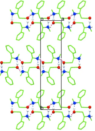
(hydroxyl-O1/phenyl-C5-C1-C2-C3), see Fig. 1. In the crystal structure, molecules are hydrogen-bonded through the
hy-droxy groups as well as the amino groups (Table 1) giving rise to a three-dimensional network. The cooperative hydrogen
bonds (alternating between hydroxy and amino groups) connect the molecules into chains down the crystallographic a
axis (Fig. 2). These chains consist of alternating centrosymmetric dimers, with each dimer further interacting through the
hydroxyl and amino groups with the adjacent one to form tetrameric units (Fig. 2). In terms of graph-set description, the
hydrogen-bonded molecules might be described as forming two types of rings (Bernstein et al., 1995), the
centrosymmetric dimers being R22(12) while R44(8) represents the descriptor for the tetramer units. These interactions
consolidate a three-dimensional network.
This amino alcohol conformation in the crystal found here is in contrast to the conformations elucidated in solution on
the basis of IR dilution experiments and extensive collections of 13C and 1H NMR data, notably coupling constants and
substituent increments - there intramolecular hydrogen bonds O—H···N prevail to form monomers with chair-like
arrangements (Jäger & Buss, 1980).
S2. Experimental
A solution of 2-methyl-3-phenylisoxazolidine-3-carbonitrile (150 mg, 0.80 mmol) in anhydrous diethylether (5 ml) at 0°C
was added to a suspension of LiAlH4 (61.0 mg, 1.6 mmol, 2 eq) in anhydrous diethylether (15 ml) and stirred for 30 min.
The mixture was allowed to warm up to room temperature and stirred for 1 h. The reaction mixture was then heated to
The organic layers were dried over Na2SO4. The solvent was evaporated in vacuo (5 mbar, 40°C) to afford the amino
alcohol I in analytically and spectroscopically pure form as a colorless solid [100 mg, 84%, m.p. 56–57°C; lit. 56–57°C
(Lubell et al., 1991)]. Crystallization of the solid from ether afforded colorless crystals suitable for crystal structure
determination. Analysis for C10H15NO, Calc.: C 72.69, H 9.15, N 8.48; Found: C 72.39, H 9.11, N 8.07.
S3. Refinement
Hydrogen atoms were located from the difference fourier map, but refined with fixed individual displacement parameters,
using a riding model with d(C—H) ranging from 0.93 to 0.98 Å and Uiso(H) = 1.2 Ueq(C) or Uiso(H) = 1.5 Ueq(Cmethyl). In
addition, the methyl group is allowed to rotate but not to tip. Hydrogen atoms attached to the hydroxy function and to the
[image:3.610.123.481.234.550.2]amino moiety are refined freely because of their relevance in hydrogen bonding.
Figure 1
Figure 2
A view down c axis showing chains of hydrogen bonding molecules along a axis. The chains consist of alternating
centrosymmetric dimers, with each dimer further interacting through the hydroxyl and amino groups with the adjacent
one to form tetrameric units.
3-Methylamino-3-phenylpropan-1-ol
Crystal data
C10H15NO
Mr = 165.23 Monoclinic, P21/n
Hall symbol: -P 2yn a = 5.9816 (8) Å b = 23.8962 (19) Å c = 7.4653 (8) Å β = 111.119 (7)°
V = 995.40 (19) Å3
Z = 4 F(000) = 360 Dx = 1.103 Mg m−3
Cu Kα radiation, λ = 1.54178 Å Cell parameters from 30 reflections θ = 21.0–22.5°
Block, colourless
Data collection
Siemens P4 diffractometer
Radiation source: fine-focus sealed tube Graphite monochromator
ω scans
3535 measured reflections 1704 independent reflections 896 reflections with I > 2σ(I)
Rint = 0.087
θmax = 67.5°, θmin = 3.7°
h = −7→7 k = −28→28 l = −8→8
3 standard reflections every 100 reflections intensity decay: 3%
Refinement
Refinement on F2
Least-squares matrix: full R[F2 > 2σ(F2)] = 0.071
wR(F2) = 0.201
S = 1.04 1704 reflections 119 parameters 0 restraints
Primary atom site location: structure-invariant direct methods
Secondary atom site location: difference Fourier map
Hydrogen site location: inferred from neighbouring sites
H atoms treated by a mixture of independent and constrained refinement
w = 1/[σ2(F
o2) + (0.0232P)2 + 0.9564P]
where P = (Fo2 + 2Fc2)/3
(Δ/σ)max < 0.001
Δρmax = 0.22 e Å−3
Δρmin = −0.22 e Å−3
Extinction correction: SHELXL97 (Sheldrick, 2008), Fc*=kFc[1+0.001xFc2λ3/sin(2θ)]-1/4
Extinction coefficient: 0.026 (2)
Special details
Experimental. 1H NMR (500.2 MHz, MeOD): d = 1.82 (dddd, 3J
1a,2a = 7.9 Hz, 3J1b,2a = 5.6 Hz, 2J2a,2b = 14.0 Hz, 3J2a,3 = 5.2
Hz, 1 H, 2-Ha), 2.06 (dddd, 3J1a,2b = 6.2 Hz, 3J1b,2b = 7.5 Hz, 2J2a,2b = 14.2 Hz, 3J2b,3 = 5.8 Hz, 1 H, 2-Hb), 2.18 (s, 3 H,
NCH3), 3.42 (ddd, 2J1a,1b = 10.8 Hz, 3J1a,2a = 8.1 Hz, 3J1a,2b = 6.1 Hz, 1 H, 1-Ha), 3.49 (ddd, 2J1a,1b = 8.0 Hz, 3J1b,2a = 5.0 Hz, 3J
1b,2b = 6.8 Hz, 1 H, 1-Hb), 3.65 (dd, 3J2a,3 = 8.3 Hz, 3J2b,3 = 6.0 Hz, 1 H, 3-H), 7.23-7.35 (m, 5 H, C6H5); 13C NMR (125.8
MHz, MeOD): d = 34.0 (q, NCH3), 40.7 (t, C-2), 60.4 (t, C-1), 63.7 (d, C-3), 128.2, 128.4, 129.6 (3 d, o-, m-, p-C of
C6H5), 143.8 (s, i-C of C6H5).
Geometry. All e.s.d.'s (except the e.s.d. in the dihedral angle between two l.s. planes) are estimated using the full covariance matrix. The cell e.s.d.'s are taken into account individually in the estimation of e.s.d.'s in distances, angles and torsion angles; correlations between e.s.d.'s in cell parameters are only used when they are defined by crystal symmetry. An approximate (isotropic) treatment of cell e.s.d.'s is used for estimating e.s.d.'s involving l.s. planes.
Refinement. Refinement of F2 against ALL reflections. The weighted R-factor wR and goodness of fit S are based on F2,
conventional R-factors R are based on F, with F set to zero for negative F2. The threshold expression of F2 > σ(F2) is used
only for calculating R-factors(gt) etc. and is not relevant to the choice of reflections for refinement. R-factors based on F2
are statistically about twice as large as those based on F, and R- factors based on ALL data will be even larger.
Fractional atomic coordinates and isotropic or equivalent isotropic displacement parameters (Å2)
x y z Uiso*/Ueq
C2 1.0300 (5) 0.02701 (12) 0.2739 (4) 0.0564 (8) H2A 1.1168 0.0238 0.1868 0.068* H2B 1.0732 −0.0049 0.3599 0.068* C3 1.1102 (5) 0.08083 (12) 0.3929 (4) 0.0565 (8) H3 1.0121 0.0848 0.4727 0.068* C4 1.4610 (8) 0.11951 (15) 0.6516 (6) 0.0922 (14) H4A 1.4654 0.1522 0.5782 0.138* H4B 1.6207 0.1102 0.7351 0.138* H4C 1.3637 0.1268 0.7270 0.138* C5 1.0643 (6) 0.13176 (12) 0.2623 (5) 0.0603 (9) C6 0.8826 (7) 0.16881 (14) 0.2513 (6) 0.0790 (11) H6 0.7948 0.1640 0.3307 0.095* C7 0.8291 (8) 0.21376 (16) 0.1211 (7) 0.0936 (14) H7 0.7072 0.2387 0.1152 0.112* C8 0.9553 (9) 0.22067 (16) 0.0047 (7) 0.0925 (14) H8 0.9167 0.2496 −0.0844 0.111* C9 1.1390 (8) 0.18531 (15) 0.0176 (6) 0.0851 (12) H9 1.2292 0.1911 −0.0595 0.102* C10 1.1927 (7) 0.14055 (14) 0.1451 (5) 0.0737 (10) H10 1.3169 0.1163 0.1511 0.088*
Atomic displacement parameters (Å2)
U11 U22 U33 U12 U13 U23
O1 0.0526 (13) 0.0690 (14) 0.0665 (15) 0.0047 (10) 0.0194 (11) 0.0030 (11) N1 0.0552 (15) 0.0620 (16) 0.0583 (17) −0.0054 (12) 0.0114 (14) −0.0037 (12) C1 0.0563 (18) 0.075 (2) 0.0562 (19) −0.0061 (15) 0.0158 (16) −0.0016 (15) C2 0.0557 (17) 0.0585 (17) 0.0554 (18) −0.0043 (13) 0.0206 (15) −0.0044 (13) C3 0.0547 (17) 0.0579 (17) 0.0563 (19) 0.0004 (13) 0.0193 (16) −0.0015 (13) C4 0.101 (3) 0.071 (2) 0.079 (3) −0.015 (2) 0.001 (2) −0.0142 (19) C5 0.0563 (18) 0.0562 (17) 0.063 (2) 0.0016 (14) 0.0145 (16) 0.0007 (14) C6 0.078 (2) 0.066 (2) 0.092 (3) 0.0102 (17) 0.030 (2) −0.0017 (19) C7 0.090 (3) 0.065 (2) 0.109 (4) 0.021 (2) 0.016 (3) 0.005 (2) C8 0.114 (3) 0.067 (2) 0.082 (3) 0.009 (2) 0.019 (3) 0.011 (2) C9 0.106 (3) 0.071 (2) 0.084 (3) −0.001 (2) 0.040 (3) 0.0100 (19) C10 0.082 (2) 0.068 (2) 0.073 (2) 0.0038 (17) 0.031 (2) 0.0077 (17)
Geometric parameters (Å, º)
C2—H2B 0.9700 C9—C10 1.390 (5) C3—C5 1.521 (4) C9—H9 0.9300 C3—H3 0.9800 C10—H10 0.9300 C4—H4A 0.9600
C1—O1—H1A 112 (2) N1—C4—H4B 109.5 C4—N1—C3 114.9 (3) H4A—C4—H4B 109.5 C4—N1—H1B 107.1 (17) N1—C4—H4C 109.5 C3—N1—H1B 106.3 (18) H4A—C4—H4C 109.5 O1—C1—C2 112.6 (3) H4B—C4—H4C 109.5 O1—C1—H1C 109.1 C10—C5—C6 118.2 (3) C2—C1—H1C 109.1 C10—C5—C3 121.2 (3) O1—C1—H1D 109.1 C6—C5—C3 120.5 (3) C2—C1—H1D 109.1 C5—C6—C7 120.6 (4) H1C—C1—H1D 107.8 C5—C6—H6 119.7 C1—C2—C3 113.9 (3) C7—C6—H6 119.7 C1—C2—H2A 108.8 C8—C7—C6 119.8 (4) C3—C2—H2A 108.8 C8—C7—H7 120.1 C1—C2—H2B 108.8 C6—C7—H7 120.1 C3—C2—H2B 108.8 C7—C8—C9 120.2 (4) H2A—C2—H2B 107.7 C7—C8—H8 119.9 N1—C3—C5 115.4 (2) C9—C8—H8 119.9 N1—C3—C2 107.7 (2) C8—C9—C10 120.5 (4) C5—C3—C2 110.6 (2) C8—C9—H9 119.8 N1—C3—H3 107.6 C10—C9—H9 119.8 C5—C3—H3 107.6 C5—C10—C9 120.7 (4) C2—C3—H3 107.6 C5—C10—H10 119.7 N1—C4—H4A 109.5 C9—C10—H10 119.7
O1—C1—C2—C3 −60.3 (4) C10—C5—C6—C7 0.9 (5) C4—N1—C3—C5 58.3 (4) C3—C5—C6—C7 −175.6 (3) C4—N1—C3—C2 −177.6 (3) C5—C6—C7—C8 0.4 (6) C1—C2—C3—N1 169.9 (3) C6—C7—C8—C9 −2.1 (7) C1—C2—C3—C5 −63.0 (3) C7—C8—C9—C10 2.5 (7) N1—C3—C5—C10 54.1 (4) C6—C5—C10—C9 −0.6 (5) C2—C3—C5—C10 −68.5 (4) C3—C5—C10—C9 175.9 (3) N1—C3—C5—C6 −129.5 (3) C8—C9—C10—C5 −1.1 (6) C2—C3—C5—C6 107.9 (3)
Hydrogen-bond geometry (Å, º)
D—H···A D—H H···A D···A D—H···A
N1—H1B···O1i 1.02 (4) 2.06 (3) 3.023 (4) 157 (2)
O1—H1A···N1ii 1.12 (4) 1.70 (4) 2.815 (3) 176 (3)