Crystal structure of (
E
)-
N
000-(4-chloro-
benzylidene)-4-methylbenzenesulfono-hydrazide: a hexagonal polymorph
J. Balaji,aJ. John Francis Xavier,bS. Prabuaand P. Srinivasana*
aDepartment of Physics, University College of Engineering Panruti, Tamil Nadu 607 106, India, andbDepartment of Chemistry, University College of Engineering Panruti, Tamil Nadu 607 106, India. *Correspondence e-mail: sril35@gmail.com
Received 14 October 2014; accepted 28 October 2014
Edited by H. Stoeckli-Evans, University of Neuchaˆtel, Switzerland
The title compound, C14H13ClN2O2S, crystallized in the enantiomorphic defining hexagonal space group P61 [Flack parameter = 0.02 (7)]. The partially hydrated form of the same compound, crystallizing in the triclinic space group P1, has been reported previously [Kiaet al.(2009b).Acta Cryst.
E65, o1119], as has the crystal structure of the bromo derivative, also crystallizing in the space groupP1 [Kia et al.
(2009a). Acta Cryst. E65, o821]. The title molecule is non-planar with the planes of the benzene rings being inclined to one another by 76.62 (13), and has anEconformation about the C N bond. In the crystal, molecules are linkedviaN— H O hydrogen bonds forming 61 helical chains running along [001]. The chains are linked via C—H O hydrogen bonds, C—H interactions and short Cl O [3.015 (3) A˚ ] interactions, forming a three-dimensional structure.
Keywords:crystal structure; hydrazones; sulfonohydrazide; Schiff base; helical chains; hydrogen bonding..
CCDC reference:1031289
1. Related literature
For the biological activities of hydrazones, see: Ajani et al.
(2010). For the crystal structure of the triclinic polymorph, which crystallized with two independent molecules in the asymmetric unit, one of which was disordered, and with 0.15 of a water molecule, see: Kia et al. (2009b). For the crystal structure of the bromo derivative, also crystallizing in space groupP1, see: Kiaet al.(2009a).
2. Experimental
2.1. Crystal data
C14H13ClN2O2S
Mr= 308.77 Hexagonal,P61 a= 10.8907 (3) A˚ c= 21.4542 (7) A˚ V= 2203.71 (11) A˚3
Z= 6
MoKradiation = 0.40 mm1
T= 293 K
0.350.300.25 mm
2.2. Data collection
Bruker Kappa APEXII CCD diffractometer
Absorption correction: multi-scan (SADABS; Sheldrick, 1996) Tmin= 0.871,Tmax= 0.910
22095 measured reflections 2586 independent reflections 2345 reflections withI> 2(I) Rint= 0.027
2.3. Refinement
R[F2> 2(F2)] = 0.029
wR(F2) = 0.072 S= 1.04 2586 reflections 186 parameters 2 restraints
H atoms treated by a mixture of independent and constrained refinement
max= 0.12 e A˚ 3
min=0.16 e A˚ 3
Absolute structure: Flack (1983), 1257 Friedel pairs
Absolute structure parameter:
0.02 (7)
Table 1
Hydrogen-bond geometry (A˚ ,).
Cgis the centroid of the C2–C7 ring.
D—H A D—H H A D A D—H A
N1—H1 O2i
0.88 (2) 2.13 (2) 2.952 (3) 156 (2) C1—H1A O1ii 0.96 2.55 3.496 (5) 169 C13—H13 Cgiii
0.93 2.94 3.823 (3) 160
Symmetry codes: (i)xy;x;zþ1
6; (ii)xy1;x1;zþ 1
6; (iii)x;yþ1;z.
Data collection:APEX2(Bruker, 2004); cell refinement:APEX2and SAINT(Bruker, 2004); data reduction:SAINTandXPREP(Bruker, 2004); program(s) used to solve structure:SIR92 (Altomareet al., 1993); program(s) used to refine structure:SHELXL97(Sheldrick, 2008); molecular graphics:ORTEP-3 for Windows(Farrugia, 2012) andMercury(Macraeet al., 2008); software used to prepare material for publication:SHELXL97andPLATON(Spek, 2009).
Acknowledgements
The authors thank the SAIF, IITM, Madras, for help with the XRD studies.
data reports
Supporting information for this paper is available from the IUCr electronic archives (Reference: SU2800).
References
Ajani, O. O., Obafemi, C. A., Nwinyi, O. C. & Akinpelu, D. A. (2010).Bioorg. Med. Chem.18, 214–221.
Altomare, A., Cascarano, G., Giacovazzo, C. & Guagliardi, A. (1993).J. Appl. Cryst.26, 343–350.
Bruker (2004).APEX2 andSAINT. Bruker Axs Inc., Madison, Wisconsin, USA.
Farrugia, L. J. (2012).J. Appl. Cryst.45, 849–854. Flack, H. D. (1983).Acta Cryst.A39, 876–881.
Kia, R., Etemadi, B., Fun, H.-K. & Kargar, H. (2009a).Acta Cryst.E65, o821– o822.
Kia, R., Fun, H.-K. & Kargar, H. (2009b).Acta Cryst.E65, o1119–o1120. Macrae, C. F., Bruno, I. J., Chisholm, J. A., Edgington, P. R., McCabe, P.,
Pidcock, E., Rodriguez-Monge, L., Taylor, R., van de Streek, J. & Wood, P. A. (2008).J. Appl. Cryst.41, 466–470.
Sheldrick, G. M. (1996).SADABS. University of Go¨ttingen, Germany. Sheldrick, G. M. (2008).Acta Cryst.A64, 112–122.
supporting information
sup-1
Acta Cryst. (2014). E70, o1250–o1251
supporting information
Acta Cryst. (2014). E70, o1250–o1251 [doi:10.1107/S1600536814023721]
Crystal structure of (
E
)-
N
′
-(4-chlorobenzylidene)-4-methylbenzenesulfono-hydrazide: a hexagonal polymorph
J. Balaji, J. John Francis Xavier, S. Prabu and P. Srinivasan
S1. Comment
The title compound was obtained by a Schiff base condensation reaction between 4-chlorobenzaldehyde and tosyl
hydrazide. Hydrazones have received much attention recently due to their biological activities (Ajani et al., 2010). The crystal structure of the triclinic polymorph, that crystallized with two independent molecules in the asymmetric unit, one
of which was disordered, and with 0.15 of a water molecule, has been reported (Kia et al., 2009b), as has the crystal structure of the bromo derivative, also crystallizing in space group P1 (Kia et al., 2009a).
The hydrazone molecule, Fig. 1, exists in a trans or E confirmation with respect to the C8═N2 bond. The dihedral angle between the (C2—C7) and (C9—C14) benzene rings is 76.62 (13) °. In the triclininc polymorph (Kia et al., 2009b) the same angle is 84.96 (11) ° (and 71.1 (3) ° for the disordered molecule), and 82.39 (13) ° for the bromo derivative (Kia et al., 2009a).
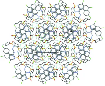
In the crystal, molecules are linked via N—H···O hydrogen bonds forming 61 helical chains running along the c axis
direction (Table 1 and Fig 2). The chains are linked via C-H···O hydrogen bonds, and a short Cl···O2i [3.015 (3) Å;
symmetry code: (i) x-y+1, x, z+1/6] interaction and a C-H···π interaction, forming a three-dimensional structure (Table 1 and Fig. 2).
S2. Experimental
4-chlorobenzaldehyde (0.140 g, 1 mmol) and tosyl hydrazide (0.186 g, 1 mmol) were dissolved in ethanol and chloroform
(4:1). The reaction mixture was heated under reflux for 3 h and cooled gradually to room temperature. Prismatic
colourless crystals were obtained by slow evaporation of an ethanol solution at room temperature.
S3. Refinement
The NH H atom was located in a difference Fourier map and freely refined. The C-bound H atoms were positioned
geometrically and treated as riding on their parent atoms with C—H = 0.93 Å (aromatic) and 0.96 Å (methyl) and with
Figure 1
The molecular structure of the title compound, with atom labelling. The displacement ellipsoids are drawn at the 30%
probability level.
Figure 2
A view along the c axis of the crystal packing of the title compound. The N—H···O and C—H···O hydrogen bonds are indicated by dashed lines (see Table 1 for details; H atoms not involved in these interactions have been omitted for
[image:4.610.127.484.325.618.2]supporting information
sup-3
Acta Cryst. (2014). E70, o1250–o1251
(E)-N′-(4-Chlorobenzylidene)-4-methylbenzenesulfonohydrazide
Crystal data
C14H13ClN2O2S
Mr = 308.77
Hexagonal, P61
Hall symbol: P 61
a = 10.8907 (3) Å
c = 21.4542 (7) Å
V = 2203.71 (11) Å3
Z = 6
F(000) = 960
Dx = 1.396 Mg m−3
Mo Kα radiation, λ = 0.71073 Å Cell parameters from 9375 reflections
θ = 2.4–25.8°
µ = 0.40 mm−1
T = 293 K Block, yellow
0.35 × 0.30 × 0.25 mm
Data collection
Bruker Kappa APEXII CCD diffractometer
Radiation source: fine-focus sealed tube Graphite monochromator
ω and φ scan
Absorption correction: multi-scan (SADABS; Sheldrick, 1996)
Tmin = 0.871, Tmax = 0.910
22095 measured reflections 2586 independent reflections 2345 reflections with I > 2σ(I)
Rint = 0.027
θmax = 25.0°, θmin = 2.2°
h = −12→12
k = −12→12
l = −25→25
Refinement
Refinement on F2
Least-squares matrix: full
R[F2 > 2σ(F2)] = 0.029
wR(F2) = 0.072
S = 1.04 2586 reflections 186 parameters 2 restraints
Primary atom site location: structure-invariant direct methods
Secondary atom site location: difference Fourier map
Hydrogen site location: inferred from neighbouring sites
H atoms treated by a mixture of independent and constrained refinement
w = 1/[σ2(F
o2) + (0.0306P)2 + 0.7106P]
where P = (Fo2 + 2Fc2)/3
(Δ/σ)max = 0.001
Δρmax = 0.12 e Å−3
Δρmin = −0.16 e Å−3
Extinction correction: SHELXL97 (Sheldrick, 2008), Fc*=kFc[1+0.001xFc2λ3/sin(2θ)]-1/4
Extinction coefficient: 0.0026 (4)
Absolute structure: Flack (1983), 1257 Friedel pairs
Absolute structure parameter: −0.02 (7)
Special details
Geometry. All e.s.d.'s (except the e.s.d. in the dihedral angle between two l.s. planes) are estimated using the full covariance matrix. The cell e.s.d.'s are taken into account individually in the estimation of e.s.d.'s in distances, angles and torsion angles; correlations between e.s.d.'s in cell parameters are only used when they are defined by crystal symmetry. An approximate (isotropic) treatment of cell e.s.d.'s is used for estimating e.s.d.'s involving l.s. planes.
Refinement. Refinement of F2 against ALL reflections. The weighted R-factor wR and goodness of fit S are based on F2,
conventional R-factors R are based on F, with F set to zero for negative F2. The threshold expression of F2 > σ(F2) is used
only for calculating R-factors(gt) etc. and is not relevant to the choice of reflections for refinement. R-factors based on F2
are statistically about twice as large as those based on F, and R- factors based on ALL data will be even larger.
Fractional atomic coordinates and isotropic or equivalent isotropic displacement parameters (Å2)
x y z Uiso*/Ueq
C1 −0.1026 (3) −0.5080 (4) 0.94696 (15) 0.0727 (9)
H1B −0.0699 −0.4482 0.9831 0.109*
H1C −0.0942 −0.5906 0.9539 0.109*
C2 −0.0151 (3) −0.4286 (3) 0.89196 (13) 0.0480 (6) C3 −0.0778 (3) −0.4059 (3) 0.84061 (13) 0.0507 (6)
H3 −0.1751 −0.4410 0.8405 0.061*
C4 0.0019 (3) −0.3321 (3) 0.78968 (12) 0.0496 (6)
H4 −0.0415 −0.3180 0.7553 0.060*
C5 0.1458 (2) −0.2795 (2) 0.79003 (11) 0.0419 (5) C6 0.2103 (3) −0.3013 (3) 0.84070 (12) 0.0483 (6)
H6 0.3077 −0.2657 0.8408 0.058*
C7 0.1296 (3) −0.3758 (3) 0.89098 (13) 0.0514 (6)
H7 0.1730 −0.3911 0.9250 0.062*
C8 0.1633 (3) 0.1027 (3) 0.74279 (11) 0.0426 (5)
H8 0.2543 0.1797 0.7474 0.051*
C9 0.0430 (3) 0.1244 (3) 0.74794 (10) 0.0415 (5) C10 −0.0947 (3) 0.0146 (3) 0.74508 (13) 0.0520 (6)
H10 −0.1127 −0.0766 0.7367 0.062*
C11 −0.2070 (3) 0.0381 (3) 0.75456 (13) 0.0579 (7)
H11 −0.3000 −0.0366 0.7526 0.069*
C12 −0.1789 (3) 0.1731 (3) 0.76682 (12) 0.0522 (6) C13 −0.0439 (3) 0.2844 (3) 0.76863 (13) 0.0574 (7)
H13 −0.0266 0.3758 0.7763 0.069*
C14 0.0664 (3) 0.2595 (3) 0.75898 (12) 0.0516 (6)
H14 0.1589 0.3353 0.7599 0.062*
O1 0.3852 (2) −0.1677 (2) 0.73010 (9) 0.0597 (5) O2 0.1652 (2) −0.2455 (2) 0.67125 (8) 0.0605 (5) S1 0.24764 (7) −0.18427 (7) 0.72562 (3) 0.04549 (16) Cl1 −0.31836 (9) 0.20068 (10) 0.78326 (4) 0.0779 (3) N1 0.2735 (2) −0.0230 (2) 0.73014 (10) 0.0445 (5) N2 0.1474 (2) −0.0187 (2) 0.73212 (9) 0.0424 (5) H1 0.337 (2) 0.033 (2) 0.7573 (10) 0.050 (8)*
Atomic displacement parameters (Å2)
U11 U22 U33 U12 U13 U23
supporting information
sup-5
Acta Cryst. (2014). E70, o1250–o1251
C14 0.0529 (16) 0.0444 (15) 0.0627 (17) 0.0282 (13) −0.0049 (12) −0.0057 (12) O1 0.0637 (12) 0.0735 (13) 0.0631 (11) 0.0502 (11) 0.0132 (9) 0.0121 (10) O2 0.0933 (15) 0.0647 (12) 0.0418 (9) 0.0533 (12) −0.0108 (10) −0.0124 (9) S1 0.0604 (4) 0.0513 (4) 0.0401 (3) 0.0394 (3) 0.0020 (3) −0.0015 (3) Cl1 0.0746 (5) 0.0945 (6) 0.0928 (6) 0.0635 (5) −0.0005 (5) −0.0118 (5) N1 0.0498 (13) 0.0489 (12) 0.0446 (11) 0.0320 (11) −0.0005 (10) −0.0030 (10) N2 0.0511 (12) 0.0469 (12) 0.0400 (11) 0.0327 (10) −0.0022 (9) −0.0032 (9)
Geometric parameters (Å, º)
C1—C2 1.491 (4) C8—H8 0.9300
C1—H1A 0.9600 C9—C10 1.374 (3)
C1—H1B 0.9600 C9—C14 1.383 (3)
C1—H1C 0.9600 C10—C11 1.384 (4)
C2—C7 1.381 (3) C10—H10 0.9300
C2—C3 1.382 (4) C11—C12 1.369 (4)
C3—C4 1.376 (4) C11—H11 0.9300
C3—H3 0.9300 C12—C13 1.360 (4)
C4—C5 1.374 (3) C12—Cl1 1.726 (3)
C4—H4 0.9300 C13—C14 1.373 (4)
C5—C6 1.378 (3) C13—H13 0.9300
C5—S1 1.751 (3) C14—H14 0.9300
C6—C7 1.372 (4) O1—S1 1.4197 (18)
C6—H6 0.9300 O2—S1 1.4189 (19)
C7—H7 0.9300 S1—N1 1.637 (2)
C8—N2 1.265 (3) N1—N2 1.399 (3)
C8—C9 1.447 (3) N1—H1 0.875 (17)
C2—C1—H1A 109.5 C10—C9—C8 122.5 (2)
C2—C1—H1B 109.5 C14—C9—C8 119.1 (2)
H1A—C1—H1B 109.5 C9—C10—C11 120.9 (2)
C2—C1—H1C 109.5 C9—C10—H10 119.6
H1A—C1—H1C 109.5 C11—C10—H10 119.6
H1B—C1—H1C 109.5 C12—C11—C10 118.8 (3)
C7—C2—C3 118.4 (2) C12—C11—H11 120.6
C7—C2—C1 121.2 (3) C10—C11—H11 120.6
C3—C2—C1 120.3 (2) C13—C12—C11 121.6 (2)
C4—C3—C2 120.9 (2) C13—C12—Cl1 119.5 (2)
C4—C3—H3 119.5 C11—C12—Cl1 118.8 (2)
C2—C3—H3 119.5 C12—C13—C14 118.8 (2)
C5—C4—C3 119.6 (2) C12—C13—H13 120.6
C5—C4—H4 120.2 C14—C13—H13 120.6
C3—C4—H4 120.2 C13—C14—C9 121.5 (3)
C4—C5—C6 120.4 (2) C13—C14—H14 119.3
C4—C5—S1 119.71 (18) C9—C14—H14 119.3
C6—C5—S1 119.89 (19) O2—S1—O1 119.65 (12)
C7—C6—C5 119.4 (2) O2—S1—N1 106.40 (11)
C5—C6—H6 120.3 O2—S1—C5 107.80 (12)
C6—C7—C2 121.2 (2) O1—S1—C5 109.72 (11)
C6—C7—H7 119.4 N1—S1—C5 107.91 (11)
C2—C7—H7 119.4 N2—N1—S1 113.13 (16)
N2—C8—C9 121.5 (2) N2—N1—H1 113.4 (17)
N2—C8—H8 119.3 S1—N1—H1 116.2 (17)
C9—C8—H8 119.3 C8—N2—N1 114.7 (2)
C10—C9—C14 118.3 (2)
C7—C2—C3—C4 −0.2 (4) C11—C12—C13—C14 −1.2 (4) C1—C2—C3—C4 179.7 (3) Cl1—C12—C13—C14 176.3 (2) C2—C3—C4—C5 −0.4 (4) C12—C13—C14—C9 −0.4 (4) C3—C4—C5—C6 0.6 (4) C10—C9—C14—C13 1.8 (4) C3—C4—C5—S1 −179.42 (19) C8—C9—C14—C13 −175.3 (2) C4—C5—C6—C7 −0.1 (4) C4—C5—S1—O2 −35.4 (2) S1—C5—C6—C7 179.9 (2) C6—C5—S1—O2 144.6 (2) C5—C6—C7—C2 −0.5 (4) C4—C5—S1—O1 −167.3 (2)
C3—C2—C7—C6 0.7 (4) C6—C5—S1—O1 12.8 (2)
C1—C2—C7—C6 −179.2 (3) C4—C5—S1—N1 79.1 (2) N2—C8—C9—C10 3.7 (4) C6—C5—S1—N1 −100.9 (2) N2—C8—C9—C14 −179.3 (2) O2—S1—N1—N2 57.70 (19) C14—C9—C10—C11 −1.6 (4) O1—S1—N1—N2 −174.64 (16) C8—C9—C10—C11 175.4 (2) C5—S1—N1—N2 −57.77 (18) C9—C10—C11—C12 0.1 (4) C9—C8—N2—N1 −178.3 (2) C10—C11—C12—C13 1.3 (4) S1—N1—N2—C8 172.28 (18) C10—C11—C12—Cl1 −176.2 (2)
Hydrogen-bond geometry (Å, º)
Cg is the centroid of the C2–C7 ring.
D—H···A D—H H···A D···A D—H···A
N1—H1···O2i 0.88 (2) 2.13 (2) 2.952 (3) 156 (2)
C1—H1A···O1ii 0.96 2.55 3.496 (5) 169
C13—H13···Cgiii 0.93 2.94 3.823 (3) 160