2,6-Diamino-4-(4-chlorophenyl)-1-
methyl-1,4-dihydropyridine-3,5-dicarbo-nitrile
Michael Purushothaman,a* Kaliyaperumal Thanigaimani,b Suhana Arshad,bSekar Silambarasan,aIbrahim Abdul Razakb‡ and Kather Mohideen Sithick Alia
aDepartment of Chemistry, Jamal Mohamed College (Autonomous), Tiruchirappalli
620 020, Tamil Nadu, India, andbSchool of Physics, Universiti Sains Malaysia,
11800 USM, Penang, Malaysia
Correspondence e-mail: purush.alpha@gmail.com
Received 10 June 2014; accepted 18 June 2014
Key indicators: single-crystal X-ray study;T= 100 K; mean(C–C) = 0.004 A˚; Rfactor = 0.072;wRfactor = 0.207; data-to-parameter ratio = 15.4.
In the title compound, C14H12ClN5, the dihydropyridine ring
adopts a shallow boat conformation. The dihedral angle between the plane of this ring and that of the chlorobenzene ring is 69.15 (15). In the crystal, molecules are linked by N— H N and N—H Cl hydrogen bonds, generating (001) sheets.
Related literature
For background to malononitrile, see: Fatiadi (1978); Raghu-kumar et al. (2003). For the stability of the temperature controller used for the data collection, see: Cosier & Glazer (1986).
Experimental
Crystal data
C14H12ClN5
Mr= 285.74 Triclinic,P1
a= 8.3893 (4) A˚
b= 8.4679 (5) A˚
c= 10.2571 (6) A˚ = 93.148 (4) = 112.478 (3)
= 93.929 (3)
V= 669.11 (6) A˚3
Z= 2
MoKradiation = 0.28 mm1
T= 100 K
0.370.280.16 mm
Data collection
Bruker SMART APEXII CCD diffractometer
Absorption correction: multi-scan (SADABS; Bruker, 2009)
Tmin= 0.904,Tmax= 0.956
9775 measured reflections 3049 independent reflections 2435 reflections withI> 2(I)
Rint= 0.053
Refinement
R[F2> 2(F2)] = 0.072
wR(F2) = 0.207
S= 1.05 3049 reflections 198 parameters
H atoms treated by a mixture of independent and constrained refinement
max= 1.17 e A˚ 3
min=0.48 e A˚ 3
Table 1
Hydrogen-bond geometry (A˚ ,).
D—H A D—H H A D A D—H A
N2—H1N2 N5i
0.86 (5) 2.21 (5) 3.043 (4) 163 (5)
N2—H2N2 Cl1ii
0.89 (4) 2.75 (4) 3.588 (3) 158 (3)
N3—H2N3 N4iii
0.83 (4) 2.27 (4) 3.071 (4) 161 (4)
Symmetry codes: (i)x;y1;z; (ii)xþ1;y;z; (iii)xþ1;y;z.
Data collection:APEX2(Bruker, 2009); cell refinement:SAINT (Bruker, 2009); data reduction:SAINT; program(s) used to solve structure: SHELXTL (Sheldrick, 2008); program(s) used to refine structure:SHELXTL; molecular graphics:SHELXTL; software used to prepare material for publication:SHELXTLandPLATON(Spek, 2009).
organic compounds
o812
Purushothamanet al. doi:10.1107/S1600536814014354 Acta Cryst.(2014). E70, o812–o813Acta Crystallographica Section E
Structure Reports
Online
ISSN 1600-5368
Universiti Sains Malaysia (USM) for the research facilities and USM Short Term Grant, No. 304/PFIZIK/6312078. MP, SS and KMSA thank the Management and the Principal, Jamal Mohamed College (Autonomous), for providing research facilities. MP thanks the UGC–SERO, Hyderabad, for a minor research project. KT thanks The Academy of Sciences for the Developing World and USM for the TWAS–USM fellowship.
Supporting information for this paper is available from the IUCr electronic archives (Reference: HB7240).
Bruker (2009).SADABS,APEX2andSAINT. Bruker AXS Inc., Madison, Wisconsin, USA.
Cosier, J. & Glazer, A. M. (1986).J. Appl. Cryst.19, 105–107. Fatiadi, A. J. (1978).Synthesis,3, 165–204.
Raghukumar, V., Thiirumalai, D., Ramakrishnan, V., Karunakara, V. & Ramamurthy, P. (2003).Tetrahedron,59, 3761–3768.
supporting information
sup-1 Acta Cryst. (2014). E70, o812–o813
supporting information
Acta Cryst. (2014). E70, o812–o813 [https://doi.org/10.1107/S1600536814014354]
2,6-Diamino-4-(4-chlorophenyl)-1-methyl-1,4-dihydropyridine-3,5-dicarbo-nitrile
Michael Purushothaman, Kaliyaperumal Thanigaimani, Suhana Arshad, Sekar Silambarasan,
Ibrahim Abdul Razak and Kather Mohideen Sithick Ali
S1. Comment
Malononitrile is a simple and versatile reagent for the synthesis of heterocyclic compounds and precursors of novel
compounds. It exhibits a unique reactivity due to the strong electron withdrawing cycano groups to activate the
methyl-ene group and the polar multiple bond suitable for nucleophilic addition (Fatiadi, 1978). Malononitrile is used as reactant
or reaction intermediate in various multicomponent reactions to prepare heterocyclic compounds. The three component
reactions of malononitrile, aldehyde and amine show very chemical diversity, from which several kinds of products were
separated (Raghukumar et al., 2003). The crystal structure of the title compound (I) is presented here.
The molecular structure of the title compound is shown in Fig. 1. The pyridine ring (N1/C7—C11) adopts a boat
conformation with puckering parameters Q= 0.402 (3) Å, Θ= 79.6 (4)° and Φ= 177.2 (4)°. The dihedral angle between
the pyridine (N1/C7—C11) and benzene (C1—C6) rings is 69.15 (15) °.
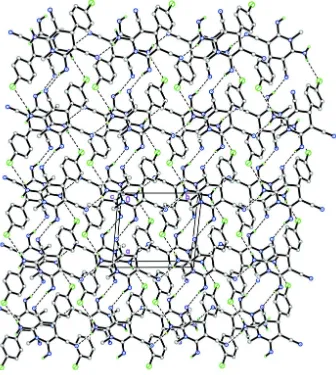
The crystal structure shown in Fig. 2 features N2—H1N2···N5i and N3—H2N3···N4iii hydrogen bonds (symmetry code
in Table 1) to result in tetrameric association of molecules, generated by inversion. These tetramers are then connected
via N2—H2N2···Cl1ii hydrogen bond (symmetry code in Table 1), forming a layer parallel to the ab plane.
S2. Experimental
Compound (I) was prepared by the reaction of p-chlorobenzaldehyde (1 mmol), malononitrile (1 mmol) and methylamine
(1 mmol) in a mixed solvent of methanol and water (5:1) was stirred at room temperature about an hour. The resulting
precipitate was collected by filtration and washed with methanol to afford pure product, m.p: 290 °C. The product was
crystallized from methanol solution as colourless plates.
S3. Refinement
N-bound H atoms were located in a difference Fourier maps and allowed to be refined freely [refined distance: N–H =
0.83 (4)–0.89 (4) Å]. The remaining hydrogen atoms were positioned geometrically [C–H= 0.95 or 0.98 Å] and were
refined using a riding model, with Uiso(H) = 1.2 Ueq(C) or 1.5 Ueq(methyl C). A rotating-group model was used for the
Figure 1
supporting information
[image:5.610.138.474.72.447.2]sup-3 Acta Cryst. (2014). E70, o812–o813
Figure 2
The crystal packing of the title compound. The H atoms not involved in the intermolecular interactions (dashed lines)
have been omitted for clarity.
2,6-Diamino-4-(4-chlorophenyl)-1-methyl-1,4-dihydropyridine-3,5-dicarbonitrile
Crystal data
C14H12ClN5 Mr = 285.74 Triclinic, P1 Hall symbol: -P 1
a = 8.3893 (4) Å
b = 8.4679 (5) Å
c = 10.2571 (6) Å
α = 93.148 (4)°
β = 112.478 (3)°
γ = 93.929 (3)°
V = 669.11 (6) Å3
Z = 2
F(000) = 296
Dx = 1.418 Mg m−3
Mo Kα radiation, λ = 0.71073 Å Cell parameters from 4444 reflections
θ = 2.6–32.3°
Bruker SMART APEXII CCD diffractometer
Radiation source: fine-focus sealed tube Graphite monochromator
φ and ω scans
Absorption correction: multi-scan
(SADABS; Bruker, 2009)
Tmin = 0.904, Tmax = 0.956
9775 measured reflections 3049 independent reflections 2435 reflections with I > 2σ(I)
Rint = 0.053
θmax = 27.5°, θmin = 2.2° h = −10→10
k = −10→9
l = −13→13
Refinement
Refinement on F2
Least-squares matrix: full
R[F2 > 2σ(F2)] = 0.072 wR(F2) = 0.207 S = 1.05 3049 reflections 198 parameters 0 restraints
Primary atom site location: structure-invariant direct methods
Secondary atom site location: difference Fourier map
Hydrogen site location: inferred from neighbouring sites
H atoms treated by a mixture of independent and constrained refinement
w = 1/[σ2(F
o2) + (0.1391P)2 + 0.3591P]
where P = (Fo2 + 2Fc2)/3
(Δ/σ)max < 0.001
Δρmax = 1.17 e Å−3
Δρmin = −0.48 e Å−3
Special details
Experimental. The crystal was placed in the cold stream of an Oxford Cryosystems Cobra open-flow nitrogen cryostat
(Cosier & Glazer, 1986) operating at 100.0 (1) K.
Geometry. All e.s.d.'s (except the e.s.d. in the dihedral angle between two l.s. planes) are estimated using the full
covariance matrix. The cell e.s.d.'s are taken into account individually in the estimation of e.s.d.'s in distances, angles and torsion angles; correlations between e.s.d.'s in cell parameters are only used when they are defined by crystal symmetry. An approximate (isotropic) treatment of cell e.s.d.'s is used for estimating e.s.d.'s involving l.s. planes.
Refinement. Refinement of F2 against ALL reflections. The weighted R-factor wR and goodness of fit S are based on F2,
conventional R-factors R are based on F, with F set to zero for negative F2. The threshold expression of F2 > σ(F2) is used
only for calculating R-factors(gt) etc. and is not relevant to the choice of reflections for refinement. R-factors based on F2
are statistically about twice as large as those based on F, and R- factors based on ALL data will be even larger.
Fractional atomic coordinates and isotropic or equivalent isotropic displacement parameters (Å2)
x y z Uiso*/Ueq
supporting information
sup-5 Acta Cryst. (2014). E70, o812–o813
H5A 0.5844 0.1308 0.1188 0.022* C6 0.7834 (3) 0.2950 (3) 0.1401 (3) 0.0136 (6) C7 0.9013 (3) 0.2604 (3) 0.2911 (3) 0.0133 (6) H7A 0.8677 0.3263 0.3587 0.016* C8 0.8816 (4) 0.0898 (3) 0.3211 (3) 0.0137 (6) C9 0.9872 (3) −0.0158 (3) 0.2979 (3) 0.0110 (6) C10 1.1955 (4) 0.1912 (3) 0.3081 (3) 0.0135 (6) C11 1.0935 (3) 0.3048 (3) 0.3278 (3) 0.0141 (6) C12 0.7568 (4) 0.0383 (3) 0.3743 (3) 0.0153 (6) C13 1.2207 (4) −0.0783 (4) 0.2184 (3) 0.0200 (7) H13A 1.1391 −0.1654 0.1568 0.030* H13B 1.3055 −0.1210 0.3008 0.030* H13C 1.2811 −0.0245 0.1657 0.030* C14 1.1690 (4) 0.4592 (4) 0.3845 (3) 0.0145 (6) H1N2 1.050 (6) −0.226 (6) 0.345 (5) 0.043 (12)* H2N2 0.873 (5) −0.220 (5) 0.322 (4) 0.022 (9)* H1N3 1.414 (5) 0.315 (6) 0.355 (4) 0.030 (11)* H2N3 1.425 (5) 0.150 (5) 0.355 (4) 0.029 (11)*
Atomic displacement parameters (Å2)
U11 U22 U33 U12 U13 U23
Cl1 0.0168 (4) 0.0188 (4) 0.0178 (4) 0.0060 (3) 0.0030 (3) 0.0034 (3) N1 0.0126 (11) 0.0132 (12) 0.0215 (12) 0.0042 (9) 0.0080 (10) −0.0010 (10) N2 0.0137 (12) 0.0082 (12) 0.0282 (14) 0.0055 (10) 0.0084 (11) 0.0015 (10) N3 0.0126 (12) 0.0132 (13) 0.0292 (14) 0.0081 (11) 0.0107 (11) 0.0041 (11) N4 0.0145 (12) 0.0168 (13) 0.0261 (14) 0.0081 (10) 0.0090 (11) 0.0030 (11) N5 0.0210 (13) 0.0136 (13) 0.0244 (13) 0.0080 (10) 0.0074 (11) 0.0020 (10) C1 0.0150 (13) 0.0141 (14) 0.0235 (15) 0.0024 (11) 0.0056 (12) −0.0009 (12) C2 0.0189 (14) 0.0164 (15) 0.0226 (15) 0.0036 (12) 0.0088 (12) 0.0034 (12) C3 0.0175 (13) 0.0154 (14) 0.0153 (13) 0.0113 (11) 0.0058 (11) 0.0010 (11) C4 0.0156 (13) 0.0162 (15) 0.0232 (15) 0.0019 (11) 0.0062 (12) 0.0023 (12) C5 0.0178 (14) 0.0135 (14) 0.0232 (15) 0.0020 (11) 0.0074 (12) 0.0048 (12) C6 0.0139 (13) 0.0130 (14) 0.0168 (13) 0.0094 (11) 0.0081 (11) 0.0002 (11) C7 0.0117 (12) 0.0118 (14) 0.0173 (14) 0.0068 (10) 0.0057 (11) 0.0005 (11) C8 0.0131 (12) 0.0112 (13) 0.0189 (14) 0.0056 (11) 0.0080 (11) 0.0016 (11) C9 0.0092 (12) 0.0084 (13) 0.0146 (13) 0.0072 (10) 0.0028 (10) −0.0003 (10) C10 0.0124 (13) 0.0136 (14) 0.0160 (13) 0.0054 (11) 0.0064 (11) 0.0022 (11) C11 0.0129 (13) 0.0131 (14) 0.0174 (14) 0.0064 (11) 0.0065 (11) 0.0003 (11) C12 0.0151 (13) 0.0130 (14) 0.0167 (14) 0.0091 (11) 0.0036 (11) 0.0022 (11) C13 0.0195 (14) 0.0142 (15) 0.0297 (17) 0.0075 (12) 0.0130 (13) −0.0041 (12) C14 0.0114 (12) 0.0164 (15) 0.0160 (13) 0.0086 (11) 0.0041 (11) 0.0032 (11)
Geometric parameters (Å, º)
N2—C9 1.379 (4) C5—H5A 0.9500 N2—H1N2 0.86 (5) C6—C7 1.543 (4) N2—H2N2 0.89 (4) C7—C8 1.505 (4) N3—C10 1.366 (4) C7—C11 1.523 (4) N3—H1N3 0.83 (5) C7—H7A 1.0000 N3—H2N3 0.83 (4) C8—C9 1.373 (4) N4—C12 1.155 (4) C8—C12 1.410 (4) N5—C14 1.162 (4) C10—C11 1.386 (4) C1—C6 1.393 (4) C11—C14 1.403 (4) C1—C2 1.395 (4) C13—H13A 0.9800 C1—H1A 0.9500 C13—H13B 0.9800 C2—C3 1.388 (4) C13—H13C 0.9800 C2—H2A 0.9500
C9—N1—C10 118.3 (2) C8—C7—C11 107.0 (2) C9—N1—C13 120.6 (2) C8—C7—C6 113.9 (2) C10—N1—C13 119.7 (2) C11—C7—C6 113.9 (2) C9—N2—H1N2 117 (3) C8—C7—H7A 107.2 C9—N2—H2N2 121 (3) C11—C7—H7A 107.2 H1N2—N2—H2N2 108 (4) C6—C7—H7A 107.2 C10—N3—H1N3 120 (3) C9—C8—C12 119.9 (3) C10—N3—H2N3 113 (3) C9—C8—C7 119.8 (3) H1N3—N3—H2N3 115 (4) C12—C8—C7 120.3 (2) C6—C1—C2 121.3 (3) C8—C9—N1 120.7 (3) C6—C1—H1A 119.4 C8—C9—N2 123.1 (3) C2—C1—H1A 119.4 N1—C9—N2 116.2 (2) C3—C2—C1 118.4 (3) N3—C10—N1 116.7 (3) C3—C2—H2A 120.8 N3—C10—C11 123.7 (3) C1—C2—H2A 120.8 N1—C10—C11 119.6 (3) C4—C3—C2 121.8 (3) C10—C11—C14 119.7 (3) C4—C3—Cl1 119.6 (2) C10—C11—C7 119.9 (3) C2—C3—Cl1 118.5 (2) C14—C11—C7 120.4 (2) C3—C4—C5 118.9 (3) N4—C12—C8 178.0 (3) C3—C4—H4A 120.5 N1—C13—H13A 109.5 C5—C4—H4A 120.5 N1—C13—H13B 109.5 C4—C5—C6 121.3 (3) H13A—C13—H13B 109.5 C4—C5—H5A 119.3 N1—C13—H13C 109.5 C6—C5—H5A 119.3 H13A—C13—H13C 109.5 C1—C6—C5 118.2 (3) H13B—C13—H13C 109.5 C1—C6—C7 121.4 (3) N5—C14—C11 177.8 (3) C5—C6—C7 120.1 (3)
supporting information
sup-7 Acta Cryst. (2014). E70, o812–o813
C3—C4—C5—C6 −0.2 (5) C10—N1—C9—N2 −157.4 (3) C2—C1—C6—C5 −1.3 (4) C13—N1—C9—N2 8.9 (4) C2—C1—C6—C7 −175.4 (3) C9—N1—C10—N3 156.2 (3) C4—C5—C6—C1 1.2 (4) C13—N1—C10—N3 −10.2 (4) C4—C5—C6—C7 175.4 (3) C9—N1—C10—C11 −25.5 (4) C1—C6—C7—C8 −152.0 (3) C13—N1—C10—C11 168.1 (3) C5—C6—C7—C8 34.0 (3) N3—C10—C11—C14 −8.3 (4) C1—C6—C7—C11 −28.9 (4) N1—C10—C11—C14 173.5 (3) C5—C6—C7—C11 157.1 (3) N3—C10—C11—C7 173.1 (3) C11—C7—C8—C9 −34.8 (3) N1—C10—C11—C7 −5.1 (4) C6—C7—C8—C9 92.0 (3) C8—C7—C11—C10 32.9 (4) C11—C7—C8—C12 144.5 (3) C6—C7—C11—C10 −93.9 (3) C6—C7—C8—C12 −88.7 (3) C8—C7—C11—C14 −145.7 (3) C12—C8—C9—N1 −170.4 (3) C6—C7—C11—C14 87.6 (3)
Hydrogen-bond geometry (Å, º)
D—H···A D—H H···A D···A D—H···A
N2—H1N2···N5i 0.86 (5) 2.21 (5) 3.043 (4) 163 (5)
N2—H2N2···Cl1ii 0.89 (4) 2.75 (4) 3.588 (3) 158 (3)
N3—H2N3···N4iii 0.83 (4) 2.27 (4) 3.071 (4) 161 (4)