Detection of Diabetic Retinopathy by Screening of
Fundus Images
Manoj Kumar S B
1, Dr. H S Sheshadri
2, Janhavi G M
3, Jayalakshmi H S
4, Madhura H R
5, Varshitha C N
61Research Scholar PET Research foundation, Mandya 2Professor, Dept .of ECE, PESCE, Mandya
Abstract: Diabetic retinopathy is diabetes complication that it affects the eyes. The diabetic retinopathy will cause blindness and vision loss. The DR is caused due to damage to the blood vessels of the eye, which contains the light sensitive tissue at the back of eye. The cause of damage to eye due to this procedure is called diabetic retinopathy. A patient who is suffering from the DR has to undergo regular or periodic screening of eye and to identify the DR. The ophthalmologists use retinal images of patients that images are acquired from digital fundus camera. There are different types of lesions they are Micro aneurysms (MAs), Haemorrhages (HEM) and Exudates (EXs). An screening of this lesions make the ophthalmologists to identify the patient who need further medical treatment. The detection of the micro aneurysms at an early stage helps us to preventing the DR. Regular screening of the DR and diabetic maculopathy diseases are necessary in order to identify the stages DR and to identify the group at risk of visual impairment. The main of this work is to increase the accuracy and sensitivity of the detection above 98% and 99%.
Keywords: Diabetic Retinopathy (DR), Exudates (EXs), Haemorrhages (HEM), Micro aneurysms (MAs,) Structure Element (SE) and Support vector network (SVM).
I. INTRODUCTION
Diabetes is a disease that occurs when your blood sugar level is too high. This is caused due to insufficient insulin in your body or does not make well use of insulin, which converts blood sugar to energy. Over time having too much glucose in your blood can cause health problems that are called diabetes. It has no cure but we can take step to manage the diabetes.
There are different types of diabetes and most common are
1) Type 1 Diabetes: if the people have type 1 diabetes your body does not make insulin, your immune system attacks and destroy the cells in your pancreas that make insulin. Type 1 diabetes are usually found in adults and you g people although it can appear at any age. People with type 1 diabetes should take insulin every day to stay alive.
2) Type 2 Diabetes: if the people have type 2 diabetes, the body does not use the insulin well. It can develop at any age. It occurs most often in middle age and older people. Type 2 is the common type of diabetes.
3) Gestational Diabetes: it develops in women when they are pregnant. Most of the time it goes away when the baby is born. If you have gestational diabetes you have greater chance of developing type 2 diabetes.
To avoid the diabetic retinopathy the regular screening of the eyes are required, due to the periodic screening we can early detect the diabetic retinopathy and avoid the vision loss. The ophthalmologists can know that patient is suffering from the diabetic retinopathy when he firstly see the presence of the micro aneurysms on the retina which is the first and most characteristic symptom of this disease. Micro aneurysms will appear as the small, round shaped and red dots on the retina.
[image:1.595.207.394.628.735.2]There are four stages which are described form the National Eye Institute (NEI).
a) Mild Non-proliferative Retinopathy (NPDR): In this stage, a few micro aneurysms, which are defined as small outpunching in the walls of the tiny blood vessels (called capillaries), appear in the retina.
b) Moderate Non-proliferative Retinopathy (NPDR): More lesions appear in this stage as more capillaries that nurture the retinal tissue become damaged, and the retina become more ischemic (lack of blood flow, and therefore lack of oxygen).
c) Severe Non-proliferative Retinopathy (NPDR): At this level of DR, many blood vessels are affected. Blood vessel supply of oxygen to the retina is severely compromised due to accumulated vessel damage. When this occurs, certain areas of the retina start sending biochemical signals to the body that they need oxygen.
d) Proliferative Retinopathy (PDR): In response to the need for oxygen, new vessels begin to grow within the retina. These new vessels are an aborted attempt of the retina to regain its oxygenation need, but these vessels are compromised and fragile. These vessels break easily causing severe bleeding into the vitreous gel of the eye and consequent loss of vision. Also, these new vessels can attach themselves into the vitreous gel and cause traction on the retinal plane, causing retinal detachments.
II. ABNORMALITIES ASSOCIATED WITH THE EYE
(a) (b)
(c) (d)
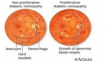
Figure 2.(a) Micro aneurysm (b) Haemorrhages (c) Hard Exudates (d) soft Exudates
1) Micro Aneurysms: These are the first clinical abnormality to be noticed in the eye. They may appear in isolation or in clusters as tiny, dark red spots or looking like tiny haemorrhages within the light sensitive retina. As their sizes ranges from 10-100 microns i.e. less than 1/12th the diameter of an average optics disc and are circular in shape, at this stage, the disease is not eye threatening.
2) Haemorrhages: Occurs in the deeper layers of the retina and are often called ‘blot’ haemorrhages because of their round shape. 3) Hard Exudates: These are one of the main characteristics of diabetic retinopathy and can vary in size from tiny specks to large
patches with clear edges. As well as blood, fluid that is rich in fat and protein is contained in the eye and this is the reason for the formation of exudates.
[image:2.595.178.447.263.581.2]III. METHODOLOGY
Figure 3.Flow of project A. Working Principle
1) Image Acquisition: Reading the eye image of diabetes patients, the input fundus image is analysed by the system and the output contains the grading and the result with the co-ordinates of the detected abnormality shown on the GUI. The input image to the Pre-Processing stage can be a colour or a grey level image. The Pre-Processing stage corrects the problem of illumination variation that occurred when taken the pictures. The output of this stage is passed to the Segmentation stage. The exudates and
the vein networks class centres also contain some noisy pixels that were over enhanced 4 during the Pre-Processing stage and
will be removed during the next stage called Disease Classifier stage.
2) Image Pre-processing: The appearance of different DR lesions differs, for example as shown in figure 4 being dark spots MAs and HEMs are mostly inseparable from the background whereas Exs are yellowish objects of high contrast. Some of sort of edge enhancement operation is therefore necessary to distinguish dark lesions from the background. On other hand, pre-processing for EX detection is more of contrast enhancement rather than edge pre-processing.
Figure 4.Curvelet Edge Enhancement Image
Data Set Lesion image extracted
from Image Image Pre-processing
Lesion Detection
SVM Classifier
[image:3.595.219.391.587.730.2]B. Lesion Detection
Matched Filtering and Laplacian of Gaussian Filtering Matched filer with a 2D Gaussian kernel produces high response for both the dark and the bright lesions that can be modelled as gaussian and step edges. On other hand at a sharp intensity transition of the bright lesions, the response of LOG shows zero crossing about its centre. Thus LOG filter along with MF, is used to detect the transient- like bright lesions and the dark lesions with Gaussian-like intensity profile.
Figure 5.Lesion Detection
Different spurious components, background pixels may also be erroneously detected as candidate lesions. Now proper post processing is necessary for each lesion type. For MA detection, if the number of neighbouring pixels less than a certain threshold, the pixel is considered to be an isolated point and that is removed. For HEM detection, the candidate region whose area is less than a certain threshold is first eliminated. For Exs, candidate regions with area above a certain threshold are first closed using disk shaped structuring element (SE).
C. SVM Classifier
[image:4.595.167.428.174.375.2]SVM are supervised learning models is also associated with the learning algorithms that analyse data used for classification and regression analysis. Each marked as belonging to one or more categories, An obtained SVM training algorithm will create a model that will provide the new examples to one or more category and making it a non-probabilistic binary linear classifier. An SVM model gives the examples of points in space and mapped so that the examples of the separate categories are divided by a clear gap that is as wide as possible. The new examples are then mapped into the similar space and identify a category based on which side of the gap they fall.
IV. RESULT
Figure 7.Selecting the Input Images
The process step performs for the Detection of Retinopathy by Screening of fundus images. The first step is to take the image from the data set. Then it completely shows all detecting stages by screening of fundus images.
A. Detection of Diseases 1) Detection of Blood Vessels
(a) (b) Figure 8.(a) Input Image (b) Blood Vessel Detected 2) Detection of Optical Disc
[image:5.595.107.513.416.568.2] [image:5.595.159.466.600.729.2]
3) Detection of Enhance Image
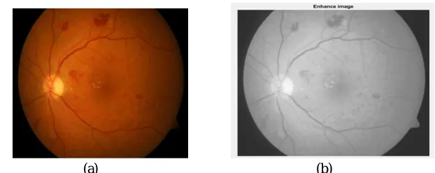
(a) (b) Figure 10. (a) Input Image (b) Enhance Image
4) Detection of Micro Aneurysms
(a) (b) Figure 11.(a) Input Image (b) Micro aneurysms Detected 5) Normal Condition
(a) (b) Figure 12.(a) Input Image (b) Normal Image Detected 6) Detection of Haemorrhages
[image:6.595.138.449.122.246.2] [image:6.595.36.460.193.741.2]7) Detection of Soft Exudates
(a) (b)
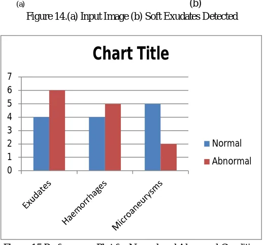
Figure 14.(a) Input Image (b) Soft Exudates Detected
Figure 15.Performance Plot for Normal and Abnormal Condition
The performance plot for normal and abnormal condition. The graph represents the comparison between abnormal and normal condition of each detected diseases like Exudates, Haemorrhages and Micro aneurysms. The Blue shade shows the Normal Condition and The Red shade shows the Abnormal Condition.
V. CONCLUSION
The development of automatic detection of diabetic retinopathy screening system is a very challenging task the system development involves not only an advanced understanding of image processing procedures, but also requires essential medical input including expert knowledge related to diabetic retinopathy and its screening procedure in addition to the eye fundus photography process. Efficient and cost-effective approaches in the field of digital retinal imaging should be established. Diabetic retinopathy and maculopathy screening is necessary to identify persons at risk of visual impairment effective screening of diabetic retinopathy therefore it is vital for early actions, alongside an effective diabetic applications preventive management.
We used a highly efficient and accurate image processing techniques in order to produce an effective screening of DR. we achieved the sensitivity and accuracy of the detector up to 93%.
VI. ACKNOWLEDGMENT
I would like sincerely thank to DR. H SSHESHADRI, Professor, Dept. of ECE, PESCE, Mandya, DR. NARENDRA B K, Principal
and DR. M B ANANDARAJU, Professor BGSIT, BG Nagara for providing the necessary facilities and support, thus making it possible for us obtains the necessary resources required to complete this Research Paper.
0 1 2 3 4 5 6 7
Chart Title
Normal
[image:7.595.168.430.248.489.2]REFERENCES
[1] Pathak D and curr Drug Deliv, Oral targeting of protein Kinase C receptor: promising route for disbetic retinopathy. [2] Anant Pai, maha m.EI Shafei and Mustafa AI hashimi, Current concepts in intravitreal drug therapy for diabetic retinopathy.
[3] M. U. Akram, S. Khalid, A. Tariq, S. A. Khan and F. Azam,Detection and classification of retinal lesions for grading of diabetic retinopathy, Computers in Biology and Medicine, vol. 45, pp. 161 – 171, 2014.
[4] I. Lazar and A. Hajdu, Retinal microaneurysm detection through local rotating cross-section profile analysis. IEEE Transactions on Medical Imaging, vol. 32, no. 2, pp. 400–407, 2013.
[5] P.R.Harper,M.G.Sayyad,V.de Senna,A.K.Shahani,C.S.Yajnik,K.M.Shelgikar,A Systems modeling approach for the prevention and treatment of diabetic retinopathy
[6] Manoj Kumar S B, Dr. H S Sheshadri and Manjunath R, Feature extraction from the fundus images for the diagnosis of diabetic retinopathy, IEEE Explore, pp. 240-245
[7] L. Donoho and M. R. Duncan, Digital Curvelet Transform: Strategy, Implementation and Experiments, Department of Statistics Stanford University,
November 1999.
[8] Manoj Kumar S B, Dr. H S Sheshadri, Classification and Detection of Diabetic Retinopathy using K-Means algorithm,IEEE Explore, pp.326-331
[9] Thomas w Gardner, David A Antonetti , Alistair and Steven w Levison , Retinal micro vascular dysfunction in diabetes, Survey of ophthalmology 47,
2002.