Int J Clin Exp Med 2019;12(6):6703-6719
www.ijcem.com /ISSN:1940-5901/IJCEM0089740
Original Article
Apatinib inhibits proliferation of different lung cancer
cells
in vitro
and
in vivo
by promoting apoptosis
and inducing cell cycle arrest
Mingtao Liu1,2, Xiuxiu Wang1, Lijun Jing1, Peng Jiang1,3, Yu Li1
1Department of Pulmonary Medicine, Qilu Hospital, Shandong University, Jinan, Shandong, China; 2Department of
Pulmonary Medicine, Binzhou People’s Hospital, Binzhou, Shandong, China; 3Department of Pulmonary Medicine,
Weihai Municipal Hospital, Weihai, Shandong, China
Received December 12, 2018; Accepted April 9, 2019; Epub June 15, 2019; Published June 30, 2019
Abstract: Apatinib is a specific VEGFR-2 inhibitor that blocks transmission of the VEGF/VEGFR-2 signaling path
-way by competitively binding to the VEGFR-2 intracellular tyrosine ATP binding site and exerting biological effects. Different levels of VEGFR-2 expression in different lung cancer cells and the cell lines with low and high expression of VEGFR-2 were selected, and the effect of apatinib on their proliferation was observed in vitro and in vivo. In vitro
studies showed that apatinib significantly inhibited the proliferation of VEGFR-2 high-expression lung cancer cell
lines H1975 and H446 in a concentration-dependent manner, mainly by promoting apoptosis and cell cycle arrest.
For the lung cancer cell line A549 with low VEGFR-2 expression, the same concentration of apatinib had no inhibi
-tory effect. In vivo studies showed that both H1975 and H446 xenografts were significantly inhibited by low- and high-dose apatinib, while in A549 cells, low-dose apatinib had no significant inhibitory effect. Therefore, apatinib has different inhibitory effects on different lung cancer cells, and the inhibitory effect on lung cancer cells with high VEGFR-2 expression is more significant by promoting apoptosis and inducing cell cycle arrest in vitro and in vivo. Keywords: Apatinib, expression level of VEGFR-2, proliferation, apoptosis, cell cycle arrest
Introduction
The incidence and mortality of lung cancer ranks first among all cancers [1], and because
its early symptoms are not obvious, combined
with the lack of effective early diagnoses, most
lung cancer patients are diagnosed in advanced
stages [2]. Currently, there is still no effective treatment for advanced lung cancer. Although
new cytotoxic drugs and molecular targeted drugs are continuously developed, the 5-year
survival rate of patients with advanced lung cancer is still low [3]. Improving the efficacy of treatments for patients with lung cancer and prolonging their survival is an urgent need for
clinicians.
Folkman proposed that tumor growth depends on the formation of tumor blood vessels and
thus proposed that anti-angiogenesis will be
an important method for the treatment of tu-mors [4]. A variety of growth factors and their
corresponding receptors are involved in tumor
angiogenesis in which vascular endothelial gr-
owth factors (VEGFs) and their vascular endo
-thelial growth factor receptors (VEGFRs) play major roles [5, 6]. The VEGFR family members comprise three types: VEGFR-1, VEGFR-2 and VEGFR-3 [7]. The VEGF/VEGFR-2 signaling path -way plays an important role in tumor
angiogen-esis, cell proliferation, invasion and migration. Therefore, blocking this signaling pathway and
inhibiting tumor angiogenesis has become a
new area of tumor-targeted therapy [8].
Apatinib is a small molecule tyrosine kinase
inhibitor that selectively inhibits the
phosphory-lation of VEGFR-2 and its tyrosine by competi
-tively binding to the VEGFR-2 intracellular tyro
-sine ATP binding site. Kinase activity blocks the transmission of the VEGF/VEGFR-2
signal-ing pathway and inhibits tumor angiogenesis,
thereby inhibiting tumor growth [9, 10].
Cur-rently, apatinib is mainly used in the treatment
cancer with apatinib is mainly focused on the clinical study of patients with non-small cell lung cancer. LIU reported that a clinical trial of
apatinib in patients with advanced non-small
cell lung cancer found that patients with apa
-tinib had a median PFS of 4 months and a dis
-ease control rate of 61.76% [12]. Cases of effective treatment of small cell lung cancer with apatinib have also been reported [13]. Apatinib has a therapeutic effect in patients
with advanced lung cancer and can improve
patient PFS and DCR to some extent. However, there are few basic studies on the effects of apatinib on lung cancer cells, especially the dif
-ferent types of lung cancer cells, and the me-chanism of action has not been reported. For this study five lung cancer cell lines were select
-ed with different expression levels of VEGFR-2 and determined the effect of apatinib on pro-liferation in vitro and in vivo and observed the
safety of apatinib. This study may provide a theoretical basis for the more rational applica
-tion of apatinib in patients with advanced lung
cancer.
Materials and methods
Cell culture
The human lung cancer cell lines A549,
HCC-827, H2228, H1975 and H446 cells were
pur-chased from the Cell Bank of the Chinese Academy of Sciences (Shanghai, China). All cell
lines were maintained in RPMI-1640 (HyClone,
USA) supplemented with 10% FBS (Gibco, NY),
100 U/mL penicillin (HyClone, USA), 50 mg/mL streptomycin (HyClone, USA), and 2 mmol/L
glutamine in a humidified CO2 incubator at
37°C. All cells were passaged for less than 3 months before renewal from frozen, early-pas
-sage stocks obtained from the indicated
so-urces.
Reagents and antibodys
Apatinib (Selleck, s2221, Shanghai, China) was dissolved in 0.1% DMSO (Sigma Aldrich) at a concentration of 10, 20 mmol/L and stored at -20°C for vitro testing. Apatinib mesylate (Heng
Rui, Jiangsu, China) were ground into powder
and dissolved 0.5% CMC (Solrbio, Shanghai, China) for vivo testing. Primary antibodies against AKT (ab8805), phosphor-AKT (ab8932), ERK (ab54230), phospho-ERK (ab201015),
el-F4E (ab205824), phosphor-elel-F4E (ab2008-58); HIF-1α (ab51608), VEGF (ab46154), CD31 (ab28364), β-actin (ab8227) were purchased from abcam (Cambridge, MA, USA). Primary
antibodies against PARP (9532), caspase3 (96-
62), cleaved-caspase3 (9664), cyclinB1 (4138), P53 (9282), VEGFR-2 (2472), Ki67 (9449) and anti-rabbit or anti-mouse IgG horseradish per
-oxidase (HRP)-linked secondary antibodies we-re purchased from Cell Signaling Technology (Boston, MA, USA).
Cell proliferation
Five lung cancer cells were plated into 96-well plates (2000~4000/well) in 100 μl of complete
medium, and six parallel wells were assigned to each group, as well as a negative control
(with-out cells). After overnight incubation, cells
we-re exposed to various concentrations (0, 1, 10,
50, 100, 250 μM) of apatinib for a further 72 hours. Cell proliferation was evaluated by Cell Counting Kit 8 (CCK8, BestBio, Shanghai, China). Briefly, the inhibitory rates of each cell line with different concentration of apatinib were calculated by comparing the OD values of the experimental groups to that of the empty group. The half maximal inhibitory concentra -tion (IC50) of apatinib that could inhibit growth by 50% were calculated by concentration-response cure fitting using GraphPad Prism 5.0 software. Each IC50 value was expressed as the mean ± SD.
Colony formation assays
For the clonogenic assays, five tumor cell lines
in the logarithmic growth phase were seeded in 6-well plates in complete culture medium either the vehicle control or two concentration
of apatinib (10 and 20 μM) for two weeks. Each group was allotted three plates. The colonies that formed on each plate were stained with crystal violet, and the number of colonies were counted by Image J software. The colony forma
-tion rate (%) was expressed as the mean ± SD.
Cell cycle analysis
For the cell cycle assays, three tumor cell lines
include A549, H1975 and H446 were
harvest-ed and washharvest-ed twice with cold PBS and then were fixed with pre-chilled 70% ethanol for 24 h at 4°C. The fixed cells were washed and resus
Effect of apatinib on lung cancer cells
A in a water bath at 37°C for 30 min. Next, the cells were filtered and incubated with 300 μL of propidium iodide at 4°C for 30 min in the dark. The stained cells were detected by flow cytom
-etry and were analyzed using ModFit LT 4.1 software.
Assessment of apoptosis
The treated cells were washed twice with PBS, resuspended in binding buffer at a density of 1
× 107 cells/mL and then were stained using an
annexin V-FITC-PI apoptosis detection kit (BD, USA) for 20 min at room temperature in the dark according to the manufacturer’s proto
-cols. Thereafter, the labeled cells were detect
-ed by flow cytometry and were analyz-ed using FlowJo 7.6 software. Next, tunel apoptosis de-tection kit was used to detect apoptosis of three kind cells treated by different concentra
-tion apatinib according to the manufacturer’s
manual (Yeasen, China).
Immunofluorescence assay
Cells were seeded on coverslips and were fixed with 4% paraformaldehyde for 30 min at room temperature. After permeabilization with 0.2% TX-100 (Sigma, × 100) for 10 minutes at room
temperature, cells were incubated with cleaved
caspase 3 antibody at 4°C for 12 h. After wash
-ing with PBS, cells were incubated with Flu-orescein-Conjugated Goat anti-Rabbit IgG (H+L) for 1 h. Immunofluorescence images were vie-wed under fluorescence microscope (Olympus I
× 81, Japan). Cleaved caspase 3 were
quanti-fied with the Image J software. Mean fluores
-cence of the nuclear staining was obtained by selecting the “measure” option. After deducting any background staining, the mean fluores
-cence of at least 50 cells from each treatment was used for statistical analysis. Data was rep -resented as mean ± SD.
Western blot analysis
Cells and tissues were lysed by radio
immuno-precipitation assay (RIPA, Beyotime, China) buf
-fer containing phenyl methane sulfonyl fluoride (PMSF, Solarbio, China) with mild sonication. The concentrations of total proteins were mea
-sured by the BCA Protein Assay Kit (Beyotime, China). Equal amounts of protein were sub-jected to 10% sodium dodecyl sulfate polya-crylamide gel electrophoresis (SDS-PAGE) and
blotted on polyvinylidene fluoride (PVDF, Milli-pore, Billerica, USA). Protein bands were visual
-ized via enhanced chemiluminescence (ECL, Millipore, USA) and were analyzed using the
western blot imaging system (AI600 images,
GE, USA), followed by measurement of the den
-sity of each band using Image J software.
RNA preparation and RT–PCR analysis
Total RNA were isolated with Trizol reagent (Invitrogen, USA). The reverse transcription re-actions were performed by use of Reverse Tr-anscription Reagent kits (Takara, Japan). Real-time PCR was conducted with SYBR Green mix (Takara, Japan). The primer for VEGFR-2 was designed as 5’-ACCCCTTGAGTCCAATCACACA-3’ (forward), 5’-CTTCCTCCAACTGCCAA TACCA-3’ (reverse); The primer for GAPDH was designed as 5’-GGAAGCT TGTCATCAATGGAAATC-3’ (for
-ward), 5’-TGATGACCCTTTTGGCTCCC-3’ (rever-se). The expression of VEGFR-2 mRNA was cal
-culated and normalized using the 2-ΔΔCt me-
thod relative to GAPDH.
Tumor xenograft mouse models
Five to six week old female BALB/c nude mice were purchased from Nanjing University
Bi-omedical Research Institute (Nanjing, China).
All animal experiments were performed in the animal research center of Shandong Medical
Academy in accordance with the National In-
stitutes of Health guide for the care and use of
laboratory animals. A549, H1975, and H446 cell lines were selected to construct three nude
mouse xenograft models. Approximately 2 ×
106 cells were suspended in 0.2 ml of PBS, sub
-cutaneously injected into the right chest wall of each nude mouse, and tumors formed in approximately 10 days. The tumor volume was
calculated as L × W2/2 (L and W are the leng-
th and width of the tumor, respectively). The nude mice with transplanted tumor volumes of
50-150 mm3 were selected for intervention
grouping. There was no significant difference in the average volume of transplanted tumors between the nude mice before intervention. Fifteen tumor-bearing nude mice from each of
the three cell lines were randomly divided into three groups according to a random number
table. The animals were treated for 21 consec -utive days once daily by oral gavage with
-ture. The body weight and the tumor volume
were recorded every 3 days by the same
per-son. At harvest, the mice were sacrificed under anesthesia. The tumor tissues were fixed in 4% paraformaldehyde for immunohistochemistry or stored at -80°C for western blotting, and blood was harvested for ELISA assays. The heart, liver, kidney, thyroid and pancreas were fixed in 4% paraformaldehyde and electron microscopy fixative for H&E staining and elec -tron microscopy specimen preparation
respec-tively. All animal experiments were perform-ed with the approval of Shandong University
Animal Care and Use Committee.
Immunohistochemical staining
For immunohistochemical staining, the trans
-planted tumor tissues were fixed in 4% parafor
-maldehyde, and paraffin blocks were prepared after dehydrating and embedding. A series of 3 mm sections were obtained from each paraffin block. Each slice was baked at 65°C for 1 hour and dewaxed by xylene. Then, the slides were
dehydrated using ethanol and antigens were
repaired by EDTA. The peroxidase was removed using 3% H2O2 and blocked using 5% BSA for
30 minutes. Incubation with the primary
anti-body was conducted overnight at 4°C. The sec -tions were incubated with a biotin secondary
antibody for 20 minutes at room temperature. Targeted proteins were visualized using the per
-oxidase substrate diaminobenzidine. Staining intensities were estimated in five random fields
per section by three independent observers individually.
Toxicological study
Hematoxylin and eosin staining: For hematoxy
-lin and eosin (H&E) staining, the heart, liver, kidney, thyroid, and pancreas tissues of the mice were fixed in 4% paraformaldehyde and embedded in paraffin and then cut into 4 μm sections. The sections were stained with both
hematoxylin and eosin and then photographed using a light microscope.
Electron microscope specimen production
The heart, liver, kidney, thyroid, and pancreas tissues of the mice were fixed in 3% glutaral-dehyde at 4°C for 4 hours and then washed with 0.1 M sodium dicarboxylate buffer and soaked in 1% citric acid at 4°C for 2 hours.
Next, the tissues were washed with 0.1 M
sodi-um diformate buffer twice and were dehydrated
by graded ethanol. The tissues were permeat -ed with propylene oxide, completely emb-edd-ed in an embedding solution, and placed in a 40°C
incubator for 12 hours. Then, the slides were transferred to an embedding plate and incubat
-ed at 60°C for 48 hours. After the fabrication, the ultrastructure of the organs were observ-ed by transmission electron microscopy (JEOL,
Japan).
Serum biochemistry
Blood samples were obtained from the vein of
the inner canthus while the mice were under
anesthesia from a 1% pentobarbital sodium intraperitoneal injection. All of the nude mice were fasted for more than 8 hours before tak
-ing blood. Blood was allowed to coagulate at room temperature for 30 minutes and was cen
-trifuged at 3,000 rpm for 10 minutes to sepa
-rate the serum. Biochemical tests for the level of ALT, AST, BUN, Cr, LDH-L, CK and glucose in serum were performed. ELISA kits (cloud clone, USA) were used to detect the concentration of FT3 and FT4 in the serum. The specific proce
-dure followed the steps provided by the ELISA kit. All tests were repeated 3 times, and the
data are represented as the mean ± SD.
Statistical analysis
All the experiments in our research were
con-ducted at least three times individually. The
results are presented as mean ± standard devi-ation (SD).
Data were analyzed using GraphPad Prism software(version 5.0). The Two-way ANOVA was used to value the overall statistical significance for the means of multiple samples. A P-value of
less than 0.05 was considered statistically
sig-nificant (*represent P values<0.05).
Results
Apatinib exerts anti-proliferative activity against lung cancer cells in vitro
CCK8 assay was performed on a panel of five
lung cancer cell lines (A549, HCC827, H2228,
H1975, ad H446). The results show that as the concentration of apatinib increased, the inhibi
-tion of prolifera-tion of the five lung cancer cell lines were increased. In five lung cancer cells, the inhibition of proliferation of H1975 and
H446 were more obvious. When the
Effect of apatinib on lung cancer cells
rate of H1975 and H446 were 54.6 ± 5.48% and 65.74 ± 2.05%, respectively, while the inhi
-bition rates of A549, HCC827 and H2228 we-re 11.36 ± 0.89%, 12.80 ± 1.26%, and 19 ± 0.75%, significantly lower than H1975 and H446. There were similar results at 48 and 72
hours (Figure 1A-C). The results showed that
the IC50 values of apatinib for the five lung can
-cer cells at 24 hours were as follows: A549: 186.5 ± 4.86 μM, HCC827: 181.86 ± 4.86 μM,
H2228: 223.85 ± 11.7 μM, H1975: 31.12 ± 1.38 μM, and H446: 13.94 ± 1.88 μM. The IC50 values of H1975 and H446 were significantly lower than that of the other three lung cancer cells (P<0.05). Over time, the IC50 values of apa
-tinib for the five lung cancer cell lines decreased
[image:5.612.93.520.67.569.2]to some extent, but the IC50 values of H1975 and H446 were still significantly lower than that of other lung cancer cell lines (P<0.05) (Table 1).
Figure 1. Effects of apatinib on proliferation and expression of VEGFR-2 of different lung can -cer cell lines. A. Inhibition rate
of apatinib on five lung cancer cell lines at 24 hours. B. Inhibi
-tion rate of apatinib on five lung
cancer cell lines at 48 hours.
C. Inhibition rate of apatinib on five lung cancer cell lines at 72
hours. D and E. Relative
expres-sion levels of VEGFR-2 protein. F. Relative mRNA levels of VEGFR-2 in five cell lines. G and H. Effect of apatinib on colony formation of five cells. As the concentration of
apatinib increased, the number
of colonies did not decrease sig
-nificantly for A549, HCC827 and
H2228 cells (P>0.05); While the
number of colonies decreased significantly compared with the
vehicle group in H1975 and
Table 1. IC50 values for apatinib of five cells at different times (μM)
Cells Times
24 h 48 h 72 h
A549 186.6 ± 5.9 161.1 ± 5.4 155.3 ± 4.8 HCC827 157.4 ± 5.7 144.2 ± 7.5 139.3 ± 6.2 H2228 233.8 ± 14.3 210.2 ± 18.4 192.4 ± 9.9 H1975 31.2 ± 1.8* 18.7 ± 2.4* 14.6 ± 1.2*
H446 13.9 ± 2.3* 11.4 ± 2.1* 9.9 ± 0.7* The IC50 values of apatinib for five lung cancer cell lines at different times, *p<0.05.
Relative VEGFR-2 protein and mRNA expres-sion levels
The CCK8 results confirmed that in these five lung cancer cell lines, the inhibitory effects of
apatinib on H1975 and H446 were more
obvi-ous. Apatinib specifically binds to VEGFR-2 to exert its biological effects, and the expression of VEGFR-2 in the five cell lines was different, leading to different inhibition effects of apa
-tinib. Expression of VEGFR-2 protein and mRNA
was observed by Western blot and qPCR. We- stern blot analysis showed that the expression
levels of VEGFR-2 protein in H1975 and H446 cell lines were significantly higher than those in
the other three cell lines (Figure 1D and 1E). Similar results were observed at the RNA level (Figure 1F). The qPCR results showed that the relative expression levels of VEGFR-2 mRNA in H1975 and H446 were significantly higher than that of the other three lung cancer cells.
Colony formation assay
To further observe the effects of different con
-centrations of apatinib on the proliferation of different lung cancer cells, colony formation as-say was performed. A549, HCC827 and H2228 cells were selected to represent VEGFR-2 low
expressing cell lines, and H1975 and H446
cells were selected to represent VEGFR-2 high expressing cell lines. A difference in the prolif
-eration of lung cancer cell lines with different expression levels of VEGFR-2 after apatinib tre-atment was observed. Following 2 weeks of cul
-ture, apatinib inhibited the number of the colo
-nies formed by H1975 and H446 cells in a
dose-dependent manner. However, apatinib did
not inhibit the formation of colonies in A 549,
HCC827 and H2228 cells (Figure 1G and 1H). Collectively, these results revealed that
apa-tinib can inhibit the formation of colonies in VEGFR-2 high-expression lung cancer cell lines.
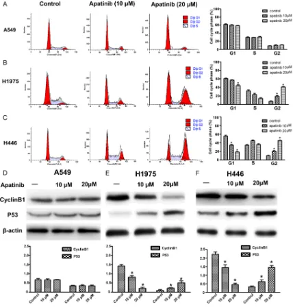
Cell cycle regulation
The capacity of apatinib to induce cell cycle
arrest was demonstrated in H1975 and H446 cells, but not in the A549 cell line. Apatinib
induced a dose-dependent G2 phase arrest in H1975 and H446 cells. At 0, 10 and 20 μM apatinib, the G2 phase ratio of the H1975 and H446 cells increased significantly (H1975: 7.45 ± 1.13%, 19.2 ± 2.12%, and 41.2 ± 0.98%,
respectively; H446: 10.09 ± 0.80, 20.33 ±
1.31%, and 45.47 ± 1.02%, respectively). There was a significant difference in the comparison
(P<0.05) (Figure 2B and 2C). However, the
effect on A549 cells was minor, as there was no significant difference compared with the
control group (Figure 2A). Western blot was
used to detect the expression of cyclin B1 and p53, which were G2 phase related proteins. The results showed that with the increase of apatinib concentration, the expression of cyclin B1 were decreased while p53 increased in H1975 and H446 cell lines. There was a sig-nificant difference compared with the control
group (P<0.05) (Figure 2E and 2F). Changes in
the expression levels of the two proteins led to an increase in the proportion of cells staying in the G2 phase, inhibiting their conversion to the M phase, thereby inhibiting the proliferation of both cell lines. For A549 cells, the expression levels of cyclin B1 and p53 were not significant
-ly different from those in the control group
(P>0.05) (Figure 2D). Therefore, apatinib can induce dose-dependent G2 phase arrest in
H1975 and H446 cell lines with high
expres-sion of VEGFR-2.
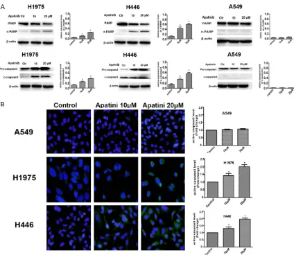
Effects on apoptosis
The effects of different concentrations of apa
-tinib on the apoptosis of A549, H1975 and H446 cells were detected by flow cytometry and a TUNEL assay. The flow cytometry results showed that the apoptosis rates of H1975 and H446 cells in the 0, 10 and 20 μM groups were as follows: H1975: 1.9 ± 1.47%, 7.8 ± 1.50%, and 15.9 ± 1.92%; H446: 0.98 ± 0.42%, 15.2 ± 2.65%, and 32.6 ± 2.68%. As the concentra
-tion of apatinib increased, the apoptosis rate of H1975 and H446 cells increased significantly
in a dose-dependent manner compared to the control group (P<0.05). In the A549 cells, the
percentage of apoptosis was lower after treat -ment with apatinib, and there was no
statisti-cally significant difference from the control
Effect of apatinib on lung cancer cells
The TUNEL test results were similar to the flow results. The results showed that the apoptosis rates of H1975 and H446 cells in the 0, 10, and 20 μM apatinib groups were as follows: H1975: 0.53 ± 0.22%, 18.6 ± 2.31%, and 34.5 ± 2.91; H446: 0.88 ± 0.57%, 28.1 ± 3.9%, and 55.9 ± 6.2%. There was a statistically significant dif-ference (P<0.05) for the H197 and H446 cells, but there was no significant difference in the percentage of apoptosis between the three
groups in A549 cells (P>0.05) (Figure 3B).
Western blot results showed that the relative
expression of c-caspase3 and c-PARP in H1975 and H446 cells increased significantly with the increase of apatinib concentration (P<0.05). However, there was no significant difference in the relative expression of c-caspase3 and
c-PARP in A549 cells (P>0.05) (Figure 4B). The results of an immunofluorescence assay show-ed that the fluorescence intensity of c-cas
-pase3 in H1975 and H446 cells was signifi
[image:7.612.92.520.73.521.2]-cantly higher than in the control group after
Figure 2. Apatinib induced cell cycle arrest in three cell lines. A-C. A549, H1975 and H446 cells were treated with 0, 10 or 20 μM apatinib for 24 hours and the cell cycle was analyzed by flow cytometry. D-F. Effect of apatinib on the expression of cyclin B1 and p53 was tested by western blotting in A549, H1975 and H446 cells, respectively. The
cells were treated with 0, 10 or 20 μM apatinib for 24 hours. Representative results are shown, and similar results
treatment with apatinib (P<0.05), while no
increase in the fluorescence intensity of c-cas -pase3 was observed in A549 cells (Figure 4A).
It was further confirmed that apatinib could
in-duce apoptosis by activating the caspase3 pathway in a dose-dependent manner in H1975
and H446 cell lines with high VEGFR-2
[image:8.612.92.520.72.598.2]expre-ssion.
Effect of apatinib on lung cancer cells
Tumor growth and immunohistochemistry
The in vivo antitumor efficacy of apatinib was
evaluated in A549, H1975 and H446 cancer
cells by utilizing BALB/c nude mouse xenograft models. The animals were repeatedly adminis -tered vehicle or apatinib once daily via oral
gavage (80 mg/kg/day and 120 mg/kg/day) for 21 consecutive days. In the nude mouse xenograft model using A549 cells, there was no significant difference in the volume of the 80 mg/kg apatinib group compared with the con
-trol group (P>0.05) while the volumes of the xenografted tumors in the 120 mg/kg group were significantly smaller than that of the con -trol group (P<0.05) (Figure 5A). In the nude
mouse xenograft model using H1975 and H446
cells, compared with the control group, the
vol-umes of the xenografted tumors of the 80 mg/ kg and 120 mg/kg groups were significantly smaller than that of the control group, and the tumors in the 120 mg/kg group were more sig
-nificantly reduced (P<0.05). Therefore, apatinib exhibited dose-dependent antitumor efficacy in the mice with xenografted H1975 and H446
lung cancer cells (Figure 5B and 5C).
Immunohistochemical staining was performed to determine the expression of CD31, Ki67 and c-caspase3 in different groups of xenografted tumor tissues. In the xenografted tumor tissu-es formed by A549 cells, there was no signifi
-cant difference in the expression of CD31, Ki67 or c-caspase3 of the apatinib 80 mg/kg group
compared with the control group (P>0.05), whi-
[image:9.612.94.518.67.435.2]le the number of CD31-positive micro-vessels
Figure 4. Effects of apatinib on the expression of apoptosis-related proteins were tested by western blotting and immunofluorescence. A. Relative expression of c-caspase3 and c-PARP in A549, H1975, and H446 cells treated
with 0, 10, or 20 μM apatinib for 24 hours was detected by Western blotting. B. Expression of c-caspase3 in A549,
H1975 and H446 cells treated with 0, 10, or 20 μM apatinib for 24 hours was analyzed by immunofluorescence
were significantly reduced, the number of Ki67-positive cells were significantly decreased, and the expression of c-caspase3 were significantly increased (P<0.05) in the 120 mg/kg group
(Figure 5D). In the xenografted tumor tissues formed by H1975 and H446 cells, compared with the control group, the number of CD31-positive micro-vessels were significantly reduc-ed, the number of Ki67-positive cells were sig
-nificantly decreased, and the expression of
c-caspase3 was significantly increased in bo-th bo-the 80 mg/kg and 120 mg/kg groups in a
dose-dependent manner (P<0.05) (Figure 5E
and 5F).
Related signaling pathways of xenograft tu-mors
Apatinib exhibited dose-dependent suppressi-
on of VEGFR-2 activation and downstream sig
-Figure 5. Apatinib inhibits tumor growth in A549, H1975 and H446 xenograft models. BALB/c nude mice xeno
[image:10.612.97.521.72.536.2]Effect of apatinib on lung cancer cells
naling in A549, H1975, and H446 tumor tis-sues. In A549 tumor tissues, the expression
levels of p-AKT, p-Erk, p-eIF4E1, HIF-1α, and VEGF of downstream signaling pathways were significantly decreased in the 120 mg/kg group (P<0.05), but there was no difference in their expression between the 80 mg/kg group and
the control group (P>0.05) (Figure 6A). In the H1975 and H446 tumor tissues, the
expres-sion levels of p-AKT, p-Erk, p-eIF4E1, HIF-1α, and VEGF protein in the 80 mg/kg and 120
mg/kg groups were significantly lower than control group, and with a dose-dependent effi -cacy (Figure 6B and 6C). All assays were repeated three times, and the images in the article were the most representative.
Toxicological study
Apatinib were well tolerated in all of the tested groups, with no mortality or significant loss of body weight (<5% relative to the
vehicle-ma-Figure 7. Toxicological study of apatinib in nude mice. A. Changes of body weight over 21 days. B. Levels of ALT, AST, BUN, Cr, CK, LDH-L, GLU, FT3 and FT4 in serum. C. Hematoxylin and eosin (H&E) staining of the heart, liver, kidney, thyroid, and pancreas tissues of mice. D. Ultrastructural observation of the heart, liver, kidney, thyroid and pancreas by transmission electron microscopy. The white arrow refers to damaged mitochondria, endoplasmic reticulum and
[image:12.612.91.524.72.508.2]Effect of apatinib on lung cancer cells
tched controls) observed during treatment (Fi- gure 7A). There was no statistically significant difference in ALT, AST, BUN, Cr, CK, or LDH-L in serum between the different groups (P>0.05), while the blood glucose levels of the high dose apatinib group were significantly higher than that of control group (P<0.05). The levels of FT3 and FT4 were significantly lower than in the
control group (P<0.05) (Figure 7B). No typical
pathological damage was found in HE staining of various organs, but the ultrastructure of vari -ous organs, such as the endoplasmic reticulum and mitochondria, under electron microscope
had different degrees of damage, and the dam -age in the high dose group were more obvious (Figure 7C and 7D).
Discussion
Inhibition of tumor neovascularization provides a new therapeutic approach for patients with advanced lung cancer [14, 15]. Since the first anti-angiogenic targeting drug bevacizumab
was used clinically, numerous clinical studies
have confirmed that bevacizumab combined
with chemotherapy or targeted therapy can
sig-nificantly improve the survival of patients with advanced non-small cell lung cancer [16, 17]. For example, studies have shown that TP che
-motherapy combined with bevacizumab can significantly prolong PFS and OS in patients with advanced non-squamous NSCLC [18]. Apatinib is a specific VEGFR-2 antagonist that competitively binds to VEGFR-2, blocks
VEGF-mediated signaling, inhibits tumor
angiogene-sis, and thus controls tumor growth [19]. There
have been more than 30 clinical studies on the
treatment of advanced lung cancer with apa
-tinib [20]. Most patients had advanced progr-essive non-small cell lung cancer, and a few patients had small cell lung cancer. Most of the
treatments were concentrated in the third line
or above treatment. In all of the clinical studies, the median PFS was more than 5 months, and the disease control rate reached 60-85% [21-23]. The clinical results of apatinib confirmed that patients can benefit in the short term, but
resistance developed rapidly, and improved
long-term OS was not obvious [24]. There is a desire to improve the efficacy of apatinib in
clinical practice.
VEGFR-2 is mainly expressed on the surface of
vascular endothelial cells and promotes the
formation of new blood vessels after binding to VEGF [25]. The increase of tumor neovascular
-ization is closely related to tumor proliferation, invasion and metastasis. Blocking tumor angio -genesis can control tumor growth and metasta-sis to some extent. Apatinib competitively binds
to VEGFR-2 on tumor vascular endothelial cells, blocks the binding of VEGF/VEGFR-2, reduces
tumor angiogenesis, and starves tumors, the- reby inhibiting tumor growth indirectly. How- ever, whether apatinib as a targeted drug can directly act on tumor cells and inhibit their
growth is unknown. For apatinib to have a direct inhibitory effect on lung cancer cells, the sur
-face of lung cancer cells must express VEGFR-2. If lung cancer cells express high VEGFR-2, such lung cancer cells can be targeted for treat
-ment with apatinib. Apatinib is less effective on cells that do not express VEGFR-2. Among the
common lung cancer cell lines are those that
highly express VEGFR-2, and the inhibitory effect of apatinib on lung cancer cells with high or low expression of VEGFR-2 is unknown. This study demonstrates that apatinib has different inhibitory effects on the proliferation of lung cancer cell lines with different VEGFR-2 expres -sion levels in vitro and in vivo, thus providing a
theoretical basis for further optimizing the tre-atment strategy of apatinib in clinical practice. This study selected common lung cancer cell
lines A549, HCC827, H2228, H1975, and H4-
46, the first four of which are lung adenocarci
-noma cells, which represent wild type, EGFR mutant, ALK mutant, and EGFR mutant with a T790M mutation cell lines, and H446 is a small cell lung cancer cell line. By detecting the expression of VEGFR-2 at both mRNA and pro -tein expression levels, we determined that H1975 and H446 lung cancer cell lines highly
expressed VEGFR-2, and the other three cell lines expressed VEGFR-2 at a lower level. A CCK-8 test confirmed that the same concen-tration of apatinib had the most obvious inhibi
-tory effect on the H1975 and H446 cell lines,
and the IC50 level was significantly lower than that of the other three lung cancer cells. Using a colony formation assay, cell cycle detection
and apoptosis assay, we determined that
apa-tinib can effectively inhibit the proliferation of
H1975 and H446 cells and promote apoptosis in a dose-dependent manner. However, in the other three lung cancer cell lines with low
-trations of apatinib could not effectively inhibit proliferation and the cell cycle arrest was not
obvious, and no obvious apoptosis was ob-
served. Apatinib has obvious inhibitory effects
on H1975 and H446 cells in vitro, and no
neo-vascularization was tested in vitro. Therefore, apatinib does not inhibit vascularization in this instance but inhibits the proliferation of lung cancer cells with high VEGFR-2 expression by directly acting on VEGFR-2 on the surface of
these two lung cancer cells and inhibiting the
activation of downstream signaling pathways. Additionally, studies have confirmed that there is cross-talk between the VEGF pathway and the downstream effectors of the EGFR pathway [26]. Once the downstream proteins of the VEGF pathway are affected, they can also af-fect proliferation via the EGFR signaling path
-way. Therefore, for lung cancer cells that hi-ghly express VEGFR-2, apatinib can significantly inhibit proliferation.
From the results of in vivo studies, apatinib has
an inhibitory effect on transplanted tumors of
A549, H1975 and H446 lung cancer cells.
However, for the transplanted A549 tumors, 80 mg/kg apatinib could not effectively inhibit tumor growth while 120 mg/kg apatinib signifi -cantly inhibited tumor growth. In the nude mice transplanted with H1975 and H446 cells, 80
and 120 mg/kg administration groups showed significant anti-tumor effects in a dose-depen
-dent manner. The inhibitory effect of apatinib on nude mice xenografts formed by high
VEGFR-2 expressing lung cancer cells is superior to
that of low expression lung cancer cell lines. However, unlike the in vitro results, apatinib showed almost no inhibition in vitro in A549
cells. In nude mouse xenograft models, howev -er, there are numerous vascular endothelial
cells, which highly express VEGFR-2, due to the presence of tumor blood vessels. Therefore,
apatinib can indirectly inhibit tumor growth, though this inhibition was not observed in the
80 mg/kg group. In the high-dose group, apa
-tinib’s indirect inhibition of tumor growth was observed. In transplanted tumors formed by H1975 and H446 cells with high VEGFR-2
ex-pression, apatinib not only inhibited tumor gr- owth by inhibiting tumor angiogenesis but also directly acted on tumor cells to directly inhibit
tumor growth. At low doses, it can significantly inhibit xenograft growth, and the antitumor effect was significantly better than in A549 ce-lls with low VEGFR-2 expression. The results of
the study partially reveal why there is a differ
-ence in the efficacy of the clinical application of apatinib in the treatment of lung cancer patients, which may be related to the differen
-tial expression of VEGFR-2 in lung cancer cells. To further investigate the mechanism by which apatinib inhibits xenografts, changes were ob-served in the levels of the relevant signaling
pathway proteins. Apatinib can reduce
neovas-cularization, inhibit proliferation, promote apop
-tosis and effectively control the growth rate of xenografts in nude mice through multiple cell
pathways. Studies have reported that the mo-
st important signaling pathways are AKT and ERK [27]. Apatinib can effectively reduce the phosphorylation level of key proteins in the ab-ove signaling pathways, thereby further reduc
-ing the phosphorylation of eukaryotic initiation factor 4E1, which plays an important role in promoting tumor cell proliferation and inhibit
-ing apoptosis and neovascularization [28]. Apatinib reduces the phosphorylation level of 4E1, thereby effectively inhibiting tumor prolif -eration and promoting apoptosis. Additionally,
the phosphorylation levels of key proteins in
the above signaling pathways were reduced,
which can reduce the expression of hypoxia-inducible factor 1α. When the level of HIF1α is decreased, the level of VEGF secreted by tumor cells decreases, effectively inhibiting the for
-mation of new blood vessels [29]. The results confirm that apatinib can inhibit tumor growth through multiple signaling pathways, and key
proteins on these pathways can become
tar-gets for new anti-tumor drugs.
This study found that apatinib has obvious ef-fects on H1975 and H446 both in vitro and in vivo. Therefore, patients with EGFR expression accompanied by L858R and T790M mutations and small cell lung cancer may benefit from
small molecule anti-angiogenic drugs acting on
VEGFR such as apatinib. At present, in patients with EGFR accompanied by L858R and T790M mutations, osimertinib is the first choice [30]. Apatinib was more effective for the H1975 cell
line, but whether osimertinib combined with
apatinib would be more effective remains to be confirmed in clinical studies. Significantly, for
patients with resistance to osimertinib with
L858R/T790M/C797S mutations [31], apa
Effect of apatinib on lung cancer cells
Small cell lung cancer accounts for approxi
-mately 15% of lung cancer [32]. Compared with
non-small-cell lung cancer, the biological be-
havior of small cell lung cancer is more aggr-essive. There are many driving genes in
non-small-cell lung cancer, and targeted therapy
can be selected under the guidance of molecu
-lar markers [33]. However, the pathogenesis and driving genes of small cell lung cancer are
not clear yet, and targeted therapy is not
appli-cable [34]. At present, relevant basic research
via genome sequencing has determined that small cell lung cancer has changes in signaling pathway genes, but it is not clear whether these
are driving genes. Treating small cell lung can
-cer remains difficult and improving the survi-val of patients with small cell lung cancer is
urgently needed in clinical practice. Currently,
the main first-line chemotherapy for small cell
lung cancer is etoposide combined with
plati-num. Although early efficacy is obvious, the long-term effect is poor, and the overall median survival time of patients is shorter [35]. Can
anti-angiogenic drugs be applied to the
treat-ment of small cell lung cancer? From the results of our study, small cell lung cancer cells express high levels of VEGFR-2, and the results confirm that apatinib had a good inhibitory effect on
small cell lung cancer in vitro and in vivo. Clinical cases have reported that apatinib has
an effect on small cell lung cancer [36]. In ongo -ing clinical research, apatinib monotherapy or combination chemotherapy and radiotherapy
for small cell lung cancer have gradually been extended to second-line or first-line clinical research, seeking a new breakthrough for the treatment of small cell lung cancer patients. Apatinib has an inhibitory effect on lung can-cer cells with high VEGFR-2 expression, but its safety and damage to major organs have not been reported. The main side effects of apa
-tinib are hypertension, proteinuria, fatigue, and hand-foot syndrome in clinical trials [37]. The overall tolerance of the animals was accept
-able, and there was no significant difference in body weight. HE staining of the heart, liver, and kidney tissues showed no obvious patho -logical damage under a light microscope. How- ever, under transmission electron microscopy,
the ultrastructure of these organs was dam -aged to some extent, and the damage was more obvious in the high dose group, including
mitochondria of cardiomyocytes, hepatocytes,
and renal tubular epithelial cells beginning to
vacuolize, mitochondrial mites becoming de-stroyed, part of the endoplasmic reticulum ex-panding, and the nucleus appearing pyknotic. The results indicated that the application of
apatinib resulted in damage to some organs, but under the current dose and times, there
was no significant pathological damage under light microscope or changes in liver or kidney function or myocardial enzymes. However, at-tention should be paid to the damage of these organs after long-term treatment. For endo -crine glands, the pancreas and thyroid were
chosen for observation. There was still no
ob-vious pathological damage under light micro-scope, but under the electron micromicro-scope, the endoplasmic reticulum system showed obvious expansion, some mitochondrial damage was obvious, and blood glucose and thyroid
hor-mone levels also changed significantly in the high dose group. These findings indicated that
endocrine gland damage by apatinib is more
obvious and worthy of clinical attention. Studies have found that anti-angiogenic therapy drugs cause different degrees of damage to different
organs, which may be related to the expression
of VEGFR in different vascular endothelial ce-lls. Endocrine glands express higher levels of VEGFR and are more likely to be damaged [38]. Clinical treatments must focus on the function of these organs.
Acknowledgements
This work was supported by Beijing Medical Reward Foundation (YXJL-2018-0164-0026) and National 13th Five-Year Major New Drug
Creation Project (2016ZX09191005).
Disclosure of conflict of interest
None.
Address correspondence to: Yu Li, Department of
Pulmonary Medicine, Qilu Hospital, Shandong Uni- versity, 107 West Wenhua Road, Jinan 250012,
Shandong, China. Tel: 15854385306; Fax:
86-053182169114; E-mail: 842764917@qq.com
References
[1] Prabhakar B, Shende P and Augustine S.
Current trends and emerging diagnostic
tech-niques for lung cancer. Biomed Pharmacother
[2] Kadir T and Gleeson F. Lung cancer prediction
using machine learning and advanced imaging
techniques. Transl Lung Cancer Res 2018; 7:
304-312.
[3] Reck M, Heigener DF, Mok T, Soria JC and Rabe KF. Management of non-small-cell lung
cancer: recent developments. Lancet 2013; 382: 709-719.
[4] Folkman J. Tumor angiogenesis: therapeutic
implications. N Engl J Med 1971; 285: 1182-1186.
[5] Li T, Kang G, Wang T and Huang H. Tumor an -giogenesis and anti-angiogenic gene therapy
for cancer. Oncol Lett 2018; 16: 687-702. [6] Kopparapu PK, Boorjian SA, Robinson BD,
Downes M, Gudas LJ, Mongan NP and Persson JL. Expression of VEGF and its receptors VEGFR1/VEGFR2 is associated with invasive
-ness of bladder cancer. Anticancer Res 2013;
33: 2381-2390.
[7] Ferrara N, Gerber HP and LeCouter J. The biol
-ogy of VEGF and its receptors. Nat Med 2003;
9: 669-676.
[8] Elaimy AL, Guru S, Chang C, Ou J, Amante JJ, Zhu LJ, Goel HL and Mercurio AM. VEGF-neuro
-pilin-2 signaling promotes stem-like traits in breast cancer cells by TAZ-mediated repres
-sion of the Rac GAP beta2-chimaerin. Sci Sig -nal 2018; 11: 528
[9] Lignet F, Benzekry S, Wilson S, Billy F, Saut O, Tod M, You B, Adda Berkane A, Kassour S, Wei MX, Grenier E and Ribba B. Theoretical investi
-gation of the efficacy of antiangiogenic drugs combined to chemotherapy in xenografted mice. J Theor Biol 2013; 320: 86-99.
[10] Tian S, Quan H, Xie C, Guo H, Lu F, Xu Y, Li J and
Lou L. YN968D1 is a novel and selective
in-hibitor of vascular endothelial growth factor receptor-2 tyrosine kinase with potent activity
in vitro and in vivo. Cancer Sci 2011; 102: 1374-1380.
[11] Scott LJ. Correction to: apatinib: a review in ad-vanced gastric cancer and other adad-vanced cancers. Drugs 2018; 78: 759.
[12] Liu Z, Ou W, Li N and Wang SY. Apatinib mono
-therapy for advanced non-small cell lung can
-cer after the failure of chemotherapy or other targeted therapy. Thorac Cancer 2018;9:
1285-1290.
[13] Zhao J, Zhang X, Gong C and Zhang J. Targeted
therapy with apatinib in a patient with relapsed small cell lung cancer: a case report and
litera-ture review. Medicine (Baltimore) 2017; 96:
e9259.
[14] Stratigos M, Matikas A, Voutsina A, Mavroudis D and Georgoulias V. Targeting angiogenesis in small cell lung cancer. Transl Lung Cancer Res
2016; 5: 389-400.
[15] Janning M and Loges S. Anti-angiogenics: their
value in lung cancer therapy. Oncol Res Treat
2018; 41: 172-180.
[16] Yamaguchi K, Masuda T, Fujitaka K, Miwata K, Sakamoto S, Horimasu Y, Hamai K, Miyamoto S, Nakashima T, Okamoto Y, Iwamoto H, Ishika
-wa N, Miyata Y, Okada M, Hamada H and Hat
-tori N. Bevacizumab with single-agent chemo -therapy in previously treated non-squamous non-small-cell lung cancer: phase II study. In
Vivo 2018; 32: 1155-1160.
[17] Zhao B, Zhang W, Yu D, Xu J and Wei Y. Erlo
-tinib in combination with bevacizumab has po
-tential benefit in non-small cell lung cancer: a systematic review and meta-analysis of ran
-domized clinical trials. Lung Cancer 2018;
122: 10-21.
[18] Zhou CC, Bai CX, Guan ZZ, Jiang GL, Shi YK, Wang MZ, Wu YL, Zhang YP and Zhu YZ. Safety and efficacy of first-line bevacizumab combina -tion therapy in Chinese popula-tion with
ad-vanced non-squamous NSCLC: data of sub
-group analyses from MO19390 (SAiL) study. Clin Transl Oncol 2014; 16: 463-468.
[19] Scott LJ. Apatinib: a review in advanced gastric cancer and other advanced cancers. Drugs 2018; 78: 747-758.
[20] Yang D, Dai R, Zhang Q and Fang P. Apatinib for
heavily treated patients with non-small cell
lung cancer: report of a case series and litera
-ture review. Saudi J Biol Sci 2018; 25:
888-894.
[21] Fang SC, Zhang HT, Zhang YM and Xie WP. Apa
-tinib as post second-line therapy in EGFR wild-type and ALK-negative advanced lung adeno
-carcinoma. Onco Targets Ther 2017; 10:
447-452.
[22] Peng Y, Cui H, Liu Z, Liu D, Liu F, Song Y, Duan H, Qiu Y and Li Q. Apatinib to combat EGFR-TKI
resistance in an advanced non-small cell lung
cancer patient with unknown EGFR status: a case report. Onco Targets Ther 2017; 10:
2289-2295.
[23] Song Z, Yu X, Lou G, Shi X and Zhang Y. Sal-vage treatment with apatinib for advanced non-small-cell lung cancer. Onco Targets Ther
2017; 10: 1821-1825.
[24] Zhang H. Apatinib for molecular targeted ther
-apy in tumor. Drug Des Devel Ther 2015; 9:
6075-6081.
[25] Comunanza V and Bussolino F. Therapy for cancer: strategy of combining anti-angiogenic and target therapies. Front Cell Dev Biol 2017;
5: 101.
Effect of apatinib on lung cancer cells
[27] Folkman J. Angiogenesis: an organizing princi
-ple for drug discovery? Nat Rev Drug Discov
2007; 6: 273-286.
[28] Keith B, Johnson RS and Simon MC. HIF1alpha and HIF2alpha: sibling rivalry in hypoxic tu -mour growth and progression. Nat Rev Cancer 2011; 12: 9-22.
[29] Sakurai T and Kudo M. Signaling pathways gov
-erning tumor angiogenesis. Oncology 2011;
81 Suppl 1: 24-29.
[30] Metro G, Baglivo S, Siggillino A, Ludovini V, Chi
-ari R, Rebonato A and Bellezza G. Successful response to osimertinib rechallenge after in
-tervening chemotherapy in an EGFR
T790M-positive lung cancer patient. Clin Drug Investig 2018; 38: 983-987.
[31] Grabe T, Lategahn J and Rauh D. C797S Resis
-tance: The undruggable EGFR mutation in non-small cell lung cancer? ACS Med Chem Lett
2018; 9: 779-782.
[32] Mishra MV, Louie AV, Gondi V and Slotman B. The evolving role of radiotherapy in the man
-agement of small cell lung cancer. J Thorac Dis
2018; 10: S2545-S2554.
[33] Gadgeel SM. Targeted therapy and immune therapy for small cell lung cancer. Curr Treat Options Oncol 2018; 19: 53.
[34] Blackhall F, Frese KK, Simpson K, Kilgour E, Brady G and Dive C. Will liquid biopsies im
-prove outcomes for patients with small-cell lung cancer? Lancet Oncol 2018; 19:
e470-e481.
[35] Tsoukalas N, Aravantinou-Fatorou E, Baxeva
-nos P, Tolia M, Tsapakidis K, Galanopoulos M, Liontos M and Kyrgias G. Advanced small cell
lung cancer (SCLC): new challenges and new
expectations. Ann Transl Med 2018; 6: 145. [36] Pan Y, Kong FW, Wang H, Wang X, Zhang H, Wu
WB and Zhang M. A recurrence-free survivor with chemotherapy-refractory small cell lung cancer after pneumonectomy: a case report and review of the literature. Medicine (Balti -more) 2017; 96: e8922.
[37] Wu F, Zhang S, Xiong A, Gao G, Li W, Cai W, Su C, Chen X, Zhou F, Zhao J, Ren S and Zhou C. A Phase II clinical trial of apatinib in pretreated
advanced non-squamous non-small-cell lung cancer. Clin Lung Cancer 2018; 19: e831-e842.
[38] Kamba T, Tam BY, Hashizume H, Haskell A, Sennino B, Mancuso MR, Norberg SM, O’Brien SM, Davis RB, Gowen LC, Anderson KD, Thur
-ston G, Joho S, Springer ML, Kuo CJ and Mc