Original Article
Clinical characteristics and serum levels of tumor
markers of connective tissue disease-associated
interstitial lung disease
Guifen Shen1, Sheng Yang3, Kejun Yao2, Wenjie Shi3, Qiuju Wang1,4, Emanuela Haxhiu5, Lingli Dong1
Departments of 1Rheumatology, 2Ophthalmology, Tongji Hospital, Tongji Medical College, Huazhong University of
Science and Technology, Wuhan 430030, Hubei, China; 3Tongji Medical College, Huazhong University of Science
and Technology, Wuhan 430030, Hubei, China; 4Department of Rheumatology, Jingzhou Central Hospital, The
Second Clinical Medical College, Yangtze University, Jingzhou 434020, Hubei, China; 5Medical Center of
Speciali-ties No. 1, Tirana, Albania
Received September 24, 2018; Accepted February 11, 2019; Epub May 15, 2019; Published May 30, 2019
Abstract: Many connective tissue diseases present with various extraarticular manifestations. One of these is in-terstitial lung disease, which should be a focus of rheumatologists and physical clinicians because of its severe complications. The current study aimed to analyze clinical manifestations of various connective tissue disease-associated interstitial lung disease (CTD-ILDs) patients, evaluating differences in the inflammatory indexes, immu-nological serologies, and alterations of tumor-associated biomarkers in CTD-ILDs patients. Retrospective analysis of 332 CTD-ILDs patients, enrolled from the ward of the Rheumatology Department in Tongji Hospital, from January 2005 through December 2013, was carried out. The study included idiopathic pulmonary fibrosis (IPF) patients as the positive control group. Gender composition in different CTD-ILDs was statistically different with female pre-dominance, while IPF patients consisted of slightly more males than females. Smoking, exertional dyspnea, and coughing were more common in patients with IPF, while velcro rales were more common in patients with RA-ILD. The percentage of patients with serum AFP, CEA, and NSE levels above upper normal limits was statistically dif-ferent (more proportion in PM/DM-ILD patients). A positive correlation was found between blood levels of CA125, CA199, CA724, and CYFRA21-1 and patient age in CTD-ILD patients. Differences in gender, age, ILD symptoms, and tumor-associated markers existed not only between CTD-ILDs and IPF patients, but also between different subtypes of CTD-ILD. For early diagnosis of ILD in CTD patients, precise physical examinations are required in patients with subclinical disease. Tumor biomarkers can be screened in ILD patients due to their prognostic potential.
Keywords: Interstitial lung disease, connective tissue disease, clinical symptom, tumor marker
Introduction
Connective tissue disease-associated intersti-tial lung diseases (CTD-ILDs) are sometimes viewed as a homogeneous entity. However, CTD-ILDs are heterogeneous diseases, com-prising different connective tissue diseases (CTDs) with various interstitial pneumonia pat-terns [1]. Interstitial lung disease (ILD) signifi-cantly increases morbidity and mortality in CTD patients and can be detected sub-clinically [2, 3]. Therefore, precisely phenotyped cohorts could be developed to better understand this disease. Moreover, respiratory symptoms are not consistent in diagnosed CTD-ILD patients. Tumor biomarkers, including CEA, CA125, SCC, NSE, and CYFRA21-1, have been widely used to
inter-stitial pneumonia (IP) and usual interinter-stitial pneumonia (UIP) [11]. Besides CEA, CA125, and CA153, other tumor biomarkers, such as CYFRA21-1, CA199, and SCC [12], are elevated in patients with interstitial lung disease.
Recent evidence supports the notion that IPF represents a risk factor for lung cancer devel-opment [13-16]. Accumulating evidence has revealed the association of malignancy [17] or lung cancer [15] with CTD-ILD. However, differ-ences in clinical manifestations and tumor bio-markers among various CTD-ILDs have been rarely reported. In this study, the common CTD-ILD types, including polymyositis/dermatomyo-sitis (PM/DM), systemic sclerosis (SSc), undif-ferentiated connective tissue disease (UCTD), mixed connective tissue disease (MCTD), rheu-matoid arthritis (RA), Sjögren’s syndrome (SS), and systemic erythematosus lupus (SLE), were selected alongside idiopathic pulmonary fibro-sis (IPF). The aim was to assess the main symp-toms of interstitial lung disease and tumor bio-markers in various CTDs, further characterizing ILD with different underlying CTDs.
Material and methods
Study subjects
This retrospective study began by reviewing the hospital’s medical records, providing access to all diagnoses of inpatients in Tongji Hospital, Tongji Medical College, Huazhong University of Science and Technology. First, patients diag-nosed with PM/DM, SSc, UCTD, MCTD, RA, SS, and SLE, from January 2005 to December 2013, with specific diagnosis criteria, were sep-arately identified. Medical records of the patients were then reviewed, consecutively, generating a longitudinal database with ILD diagnosis. Data of IPF patients were collected for the same time period. Medical documents of IPF patients were reviewed, individually, to ensure that they did not have evidence of CTD. The present study was approved by the HUST Committee on Human Research of Tongji Medical College and all subjects provided informed consent.
Patients with a defined CTD according to cur-rent respective criteria (e.g. PM [18]/DM [19], SSc [20], MCTD [21], RA [22], SS [23], and SLE [24]) were diagnosed with specific CTD-related ILDs. Patients were considered to have UCTD
with signs or symptoms and laboratory findings meeting UCTD criteria [25]. Patients included as IPF, according to published guidelines [26], were assessed as positive controls. ILD was diagnosed according to BTS Interstitial Lung Disease Guidelines [27]. Patients suffering from ILD with specific underlying causes (e.g., hypersensitivity pneumonitis, drug-induced lung disease, and environmental exposures) were excluded. Patients with tumors or any other severe pulmonary diseases, including bronchial asthma, chronic obstructive pulmo-nary disease, arterial pulmopulmo-nary hypertension, pleural effusion/pneumothorax, and sleep apnea hypopnea syndrome, were also ex- cluded.
Study plan
All medical records were collected based on the diagnosis code in the Inpatient Department of the hospital, identifying 472 PM/DM, 133 SSc, 486 UCTD, 77 MCTD, 811 RA, 230 SS, and 2,331 SLE patients. There were, respec-tively, 49, 35, 32, 61, 52, 53, and 50 patients with accompanying interstitial lung disease (Figure 1). Detailed medical information of both IPF and CTD-ILD patients was carefully re- viewed. Lung biopsies were not performed and, therefore, histopathologic analysis was not possible.
Clinical and radiologic characteristics
Manually collected clinical baseline data includ-ed age at the time of enrollment, gender, smok-ing status, ILD symptoms or signs, physical exam findings, and serological test results. Acute-phase reactants, such as erythrocyte sedimentation rate (ESR) and C reactive protein (CRP), were recorded. Levels of serum tumor-related biomarkers were obtained routinely. All laboratory tests were collected as part of a clinical evaluation and not for the purposes of this study.
Statistical analysis
SPSS 22.0 (SPSS Inc., Chicago, IL, USA (www. spss.com)) and GraphPad Prism version 6.02 (GraphPad Software, Inc., San Diego, CA, USA (www.graphpad.com)) were used for statistical analysis. Descriptive data for continuous vari-ables are reported as mean ± SEM. Two-tailed parametric ANOVA, nonparametric Mann-Whitney U-tests, and Kruskal Wallis tests were performed to assess levels of different bio-markers in various patient groups divided based on various CTD types. This was followed by the LSD test for differences in group pairs. Chi-squared test was performed for categorical data. Pearson’s correlation test was utilized to analyze correlation between age and levels of tumor markers. P < 0.05 indicates statistical significance.
Results
Patient characteristics
[image:3.612.91.522.74.379.2]ILD occurred in younger patients (41.18 ± 11.8 years old). Similar differences appeared in age at CTD diagnosis (Supplementary Table 1). Ever smokers were predominately in IPF, PM/DM, and RA patients (P < 0.05). Interval between diagnosis of CTD and onset of ILD in various CTDs showed no statistical differences (P < 0.069). However, after pairwise comparison (Kruskal-Wallis test), ILD occurred in PM-DM patients earlier than in SSC, CTD, MCTD, and SLE patients (P < 0.05).
ILD symptoms
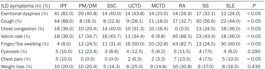
Main ILD symptoms, including exertional dys-pnea, cough, chest congestion, Velcro rale, fin-ger/toe swelling, cyanosis, chest pain, and weight loss, were evaluated in different ILDs (Table 2). Significant differences were found in occurrence rates of exertional dyspnea, cough, chest congestion, Velcro rale, finger/toe swell-ing, and chest pain in various CTD types (P <
0.05). Incidence rates of exertional dyspnea and cough were 82% and 88% in IPF, respec-tively, and 40.8% and 16.3% in PM/DM patients, respectively. The Velcro rale was aus-cultated in 86.5% of RA-ILD patients, but only in 36% of IPF patients.
Acute-phase reactants and immunological parameters
CRP and ESR were assessed in ILD patients (Table 3). In all CTD-ILD types, CRP and ESR were elevated, but only ESR showed statisti-cally significant differences. CRP levels were 11.69 ± 13.08 mg/dl and 43.67 ± 79.16 mg/dl in SSc-ILD and SS-ILD patients, respectively (P
[image:4.612.91.523.85.228.2]> 0.05). Multiple comparisons indicated that ESR levels in IPF patients were lower than those of other CTD-ILD types, except DM/PM and SSc. ESR was lower in SSc-ILD than in other CTD-ILD types and lower in PM/DM-ILD patients than in individuals with SS-ILD and SLE-ILD. Table 2. Comparison of ILD symptoms between IPF and different CTD-ILDs
ILD symptoms (n) (%) IPF PM/DM SSC UCTD MCTD RA SS SLE P
[image:4.612.91.523.294.408.2]Exertional dyspnea (%) 41 (82.0) 20 (40.8) 14 (40.0) 14 (43.8) 14 (23.0) 14 (26.9) 17 (32.1) 12 (24.0) < 0.05 Cough (%) 44 (88.0) 8 (16.3) 8 (22.9) 9 (28.1) 11 (18.0) 17 (32.7) 30 (56.6) 22 (44.0) < 0.05 Chest congestion (%) 18 (36.0) 10 (20.4) 14 (40.0) 10 (31.3) 10 (16.4) 0 (0.0) 13 (24.5) 18 (36.0) < 0.05 Velcro rale (%) 18 (36.0) 17 (34.7) 16 (45.7) 11 (34.4) 6 (9.8) 45 (86.5) 23 (43.4) 18 (36.0) < 0.05 Finger/Toe swelling (%) 4 (8.0) 12 (24.5) 11 (31.4) 16 (50.0) 20 (32.8) 43 (82.7) 13 (24.5) 30 (60.0) < 0.05 Cyanosis (%) 5 (10.0) 11 (22.4) 3 (8.6) 4 (12.5) 5 (8.2) 6 (11.5) 4 (7.5) 4 (8.0) 0.290 Chest pain (%) 5 (10.0) 0 (0.0) 0 (0.0) 2 (6.3) 2 (3.3) 7 (13.5) 4 (7.5) 5 (10.0) < 0.05 Weight loss (%) 10 (20.0) 10 (20.4) 5 (14.3) 8 (25.0) 9 (14.8) 16 (30.8) 9 (17.0) 8 (16.0) 0.439 ILD = interstitial lung disease, IPF = idiopathic pulmonary fibrosis, MCTD = mixed connective tissue disease, PM/DM = polymyositis/dermatomyo-sitis, RA = rheumatoid arthritis, SLE = systemic erythematosus lupus, SS = Sjögren syndrome, SSc = systemic sclerosis, UCTD = undifferentiated connective tissue disease. Chi-squared test was performed for categorical data.
Table 1. Baseline characteristics at the time of diagnosis as interstitial lung disease
Type of ILD Case (n) Gender n (M/F) Age (year) mean (min, max) Ever Smoker (n) (month) Mean (min, max)Course of CTD to ILD
IPF 50 28/22 51.0 (19, 86) 17
PM/DM 49 17/31 46.0 (22, 65) 11 5.5 (-12.0, 36.0)
SSC 35 7/28 48.9 (26, 67) 0 37.3 (0, 192.0)
UCTD 32 7/25 54.8 (31, 79) 0 41.8 (-36.0, 778.0)
MCTD 61 7/54 50.5 (23, 75) 0 31.1 (0, 360.0)
RA 52 13/39 60.6 (40, 80) 11 17.5 (0, 228.0)
SS 53 1/52 50.5 (29, 77) 0 14.5 (-110, 108.0)
SLE 50 2/48 41.1 (20, 65) 1 36.9 (0, 275.0)
P < 0.05 < 0.05 < 0.05 0.069
Table 3. Analysis of acute-phase inflammatory reactants and immunologic panel in CTD-ILDs
Mean (SD) IPF PM/DM SSC UCTD MCTD RA SS SLE P
CRP (mg/dl) 23.62 (25.13) 18.25 (19.16) 11.69 (13.08) 33.91 (40.86) 33.85 (55.97) 31.64 (30.79) 43.67 (79.16) 36.16 (52.19) 0.086 ESR (mm/1 h) 27.39 (23.42) 37.61 (27.49) 19.48 (19.15) 46.52 (29.23) 50.32 (34.58) 47.94 (32.66) 54.98 (39.69) 60.61 (41.84) < 0.05 IgG (g/L) 12.88 (3.18) 15.30 (5.76) 12.82 (6.22) 16.75 (8.87) 21.06 (8.50) 12.95 (9.98) 20.37 (7.57) 19.04 (8.70) < 0.05 IgA (g/L) 2.61 (1.05) 3.02 (1.66) 3.43 (4.74) 3.07 (2.53) 3.21 (1.54) 2.42 (1.00) 3.95 (3.80) 2.97 (2.92) < 0.05 IgM (g/L) 1.48 (0.95) 1.90 (1.29) 1.49 (0.91) 1.70 (0.99) 1.77 (1.06) 1.72 (1.35) 1.65 (1.06) 1.56 (0.93) 0.786 C3 (g/L) 1.19 (0.38) 0.97 (0.24) 1.03 (0.23) 1.09 (0.24) 0.99 (0.31) 1.00 (0.23) 0.93 (0.28) 0.73 (0.38) < 0.05 C4 (g/L) 0.24 (0.10) 0.29 (0.09) 0.28 (0.17) 0.31 (0.19) 0.23 (0.09) 0.23 (0.10) 0.22 (0.11) 0.16 (0.09) < 0.05
Both ESR and CRP were detected in different CTD-ILDs, but only ESR values were found to be statistically significant. Besides, IgG, C3, and C4 values alterations were also significant. C3 = complement 3, C4 = complement 4, CRP = C reactive protein, ESR = erythrocyte sedimentation rate, IgA = immunoglobulin A, IgG = immunoglobulin G, IgM = immunoglobulin M, ILD = interstitial lung disease, IPF = idiopathic pulmonary fibrosis, MCTD = mixed connective tissue disease, PM/DM = polymyositis/derma-tomyositis, RA = rheumatoid arthritis, SLE = systemic erythematosus lupus, SS = Sjögren syndrome, SSc = systemic sclerosis, UCTD = undifferentiated connective tissue
disease. Two-tailed parametric ANOVA was performed to assess levels of different biomarkers in various patient groups divided based on various CTD types. This was followed by the LSD test for differences in group pairs.
IgG, C3, and C4 levels were significantly differ-ent in various CTD-ILD types. For example, IgG levels were significantly higher in MCTD-ILD patients than in individuals with SSc-ILD (21.06 ± 8.87 g/L vs. 12.82 ± 6.22 g/L, P < 0.05, according to multiple comparison test). C3 and C4 levels were lower in patients with SLE-CTD than those with other ILDs (P < 0.05, according to multiple comparison test).
Tumor biomarkers related to age
ILD data, including CTD-ILDs and IPF, assessed together, demonstrated that individual blood
normal limits were significantly different in vari-ous CTD-ILDs (especially more in PM/DM-ILD patients). For example, blood AFP levels were higher than normal in 37.1% of DM/PM-ILD and 12.5% of UCTD-ILD patients, while remaining normal in other CTD-ILDs. Elevated CYFRA21-1 and NSE were found in various CTD-ILD pati- ents. NSE showed statistically significant dif- ferences, while CYFRA21-1 did not. Elevated NSE and CYFRA21-1 levels were prominent in DM/PM and IPF patients, respectively. Similar to NSE, no CYFRA21-1 was detected in SLE-ILD patients, requiring further studies of CYFRA21-1 and NSE detection in SLE-ILD.
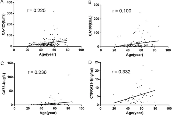
Figure 2. Correlation between age and CA125, CA199, CA724, or CYFRA21-1 in all patients. The relationship between age and values of tumor biomarkers of in-dividual ILD patients was analyzed by Pearson’s correlation test. R values for the correlation between age and CA-125, CA-199, CA724, or CYFRA21-1 are 0.225, 0.100, 0.236, and 0.332, respectively. Linear correlation between patient age and level of different tumor biomarkers was tested by linear regression analy-sis. Data shows the ages of individual patients against levels of CA125, CA199, CA724, or CYFRA21-1. Pearson’s correlation test was utilized to analyze the cor-relation between age and levels of tumor markers.
levels of CA125, CA1- 99, CA724, and CYF- RA21-1 were correlated with age, respectively. As shown in Figure 2, blood CA125, CA199, CA724, and CYFRA21-1 levels increased with age.
Discrepant tumor bio-markers
[image:5.612.91.392.250.453.2]Table 4. Proportion of CTD-ILD patients with blood tumor markers above upper normal limits
% IPF DM/PM SSC UCTD MCTD RA SS SLE p value
AFP 0.0 37.1 0.0 12.5 0.0 0.0 0.0 0.0 < 0.001
CA-125 46.2 8.7 27.3 20.0 33.3 41.7 29.0 44.4 0.235 CA-199 30.8 12.5 16.7 11.1 21.4 23.8 10.7 33.3 0.474
CA72-4 8.0 28.0 20.0 6.7 14.3 16.7 16.0 0.0 0.549
CA-153 42.9 25.0 31.3 30.0 25.0 18. 12.5 0.0 0.402
CEA 32.5 37.1 15.2 8.7 20.0 24.0 3.0 11.1 0.009
SCC 12.9 3.2 3.2 5.3 11.1 4.3 6.9 0.0 0.827
CYFRA21-1 68.0 50.0 57.1 25.0 40.0 40.0 25.0 0.537
NSE 20.0 80.0 40.0 37.5 57.1 40.0 16.7 0.03
The proportion of positive patients is presented as percentage. The difference of positive ratio between different CTD-ILD was analyzed by Pearson’s Chi-squared test, with P value less than 0.05 indicating statistical differences. AFP = alpha fetal protein, CA (125, 199, 72-4, 193) = cancer antigen (125, 199, 72-4, 193), CEA = carcinoembryonic antigen, CYFRA21-1 = Cytokeratin fragment antigen21-1, ILD = interstitial lung disease, IPF = idiopathic pulmonary fibrosis, MCTD = mixed connective tissue disease, NSE = neuron-specific enolase, PM/DM = polymyositis/dermatomyositis, RA = rheumatoid arthritis; SS = Sjögren syndrome, SCC Squamous cell carcinoma associated antigen, SLE = systemic erythematosus lupus, SSC = systemic sclerosis, UCTD = undifferentiated connective tis-sue disease. Chi-squared test was performed for categorical data.
Age correlated with levels of tumor biomarkers, as shown above, constituting a significant con-founding factor in analyzing level differences of tumor biomarkers in various connective tissue
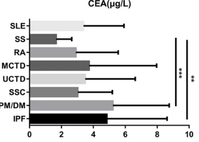
diseases. To eliminate the confounding effects of age, patients were divided into three groups based on age, 20~40, 40~60, and 60~80, res- pectively. Since most in- cluded patients were be- tween 40 and 60, this study deleted the data of patients younger than 20 or older than 60, largely controlling the confound-ing effects of age. In this way, the confounding eff- ects of age can be largely controlled. Blood CEA levels were significantly lower in SS-ILD patients than in individuals with PM/DM-ILD and IPF (P < 0.01 and P < 0.001 res- pectively, Figure 3). Bl- ood levels of the remain-ing tumor biomarkers showed no statistically significant differences after age-adjustment (Supplementary Figure 1).
Discussion
CTD-ILD is one of the major components of in- terstitial lung disease. While CTD-ILD is differ-ent from idiopathic interstitial lung disease, such as IPF, this study revealed that gender, age, respiratory symptoms, and tumor biomark-ers showed differences not only between CTD-ILD and IPF, but also among CTD-CTD-ILD subtypes. Moreover, some tumor biomarkers found not to significantly differ in various CTD-ILDs were positively correlated with age.
Since CTD incidence is higher in female patients than in males [28, 29], it is not surprising that CTD-ILD patients were mostly female, especial-ly SLE-ILD and SS-ILD patients. IPF patientshad similar proportions between the two genders. Age at onset in different CTD-ILD subtypes is certainly influenced by age at primary disease onset. However, this study did not compare ages of CTD patients without ILD with those of individuals with CTD-ILD. This deserves further attention.
Common symptoms, such as exertional dys-pnea and coughing, frequently occurred in IPF, but not so often in CTD-ILD. In some CTD-ILD Figure 3. CEA levels were detected in ILD patients
[image:6.612.95.290.370.509.2]subtypes, such as RA-ILD, incidence rates of exertional dyspnea and coughing were low, while Velcro rale was auscultated frequently. This finding indicates that, in CTD diseases, ILD cannot be excluded by the nonexistence of exertional dyspnea and coughing and thorough physical examinations (including auscultation) could provide clues for ILD. Although pu- lmonary function tests, HRCT, and lung biop-sies are not routinely used, they can be helpful in ILD diagnosis. Some common respiratory symptoms did not occur as frequently in CTD-ILD as in IPF. Therefore, such examinations are recommended for early detection and diagno-sis of CTD-ILD.
CRP and ESR are disease activity indicators, but elevated CRP and ESR may also indicate infections, tumors, and inflammatory condi-tions. Th-erefore, CRP and ESR are not specific for CTD-ILD diagnosis or activity detection. CRP and ESR levels were elevated mostly in the IPF and CTD-ILD subtypes. No significant differenc-es in CRP levels were obtained between IPF and CTD-ILD patients, but ESR was lower in SSc-ILD patients than in individuals with other CTD-ILD types. Blood IgG, IgA, IgM, and com-plement C3 and C4 levels were also detected. Except for IgA and IgM, the remaining immuno-logical parameters were significantly different between IPF and CTD-ILD subtypes. Different blood IgG, C3, and C4 levels might be due to the underlying CTD type. For instance, SLE-ILD patients showed higher IgG levels and lower C3 and C4 amounts, compared with other ILDs. Lung cancer patients with CTD have a signifi-cantly higher incidence of ILD, as a complica-tion, compared with those without CTD [30], indicating a potential relationship between lung cancer and CTD-ILD. In this study, besides lung cancer-associated tumor biomarkers (CEA, SCC, NSE, and CYFRA21-1), other tumor bio-markers (CA125 and CA153) were analyzed. Pathophysiological mechanisms of high levels of tumor biomarkers in patients with interstitial lung disease, with no evidence of malignancy, remain unclear. In this work, CEA levels were increased in 32.5% of IPF patients, far below previously reported values, with serum CEA amounts elevated in more than half of IPF pa- tients [9]. Blood CEA levels were elevated not only in IPF patients, but also in individuals with PM/DM-ILD, as shown above. It is well-estab-lished that PM/DM is related to malignancy.
Cancer risk was unchanged in patients with CTD without dermatomyositis or polymyositis, but increased after inclusion of individuals with dermatomyositis or polymyositis [31]. Whether blood CEA levels are increased in IPF or PM/ DM-ILD patients for disease exacerbation or if they lead to cancer remains unclear, but such changes may result in poor prognosis. Other tumor biomarkers are related to the severity of IPF, including CA153 [32]. Additionally, serum CYFRA21-1 levels reflect the severity of lung injury in nonmalignant respiratory diseases and might be associated with prognosis in patients with IPF and collagen disease associated pul-monary fibrosis [33]. CYFRA21-1 has been con-sidered a useful parameter in evaluating lung epithelial cell damage and repair [34], since cytokeratins are cytoskeletal structures ex-pressed only in epithelial cells. Evidence indi-cates that cytokeratin fragment 19 levels con-stitute a useful variable for lung injury evaluation in interstitial pneumonia associated with PM/ DM [35]. Other findings support this conclusion as well, demonstrating that the survival of IPF patients with high CYFRA 21.1 levels in BAL is worse, compared with that of the low CYFRA 21.1 group [36]. The usefulness of serum CA199 has been well-recognized for pancreatic cancer diagnosis. However, increased CA199 has also been found in other pathologies, such as bronchiectasis, bronchiolitis, emphysema, and interstitial fibrosis [37]. Interestingly, sur-vival rates are significantly lower in patients with interstitial lung diseases (idiopathic inter-stitial lung disease and CDPF) and elevated serum CA199 than in counterparts with CA199 levels in the normal range [7]. NSE, a bioactive peptide, is produced and secreted by neuroen-docrine cells of the lungs. To the best of our knowledge, the relationship between NSE and interstitial lung disease has not been reported yet.
Several limitations of this study should be men-tioned. First, biopsies or histopathologic exami-nations were not performed. Therefore, a com-parative assessment of ILD pathology with various underlying CTDs is needed. Second, normal healthy individuals and CTD patients without ILD are needed as negative controls in such studies, excluding the effects of baseline differences. Third, long-term follow-ups are required to assess the prognostic values of clinical manifestations and tumor biomarkers for CTD-ILD patient survival, as well as the pos-sibility of cancer occurrence.
Conclusion
This research revealed that gender, age, respi-ratory symptoms, and tumor biomarkers differ between CTD-ILD and IPF. Tumor biomarkers CA125, CA199, CA724, and CYFRA21-1 in- creased in blood levels with age in the whole ILD patient population. Assessment of tumor biomarkers elevated, to varying degrees, in various CTD-ILDs demonstrated that only CEA concentrations were significantly different after age-adjusted comparison. They were much lower in SS-ILD patients than in individuals with PM/DM-ILD or IPF. For early diagnosis of ILD in CTD patients, precise physical examinations are required in patients with subclinical dis-ease. Tumor biomarkers can be screened in ILD patients due to their prognostic potential.
Acknowledgements
This study was supported by the National Natural Science Foundation of China (81- 401354).
Disclosure of conflict of interest
None.
Address correspondence to: Dr. Lingli Dong, De- partment of Rheumatology, Tongji Hospital, Tongji Medical College, Huazhong University of Science and Technology, 1095 Jiefang Avenue, Wuhan 430030, Hubei, China. Tel: +86-27-83663614; Fax: +86-27-83663614; E-mail: [email protected]
References
[1] Chartrand S and Fischer A. Management of connective tissue disease-associated intersti-tial lung disease. Rheum Dis Clin North Am 2015; 41: 279-294.
[2] Fischer A and du Bois R. Interstitial lung dis-ease in connective tissue disorders. Lancet 2012; 380: 689-698.
[3] Doyle TJ, Hunninghake GM and Rosas IO. Sub-clinical interstitial lung disease: why you should care. Am J Respir Crit Care Med 2012; 185: 1147-1153.
[4] Molina R, Auge JM, Bosch X, Escudero JM, Vi-nolas N, Marrades R, Ramirez J, Carcereny E and Filella X. Usefulness of serum tumor mark-ers, including progastrin-releasing peptide, in patients with lung cancer: correlation with his-tology. Tumour Biol 2009; 30: 121-129. [5] Fang SC, Zhang HT, Wang CY and Zhang YM.
Serum CA125 and NSE: biomarkers of disease severity in patients with silicosis. Clin Chim Acta 2014; 433: 123-127.
[6] Fujita J, Ohtsuki Y, Bandoh S, Takashima H, Ueda Y, Wu F, Tojo Y, Kubo A and Ishida T. Ele-vation of cytokeratin 19 fragment (CYFRA 21-1) in serum of patients with radiation pneumo-nitis: possible marker of epithelial cell damage. Respir Med 2004; 98: 294-300.
[7] Kodama T, Satoh H, Ishikawa H and Ohtsuka M. Serum levels of CA19-9 in patients with nonmalignant respiratory diseases. J Clin Lab Anal 2007; 21: 103-106.
[8] Collazos J, Esteban C, Fernandez A and Genol-la J. Measurement of the serum tumor marker neuron-specific enolase in patients with be-nign pulmonary diseases. Am J Respir Crit Care Med 1994; 150: 143-145.
[9] Fahim A, Crooks MG, Wilmot R, Campbell AP, Morice AH and Hart SP. Serum carcinoembry-onic antigen correlates with severity of idio-pathic pulmonary fibrosis. Respirology 2012; 17: 1247-1252.
[10] Dai H, Liu J, Liang L, Ban C, Jiang J, Liu Y, Ye Q and Wang C. Increased lung cancer risk in pa-tients with interstitial lung disease and elevat-ed CEA and CA125 serum tumour markers. Respirology 2014; 19: 707-713.
[11] Kruit A, Gerritsen WB, Pot N, Grutters JC, van den Bosch JM and Ruven HJ. CA 15-3 as an alternative marker for KL-6 in fibrotic lung dis-eases. Sarcoidosis Vasc Diffuse Lung Dis 2010; 27: 138-146.
[12] Calabrese F, Lunardi F, Giacometti C, Marulli G, Gnoato M, Pontisso P, Saetta M, Valente M, Rea F, Perissinotto E and Agostini C. Overex-pression of squamous cell carcinoma antigen in idiopathic pulmonary fibrosis: clinicopatho-logical correlations. Thorax 2008; 63: 795-802.
[13] Archontogeorgis K, Steiropoulos P, Tzouvelekis A, Nena E and Bouros D. Lung cancer and in-terstitial lung diseases: a systematic review. Pulm Med 2012; 2012: 315918.
lung cancer: a clinical and pathogenesis up-date. Curr Opin Pulm Med 2015; 21: 626-633. [15] Enomoto Y, Inui N, Yoshimura K, Nishimoto K,
Mori K, Kono M, Fujisawa T, Enomoto N, Naka-mura Y, Iwashita T and Suda T. Lung cancer development in patients with connective tis-sue disease-related interstitial lung disease: a retrospective observational study. Medicine (Baltimore) 2016; 95: e5716.
[16] Choi WI, Park SH, Park BJ and Lee CW. Intersti-tial lung disease and lung cancer develop-ment: a 5-year nationwide population-based study. Cancer Res Treat 2018; 50: 374-381. [17] Bouros D, Hatzakis K, Labrakis H and
Zeibeco-glou K. Association of malignancy with diseas-es causing interstitial pulmonary changdiseas-es. Chest 2002; 121: 1278-1289.
[18] Milisenda JC, Selva-O’Callaghan A and Grau JM. The diagnosis and classification of poly-myositis. J Autoimmun 2014; 48-49: 118-121. [19] Iaccarino L, Ghirardello A, Bettio S, Zen M,
Gatto M, Punzi L and Doria A. The clinical fea-tures, diagnosis and classification of dermato-myositis. J Autoimmun 2014; 48-49: 122-127. [20] van den Hoogen F, Khanna D, Fransen J, John-son SR, Baron M, Tyndall A, Matucci-Cerinic M, Naden RP, Medsger TA Jr, Carreira PE, Rieme-kasten G, Clements PJ, Denton CP, Distler O, Allanore Y, Furst DE, Gabrielli A, Mayes MD, van Laar JM, Seibold JR, Czirjak L, Steen VD, Inanc M, Kowal-Bielecka O, Muller-Ladner U, Valentini G, Veale DJ, Vonk MC, Walker UA, Chung L, Collier DH, Ellen Csuka M, Fessler BJ, Guiducci S, Herrick A, Hsu VM, Jimenez S, Ka-haleh B, Merkel PA, Sierakowski S, Silver RM, Simms RW, Varga J and Pope JE. 2013 classifi-cation criteria for systemic sclerosis: an Ameri-can college of rheumatology/European league against rheumatism collaborative initiative. Ann Rheum Dis 2013; 72: 1747-1755.
[21] Ortega-Hernandez OD and Shoenfeld Y. Mixed connective tissue disease: an overview of clini-cal manifestations, diagnosis and treatment. Best Pract Res Clin Rheumatol 2012; 26: 61-72.
[22] Kay J and Upchurch KS. ACR/EULAR 2010 rheumatoid arthritis classification criteria. Rheumatology (Oxford) 2012; 51 Suppl 6: vi5-9.
[23] Shiboski SC, Shiboski CH, Criswell L, Baer A, Challacombe S, Lanfranchi H, Schiodt M, Ume-hara H, Vivino F, Zhao Y, Dong Y, Greenspan D, Heidenreich AM, Helin P, Kirkham B, Kitagawa K, Larkin G, Li M, Lietman T, Lindegaard J, Mc-Namara N, Sack K, Shirlaw P, Sugai S, Vollen-weider C, Whitcher J, Wu A, Zhang S, Zhang W, Greenspan J, Daniels T; Sjögren’s Internation-al Collaborative ClinicInternation-al Alliance (SICCA) Re-search Groups. American College of Rheuma-tology classification criteria for Sjogren’s
syndrome: a data-driven, expert consensus approach in the Sjogren’s International Collab-orative Clinical Alliance cohort. Arthritis Care Res (Hoboken) 2012; 64: 475-487.
[24] Yu C, Gershwin ME and Chang C. Diagnostic criteria for systemic lupus erythematosus: a critical review. J Autoimmun 2014; 48-49: 10-13.
[25] Kinder BW, Collard HR, Koth L, Daikh DI, Wolt-ers PJ, Elicker B, Jones KD and King TE Jr. Idio-pathic nonspecific interstitial pneumonia: lung manifestation of undifferentiated connective tissue disease? Am J Respir Crit Care Med 2007; 176: 691-697.
[26] Raghu G, Collard HR, Egan JJ, Martinez FJ, Behr J, Brown KK, Colby TV, Cordier JF, Flaherty KR, Lasky JA, Lynch DA, Ryu JH, Swigris JJ, Wells AU, Ancochea J, Bouros D, Carvalho C, Costabel U, Ebina M, Hansell DM, Johkoh T, Kim DS, King TE Jr, Kondoh Y, Myers J, Muller NL, Nicholson AG, Richeldi L, Selman M, Dud-den RF, Griss BS, Protzko SL and Schunemann HJ. An official ATS/ERS/JRS/ALAT statement: idiopathic pulmonary fibrosis: evidence-based guidelines for diagnosis and management. Am J Respir Crit Care Med 2011; 183: 788-824. [27] Bradley B, Branley HM, Egan JJ, Greaves MS,
Hansell DM, Harrison NK, Hirani N, Hubbard R, Lake F, Millar AB, Wallace WA, Wells AU, Whyte MK, Wilsher ML; British Thoracic Society Inter-stitial Lung Disease Guideline Group BTSSoCC, Thoracic Society of A, New Zealand Thoracic S and Irish Thoracic S. Interstitial lung disease guideline: the British Thoracic Society in col-laboration with the Thoracic Society of Austra-lia and New Zealand and the Irish Thoracic Society. Thorax 2008; 63 Suppl 5: v1-58. [28] Margery-Muir AA, Bundell C, Nelson D, Groth
DM and Wetherall JD. Gender balance in pa-tients with systemic lupus erythematosus. Au-toimmun Rev 2017; 16: 258-268.
[29] Piram M, Maldini C and Mahr A. Effect of race/ ethnicity on risk, presentation and course of connective tissue diseases and primary sys-temic vasculitides. Curr Opin Rheumatol 2012; 24: 193-200.
[30] Saijo A, Hanibuchi M, Goto H, Toyoda Y, Tezuka T and Nishioka Y. An analysis of the clinical fea-tures of lung cancer in patients with connec-tive tissue diseases. Respir Investig 2017; 55: 153-160.
[31] Xu W, Guo H, Liu Z, Chen C and Lei CC. The risk of cancer in patients with connective tissue diseases but without dermatomyositis or poly-myositis: a multicenter cohort study conducted over 15 years in China. Immunol Lett 2016; 177: 70-74.
idio-pathic pulmonary fibrosis before and after lung transplantation. Chest 2012; 141: 1047-1054. [33] Nakayama M, Satoh H, Ishikawa H, Fujiwara M,
Kamma H, Ohtsuka M and Sekizawa K. Cyto-keratin 19 fragment in patients with nonmalig-nant respiratory diseases. Chest 2003; 123: 2001-2006.
[34] Dobashi N, Fujita J, Ohtsuki Y, Yamadori I, Yo-shinouchi T, Kamei T and Takahara J. Elevated serum and BAL cytokeratin 19 fragment in pul-monary fibrosis and acute interstitial pneumo-nia. Eur Respir J 1999; 14: 574-578.
[35] Fujita J, Dobashi N, Tokuda M, Bandoh S, Oht-suki Y, Yamadori I, Yoshinouchi T, Ueda Y and Takahara J. Elevation of cytokeratin 19 frag-ment in patients with interstitial pneumonia associated with polymyositis/dermatomyositis. J Rheumatol 1999; 26: 2377-2382.
[36] Vercauteren IM, Verleden SE, McDonough JE, Vandermeulen E, Ruttens D, Lammertyn EJ, Bellon H, De Dycker E, Dooms C, Yserbyt J, Verleden GM, Vanaudenaerde BM and Wuyts WA. CYFRA 21.1 in bronchoalveolar lavage of idiopathic pulmonary fibrosis patients. Exp Lung Res 2015; 41: 459-465.
Supplementary Table 1. Age of CTD onset Type of disease Age (year) of onsetmean (sd)
PM/DM 45.2 (9.2)
SSC 44.6 (12.7)
UCTD 51.1 (14.9)
MCTD 47.8 (11.1)
RA 54.1 (15.7)
SS 48.4 (10.9)
SLE 38.4 (12.9)
P < 0.05
MCTD = mixed connective tissue disease, PM/DM = polymyositis/dermatomyositis, RA = rheumatoid arthritis, SLE = systemic erythematosus lupus, SS = Sjögren syndrome, SSC = systemic sclerosis, UCTD = undifferentiated connective tissue disease.
Supplementary Table 2. The number of ILD patients whose serum tumor markers were tested
ILD Patient (n) AFP CA-125 CA-199 CA72-4 CA-153 CEA SSC CFRA 21-1 NSE
IPF 15 13 26 25 14 40 31 19 30
PM 33 23 30 28 21 39 33 6 14
SSC 20 15 18 15 10 23 19 4 8
CTD 16 12 14 14 12 20 18 5 7
MCTD 28 22 24 15 16 33 17 7 10
RA 19 12 21 18 11 25 23 5 10
SS 28 31 28 25 24 33 29 4 6
SLE 9 10 6 4 6 10 5
Total 168 138 167 144 114 223 175 50 85