Case Report
An uncommon diagnosis and treatment challenge in a
patient with primary hepatic lymphoma: a case report
Lina Yan1*, Bo Tang2*, Jing Zhang1, Zhenqiang Wang1, Dan Zhang1, Lihua Cao1
Departments of 1Respiratory Medicine, 2Hematology, The Second Hospital of Dalian Medical University, Dalian 116023, Liaoning, P.R. China. *Equal contributors.
Received December 29, 2018; Accepted May 7, 2019; Epub July 15, 2019; Published July 30, 2019
Abstract: Primary hepatic lymphoma (PHL) is a rare condition that is likely to pose a diagnostic challenge. Here is re-ported a 71-year-old man with PHL who presented with a history of night sweats, low-grade fever, dizziness, nausea, and weight loss in 1 month. The patient was initially misdiagnosed as a case of liver abscess. A histopathological examination of a percutaneous ultrasound-guided liver biopsy specimen revealed that the pathologic diagnosis of the tissue was diffuse large B cell lymphoma (NOS, non-GCB); and did not exclude follicular lymphoma.On immuno-histochemical examination, the tumor tested positive for CD20, BCL-6, MUM-1, BCL-2, and c-myc; CD3, CD10, CD23, CyclinD1, AE1/AE3, but CD56 were negative.The Ki-67 labeling index was approximately 70%. He was treated with six cycles of the R-CHOP regimen (cyclophosphamide, doxorubicin, vincristine, and prednisone with rituximab injec-tion). As of the 2-year follow-up, there were no signs of relapse or metastasis. Based on this experience and review of relevant literature, due caution should be exercised with respect to the diagnosis of PHL. Chemotherapy is the main treatment option for these patients.
Keywords: Primary hepatic lymphoma, misdiagnosis, liver biopsy, immunohistochemistry, chemotherapy
Introduction
Primary hepatic lymphoma (PHL) is an unusual type of lymphoma characterized by no evidence of lymphomatous involvement of the other lymphoid structures. Extra-nodal lymphomas account for 10-25% of non-Hodgkin lymphoma cases [1]. PHL represents 0.016% of all non-Hodgkin lymphomas and 0.4% of all extra-nod-al lymphomas [2]. Therefore, PHL is a rare malignancy. In patients with multiple liver lesions, discriminating between primary and secondary is a key diagnostic challenge in the context of PHL. PHL is considered an early stage disease. However, secondary hepatic involvement indicates that the disease has developed into an advanced stage. Therefore, a diagnosis of primary non-Hodgkin disease is not straightforward in patients with a solitary liver lesion. PHL is frequently diagnosed intra-or-post operatively or based on a liver biopsy, therefore it is liable to be misdiagnosed as some other disease, such as liver abscess, liver cancer, or hepatic tuberculosis. No large stud-ies have described a treatment for PHL.
Surgery, chemotherapy and local radiotherapy are often chosen. This is the report of a PHL case that was difficult to discriminate from liver abscess. In addition, a review of the literature is presented, including studies on the clinical, radiological, histological, and therapeutic fea-tures of this disease. Written informed consent from the patient was obtained for the publica-tion of this case report.
Case presentation
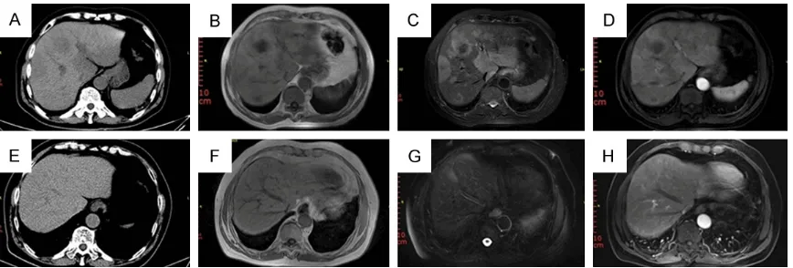
investiga-tions, including blood and bone marrow culture, were normal. The serology results were nega-tive for hepatitis B and C viruses. The serum levels of alfa fetoprotein (AFP), carcinoembry-onic antigen (CEA), and other tumor markers were normal. However, serum lactate dehydro-genase (LDH) (243 U/L; reference range, 135-226 U/L), hypersensitive C-reactive protein (hs-CRP) (75.91 mg/L; reference range, 0-5 mg/L), erythrocyte sedimentation rate (41 mm/h; ref-erence range, 0-20 mm/h), and procalcitonin levels (0.08 ng/mL; reference range, 0-0.05 ng/mL) were elevated. Elevated inflammatory indices were indicative of infection. A com- puted tomography (CT) of the chest, brain, and pelvis revealed no abnormalities. Three-dimen- sional CT imaging of the liver showed multiple hypodense lesions with unclear boundaries. The size of the largest lesion was approximately 58 × 53 mm, and the CT value was about 45 HU (Figure 1A). Magnetic resonance imaging (MRI) of the liver showed multiple diffuse sli- ght hypo-intense lesions on T1-weighted imag-es (Figure 1B) and slight hyper-intense on T2-weighted images enhanced in the arterial phase after contrast injection. No significant lesion enhancement was observed in the venous and delayed phases (Figure 1C, 1D). There were no signs of lymphadenopathy on the MRI. Collectively, the radiographic charac-teristics were consistent with potential inflam -matory lesions. On the basis of these findings, liver abscess or hepatocellular carcinoma was suspected. Based on a presumptive diagnosis of liver abscess, the patient was initially treated with amoxicillin sulbactam, imipenem, and cila- statin sodium for twenty days. However, the
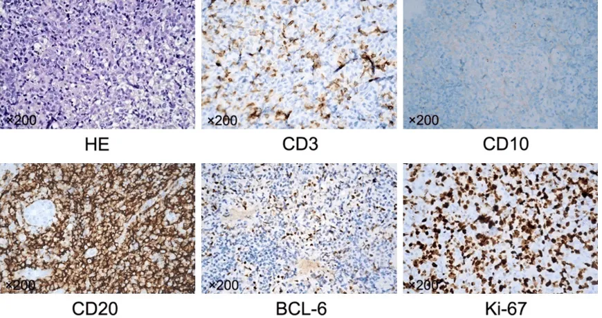
patient did not respond to antibiotic therapy; his body temperature rose to 38.2-38.8°C, accompanied by sweating, fatigue, and a poor appetite. Therefore, a percutaneous, ultra-sound-guided liver biopsy was performed. Hi- stopathological examination showed that the pathologic diagnosis of the tissue was diffuse large B cell lymphoma (NOS, non-GCB) and did not exclude follicular lymphoma. On im- munohistochemical examination, CD20, BCL-6, MUM-1, BCL-2, and c-myc were positive; CD3, CD10, CD23, CyclinD1, AE1/AE3, CD56 were negative. Ki-67 labeling index was approxi-mately 70% (Figure 2). Subsequently, the pa- tient was transferred to the department of hematology for further treatment. He was treat-ed with six cycles of a R-CHOP regimen (cyclo-phosphamide, doxorubicin, vincristine, and prednisone with rituximab injection). His body temperature returned to a normal level after 1 cycle. The low-density lesions disappeared after 6 chemotherapy cycles (Figure 1E-H). As of his 2-year follow-up, no new lesions have been noted, and the patient has survived with no signs of recurrence.
Discussion
Owing to the non-specific clinical manifesta -tions, patients with PHL are liable to be misdi-agnosed. This patient was initially misdiag-nosed as a case of liver abscess. Currently, there is no consensus on the precise definition of PHL. In some reports, cases were consid-ered as primary if the disease was confined to the liver at presentation. Edgardo et al [3]. pro-posed the following criteria for diagnosis:
[image:2.612.91.525.70.217.2]toms explained predominantly by liver involve-ment, an absence of lymph node involveinvolve-ment, normal hematological parameters, and no spleen or bone marrow involvement for at least 6 months since the appearance of the hepatic lesion. Accordingly, this definition is cited in some cases. In the present case, the diagnosis of PHL was made after exclusion of other sites of disease by imaging studies, bone marrow biopsy, and liver biopsy.
PHL can occur at any age, and the average age of the reported patients is 55 years old. The condition is more common in males (male to female ratio, 3:1) [4]. The etiology of PHL is uncertain, and it may be associated with HIV, AIDS, hepatitis B and C, the Epstein-Barr virus, liver cirrhosis, primary biliary cirrhosis, immu-nosuppressive therapy, and autoimmune dis-eases. Its mechanism potentially involves loss of inherent immune surveillance by T lympho-cytes and impaired function after viral infection or the use of immunosuppressive agents. This may result in unrestrained lymphopoiesis, thereby forming lymphoma [5].
The clinical manifestations of PHL are usually non-specific and include hepatomegaly, gastro -intestinal symptoms (abdominal pain, vomiting, loss of appetite), typical lymphoma symptoms (such as fever, night sweats), and weight loss.
Other rare clinical manifestations include pleu-ral effusion, jaundice, thrombocytopenia, meta-bolic acidosis, and hypercalcemia [4]. Patients with PHL tend to have abnormal liver enzymes (elevation of aspartate aminotransferase, ala-nine aminotransferase, bilirubin, gamma glu-tamyl transferase, alkaline phosphatase, and LDH). In particular, elevated LDH and ALP levels are more frequently encountered. Tumor mark-ers such as AFP and CEA are typically normal in patients with PHL [6]. In this case, the patient experienced weight loss, night sweats, and a sustained fever, which are typical lymphoma symptoms. The patient’s LDH level was in- creased, but his AFP and CEA levels were nor-mal. The clinical characteristics were consis-tent with those of primary hepatic non-Hodgkin lymphoma. In a previous study, LDH levels in patients with PHL were normal or significantly decreased after surgery or chemotherapy. The recurrence of PHL was associated with an increase in the LDH level again. Dynamic changes in serum LDH levels may serve as a marker of diagnostic and prognostic relevance [7].
The imaging features of hepatic lymphoma are non-specific and difficult to distinguish from those of focal nodular hyperplasia, primary hepatic tumor, hepatic-metastases, or system-ic lymphoma with secondary hepatsystem-ic
involve-Figure 2. Histopathological examination showing diffuse large B-cell lymphoma (H&E); On the immunohistochemis-try examination, the tumor cells tested positive for CD20 and Bcl-6; the tumor cells were negative for CD3 and CD10.
[image:3.612.90.525.73.303.2]ment [8]. PHL may appear as a solitary lesion (39-60%), multiple lesions (25-40%), or as a dif-fuse infiltration (rare) in the liver [9, 10]. Our patient exhibited multiple lesions on his imag-ing examination. PHL appears as a low-density lesion on CT and exhibits no enhancement or minimally patchy or ring-like enhancement on contrast-enhanced CT. On MRI, the lesion appears hypo-intense on T1-weighted and mild-ly hyper-intense on T2-weighted images. On dif-fusion weighted MRI, PHL lesions showed a signal restriction [11]. Contrast enhancement was higher in lesions with higher T2 signals in our patient. Furthermore, peripheral enhance-ment was occasionally seen. Similar to lympho-ma at other sites, PHL also shows a diffuse restriction owing to its cellularity [12]. The imaging features of PHL may mimic those of hepatic tuberculosis or primary hepatic tumors among other conditions. Radiological and labo-ratory investigations are extremely useful in dif-ferentiating PHL from other diseases. Patients with hepatic tuberculosis typically have a his-tory of pulmonary tuberculosis. In 80% of cases, primary hepatic tumor develops in the cirrhotic liver; these patients are HBV- or HCV-positive and often have elevated AFP levels. In addition, a primary hepatic tumor usually exhib-its hyper-vascularity with marked enhancement in the arterial phase and washout in the delayed phase [13]. On CT images, hepatic miliary tuberculosis shows liver volume increased, dif-fuse miliary or nodular lesions with low density, uniform size, < 2 cm in diameter, with clear boundaries, calcification, and enhancement in the arterial and venous phases. Hepatic lym-phoma lacks a blood supply and has no obvi-ous enhancement ring.
This combination of clinical and laboratory fea-tures allows a speculative diagnosis of PHL. However, a definite diagnosis requires histologi -cal results (liver biopsy or surgi-cal specimen)
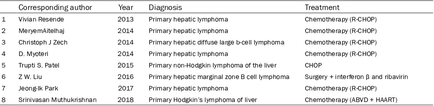
and the absence of lymphoproliferative disease outside the liver. An immunohistochemical examination is essential for the identification of the type and for the differential diagnosis of PHL. The currently reported PHL are all non-Hodgkin’s lymphoma, and there are no Hodgkin lymphoma reports. Most PHL cases (46-68%) reported in the literature are diffuse large B-cell lymphoma. Other types have been described in less than 5% of cases including diffuse mixed large and small cell, lymphoblastic, diffuse immunoblastic, diffuse histiocytic, mantle cell, and small non-cleaved or Burkitt lymphoma [7]. PHL treatment options include surgery, chemo-therapy and local radiochemo-therapy; these options have been used alone or in combination. The treatment of similar diseases from recent years is enumerated in Table 1. However, with the continuous development of chemotherapeutic regimens, especially the application of targeted therapeutic drugs, chemotherapy combined targeted therapeutic drugs (rituximab) has become the first-line treatment of PHL [14]. In this case, a single course treatment with R-CHOP (rituximab, cyclophosphamide, doxoru-bicin, vincristine, and prednisone) regimen to remarkable attenuation of low-density lesions. Conclusion
Cases of primary hepatic lymphoma initially presenting with liver mass or liver infiltration with normal levels of alpha-fetoprotein and CEA are indeed rare. The clinical and imaging mani-festations of PHL are non-specific. The diagno -sis of PHL should be strictly based on histopa-thology and immunophenotype. Owing to the rarity of the disease, the diagnosis and man-agement of PHL is typically challenging. Ne- vertheless, chemotherapy is still the main treat-ment option for PHL. The patient rapidly responded to 6 cycles of a R-CHOP chemother-Table 1. Published case reports of primary hepatic lymphoma in the contemporary literature
Corresponding author Year Diagnosis Treatment
1 Vivian Resende 2013 Primary hepatic lymphoma Chemotherapy (R-CHOP) 2 MeryemAitelhaj 2014 Primary hepatic lymphoma Chemotherapy (R-CHOP) 3 Christoph J Zech 2014 Primary hepatic diffuse large b-cell lymphoma Chemotherapy (R-CHOP) 4 D. Myoteri 2014 Primary hepatic lymphoma Chemotherapy (R-CHOP) 5 Trupti S. Patel 2015 Primary non-Hodgkin lymphoma of the liver CHOP
6 Z W. Liu 2016 Primary hepatic marginal zone B cell lymphoma Surgery + interferon β and ribavirin
7 Jeong-Ik Park 2017 Primary hepatic lymphoma Chemotherapy (R-CHOP) 8 Srinivasan Muthukrishnan 2018 Primary Hodgkin’s lymphoma of liver Chemotherapy (ABVD + HAART)
[image:4.612.91.523.84.190.2]apy regimen. Therefore, a confirmation of the diagnosis and appropriate therapy is neces-sary for such a challenging case.
Disclosure of conflict of interest
None.
Abbreviations
AFP, alpha fetoprotein; ALP, alkaline phospha-tase; ALT, alanine aminotransferase; AST, aspartate aminotransferase; GGT, glutamate transpeptidase; CEA, carcinoembryogenic anti-gen; LDH, lactate dehydrogenase; hs-CRP, hypersensitive c-reactive protein; CT, comput-ed tomography; MRI, magnetic resonance imaging.
Address correspondence to: Lihua Cao, Department
of Respiratory Medicine. The Second Hospital of Dalian Medical University, 467 Zhong Shan Road, Dalian 116023, Liaoning, P.R. China. Tel: +86-17709875757; E-mail: ydeyclh@163.com
References
[1] Aitelhaj M, Akaaboun S, Lkhouyaali S, Ait Ali M, Benhmida S, Latib R, Chami I, Boujida N, M’rabti H and Errihani H. Primary hepatic lym-phoma: a case report. J Gastrointest Cancer 2014; 45: 212-5.
[2] Ugurluer G, Miller RC, Li Y, Thariat J, Ghadjar P, Schick U and Ozsahin M. Primary hepatic lym-phoma: a retrospective, multicenter rare can-cer network study. Rare Tumors 2016; 8: 6502.
[3] Santos ES, Raez LE, Salvatierra J, Morgensz-tern D, Shanmugan N and Neff GW. Primary hepatic non-Hodgkin’s lymphomas: case re-port and review of the literature. Am J Gastro-enterol 2003; 98: 2789-2793.
[4] Gatselis NK and Dalekos GN. Education and imaging. Hepatobiliary and pancreatic: prima-ry hepatic lymphoma. J Gastroenterol Hepatol 2011; 26: 210.
[5] Liu Y, Jiang J, Wu Q, Zhang Q, Xu Y, Qu Z, Ma G, Wang X, Wang X, Jin W and Fang B. A case of primary hepatic lymphoma and related litera-ture review. Case Reports Hepatol 2016; 2016: 6764121.
[6] Lee JA, Jeong WK, Min JH and Kim J. Primary hepatic lymphoma mimicking acute hepatitis. Clin Mol Hepatol 2013; 19: 320-323.
[7] Page RD, Romaguera JE, Osborne B, Medeiros LJ, Rodriguez J, North L, Sanz-Rodriguez C and Cabanillas F. Primary hepatic lymphoma: favor-able outcome after combination chemothera-py. Cancer 2001; 92: 2023-2029.
[8] Zentar A, Tarchouli M, Elkaoui H, Belhamidi MS, Ratbi MB, Bouchentouf SM, Ali AA, Boun-aim A and Sair K. Primary hepatic lymphoma. J Gastrointest Cancer 2014; 45: 380-382. [9] Levy AD. Malignant liver tumors. Clin Liver Dis
2002; 6: 147-164.
[10] Cerban R, Gheorghe L, Becheanu G, Serban V and Gheorghe C. Primary focal T-cell lympho-ma of the liver: a case report and review of the literature. J Gastrointestin Liver Dis 2012; 21: 213-216.
[11] Colagrande S, Calistri L, Grazzini G, Nardi C, Busoni S, Morana G and Grazioli L. MRI fea-tures of primary hepatic lymphoma. Abdom Radiol (NY) 2018; 43: 2277-2287.
[12] Dhamija E, Madhusudhan KS, Shalimar, Das P, Srivastava DN and Gupta AK. Primary hepatic diffuse large B-cell lymphoma: unusual pre-sentation and imaging features. Curr Probl Di-agn Radiol 2015; 44: 290-293.
[13] Hussain SM, Semelka RC and Mitchell DG. MR imaging of hepatocellular carcinoma. Magn Reson Imaging Clin N Am 2002; 10: 31-52. [14] Ma YJ, Chen EQ, Chen XB, Wang J and Tang H.