443
THE PHYSIOLOGY OF EXCRETION IN A BLOOD
SUCKING INSECT, RHODNIUS PROLIXUS
(HEMIPTERA, REDUVIIDAE)
III. THE MECHANISM OF URIC ACID EXCRETION
BY V. B. WIGGLESWORTH, M.A., M.D.
(From the Department of Entomology, London School of Hygiene and Tropical Medicine.)
(Received i^thjtaie, 1931.)
(With Three Text-figures.)
CONTENTS.
PAGE
I n t r o d u c t i o n . . . 4 4 3 1. G e n e r a l considerations . . . 4 4 4 2. E x c r e t i o n of n e u t r a l r e d a n d o t h e r dyes . . 445 3 . Effects of o b s t r u c t i n g t h e M a l p i g h i a n t u b e s . 446 4 . R e a c t i o n of t h e c o n t e n t s of t h e M a l p i g h i a n t u b e s 4 4 8 Discussion 449
S u m m a r y . . . . . . . . 4 5 O
References 451
INTRODUCTION.
THE first two papers of this series (Wigglesworth, 1931 a, b) have made it clear that the main function of the Malpighian tubes in Rhodnius is the elimination of free uric acid, and that the greater part of this elimination takes place with a minimum loss of water. The present paper deals with the mechanism by which this is accomplished.
It has been shown that the uric acid appears in solid form (uratic spheres) in the lower third of the Malpighian tube, and that this distribution is associated with well-marked differences in the structure of the epithelium. It is probable, therefore, that these structural differences denote a difference in function, and on this basis the following hypothesis has been constructed (Fig. 1).
It is supposed that in the upper segment of the tube a solution of potassium or sodium acid urate is secreted from the blood into the lumen. In the lower segment both water and alkali are reabsorbed, until, at a given concentration, the uric acid, carrying with it perhaps a small amount of urate, crystallises out. The alkali is then used to combine with more uric acid produced in the tissues, and the process is repeated. Thus there is a continuous circulation of both water and base.
444 V. B. WlGGLESWORTH
In Fig. i the reabsorption of base is pictured as occurring in the form of bicarbonate; but there are, of course, many other possible mechanisms by which this could be accomplished, such as the absorption of alkaline phosphate from a mixture of urate and acid phosphate. That is a detail which does not affect the general principle.
The evidence in support of this hypothesis will be presented under four headings.
KHU + HaO
<-• r i
KHD + HjO " * ) KHC03+Hj0 +
Fig. I.
i. GENERAL CONSIDERATIONS.
It is quite evident from the observations recorded in paper II (Wigglesworth, 1931 b) that uric acid is discharged into the lumen in solution, for solid urates are never present within the cells. Yet the excreta, in the later stages of digestion, contain comparatively little fluid. It is clear, therefore, that water must have been reabsorbed.
Now the greater part (the upper two-thirds) of the Malpighian tube never contains solid uric acid, and yet the very extent of this region suggests that it is important in excretion. It is natural, therefore, to conclude that this segment is secreting uric acid, and the lower segment, where the solid granules occur, is reab'sorbing water. This idea is supported by the fact that when solid urates first appear, the clear fluid extends some way into the lower segment, but gradually the uratic spheres extend higher and higher until they reach the junction between the two segments.
Now it was shown in paper I (Wigglesworth, 1931 a) that the output of uric acid during the first fortnight or three weeks after a meal is at the rate of 0-5 to o-6 mg. per diem. Since the solubility of uric acid is given as 1 part in 39,500 of water (Hoppe-Seyler and Thierfelder (1924)), this would require about 20 c.c. for its solution; in other words, if the insect were secreting a concentrated solution of uric acid and reabsorbing water, its four Malpighian tubes would have to deal with 20 c.c. of fluid in twenty-four hours. Yet, insect A of Fig. 1 in paper I (Wiggles-worth, 1931 a) at the most rapid stage of the elimination of water was excreting at the rate of only O'8 c.c. per diem.
re-Physiology of Excretion in a Blood-sticking Insect, Rhodnius prolixus 445
absorbed, and its reabsorption will favour the precipitation of the highly insoluble free acid.
Uric acid and its salts are, of course, very prone to form supersaturated solutions, especially in biological fluids, so that the figures quoted above have little real significance; but this does not impair the general argument, which affords con-siderable a priori support for the hypothesis.
2. EXCRETION OF NEUTRAL RED AND OTHER DYES.
Neutral red. As being one of the least toxic and most diffusible of dyes, neutral red was employed as an index of the flow of water through the excretory system. The course of its excretion may be very clearly seen if several loops of the Malpighian tubes in the living insect are exposed on a slide and examined by transmitted light (as described in paper II). The following is a shortened account of an actual ex periment.
10.5 a.m. Malpighian tubes exposed in insect's own blood and drop of Ringer's solution containing neutral red added.
10.6 a.m. Cells of upper segment diffusely red. Lumen filled with dye. Cells of lower segment entirely colourless.
10.10 a.m. Faint pink flush appearing in cells of lower segment.
10.15 a.m. Outer part of cells of upper segment becoming colourless. Minute red granules appearing in cells of lower segment, especially towards lower end.
10.30 a.m. Cells of upper segment almost colourless. Granules in lower segment more marked.
1045 a.m. Dye in lumen becoming less throughout. Granules in lower cells very pronounced.
11.0 a.m. Dye has almost disappeared from lumen. Cells almost colourless in upper segment, full of intense red granules in lower segment. These granules most conspicuous in the lower extremity and in the ampullae1. Rectal gland colourless. These observations strongly suggest that there is a flow of water into the Malpighian tube in the upper segment, and out of the tube in the lower segment.
Other vital dyes. A number of experiments have been performed with various other dyes. Most of these experiments have consisted in replacing the abdominal tergites with a coverglass (as described in paper II), adding a small quantity of the dye to the insect's blood, and observing the Malpighian tubes under a binocular dissecting microscope. But the observations have been supplemented by dissecting out the tubes at various stages. In general, the results confirm those obtained with neutral red and may be described very briefly.
Basic fuchsin is rapidly taken up by the cells of the upper segment, staining the striated border intense red. Within an hour the cells of the lower segment are becoming faintly pink. At the end of twenty-four hours, the upper cells are almost colourless; lower cells and ampullae full of fine red granules.
1
It is worth noting that the red granules in the cells of the ampullae tend to lie in longitudinal rows. This confirms the impression given by sections (Wigglesworth, 1931 b) that the cytoplasm of these cells is made up of longitudinal fibrillae.
446 V. B. WlGGLESWORTH
Janus green B is very actively taken up by the upper segment and discharged into the lumen. The cells of the lower segment are almost colourless at all stages of excretion. If much dye is present it may be segregated in the upper cells in the form of dark granules.
Meihylene blue behaves just like Janus green B.
Bismarck brown, congo red and trypan blue, like ammonia carmine, are slowly taken up by the pericardial cells, but do not seem to be eliminated at all by the Malpighian tubes.
Excretion of indigocarmine. It is well known that indigocarmine is actively taken up by many of the excretory organs of animals and that it is frequently deposited in the lumen in the form of solid granules or crystals. According to current notions of renal secretion (see v. Mollendorff, 1930), this precipitation is the result of reabsorption of water. It was therefore expected that if indigocarmine were added to the Malpighian tubes of Rhodnius, it would appear in solution in the upper segment of the tube, and be precipitated, Like the urate, in the lower segment.
This, however, did not happen. The dye was rapidly taken up by the tubes and, as usual, after traversing the cells in the colourless form, it appeared in the lumen, in a few minutes, as a blue solution. But at the end of an hour, whereas the lower segment still contained a bright blue solution, solid crystals of the dye had appeared in the lumen of the upper segment, and after twenty-four hours, the entire upper segment was filled with needle-shaped crystals1.
At first sight this result does not appear to tally with the hypothesis that re-absorption of water is confined to the lower segment. But it is possible that the precipitation of the dye in the upper segment is due to causes other than re-absorption. The following is a possible explanation. The dye traverses the cells in the "leuco" form and reverts to the coloured form immediately it is discharged into the lumen. Now it is well known that the "leuco" form of indigoid dyes is far more soluble than the coloured form. If, therefore, the cells are discharging a saturated solution of the "leuco" form, when this reverts to the coloured form in the lumen it will be supersaturated and will immediately crystallise out without the necessity for any absorption of water.
3. EFFECTS OF OBSTRUCTING THE MALPIGHIAN TUBES.
An attempt was then made to compare the function of the different segments by obstructing the Malpighian tube at different levels.
Technique.
After many failures at ligating the tubes with fine threads, the following pro-cedure has been adopted. The abdominal tergites are removed, as already described, and the insect secured to a glass slide. The Malpighian tubes, though freely visible, lie deep to a delicate pericardial fascia, and this must therefore be incised. A small
1
Physiology of Excretion in a Blood-sucking Insect, Rhodnius prolixus 447
piece of soft wax (" Sira" adhesive wax, as supplied by British Drug Houses, Ltd., has been used) is then fixed on the tip of a dissecting needle, drawn out at the end into a fine filament and bent to form a minute hook (Fig. 2 D). The Malpighian tube is grappled with this hook, which is thereupon flattened and pinched off with the finest watchmaker's forceps (Fig. 2 E and F). The abdomen is then closed with a coverslip and the insect kept in a moist chamber.
[image:5.451.63.400.233.459.2]In order to decide which part of the tube is responsible for the production of uric acid, it has been necessary to use insects with no solid urates in the Malpighian tubes at the commencement of the experiment. In each case, therefore, the obstructions were made three or four hours after feeding, i.e. when the discharge of clear fluid has ceased, but before any uratic granules have appeared in the tubes.
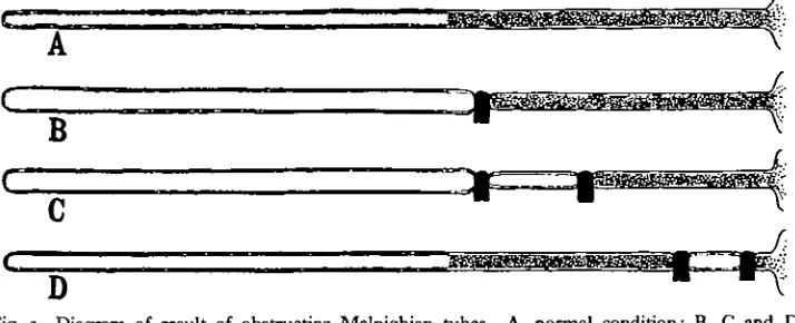
Fig. 2. A, B and C, portions of Malpighian tubes 12 hours after obstruction at various levels. Arrows point down the tube. D, E and F, method of obstructing the tubes. *, junction of upper and lower segmente of tube.
Results.
It was expected that after a single ligature on the lower segment, uratic granules would appear above the obstruction (as the result of reabsorption) but not below. In fact, this was not so. Fig. 2 A shows a typical experiment, 12 hours after the obstruction, and here, as in every case, there were granules below the ligature but none above.
44$ V. B. WlGGLESWORTH
In the next series of experiments, therefore, two ligatures were applied in the course of the same tube. Fig. 2 B shows a typical example, 12 hours after ligation. There is again much distension and no uric acid above the upper ligature, and plenty of uric acid below the lower ligature, but between the ligatures the tube is clear and contracted. Fig. 2 C shows an experiment in which the two ligatures were applied near the lower end of the tube. In this case there were granules of uric acid both above and below the obstructed region, but none within it.
These results strongly support the idea that the formation of uratic granules in the lower segment of the tube is due to the reabsorption of water. The absence of distension in the section of the tube between the ligatures (Fig. 2 B and C) shows that there is no secretion in this part, and, as already suggested, the absence of granules above the ligature in Fig. 2 A and B is probably due to over-distension of the tube. The presence of uric acid above the upper obstruction in Fig. 2 C is
"" ' fe
A V
_ _ _ . _ f,
(
[image:6.451.49.406.256.401.2]B
Fig. 3. Diagram of result of obstructing Malpighian tubes. A, normal condition; B, C and D correspond respectively with A, B and C of Fig. 2.
probably to be explained by the fact that the extent of the reabsorbing lower segment was here sufficient to cope with the secretion of the upper segment; for in this case there was no distension of the tube. These results are shown diagram-matically in Fig. 3.
4. REACTION OF THE CONTENTS OF THE MALPIGHIAN TUBES.
The evidence presented under sections 2 and 3 supports the idea of a reabsorption of water. Section 1 gives strong reasons for supposing that there is also a re-absorption of base, and experiments with indicators have yielded more evidence for this view.
Physiology of Excretion in a Blood-sucking Insect, Rhodnius prolixus 449
insect. The best results have been obtained when the uric acid is appearing for the first time, say, four or five hours after feeding. The results are shown in Table I.
Table I.
Indicator*
Neutral red Phenol red Chloro-phenol red
pH range and
colour change
6'6 pink—80 orange 6-6 yellow-8-4 red 4-6 yellow—6-s pink
Colour and approximate pK of contents of Malpighian tubes
Upper segment
Orange red 7-2 Flesh colour 7-2 Bright pink >6-8
Lower segment
Bright pink 6-6 Bright yellow 6-6 Pink 6-6
• Bromo-thymol blue was not taken up by the Malpighian tubes.
These results show that the contents of the tube are very weakly alkaline in the upper segment and definitely acid in the lower segment. Now the dissociation constant of uric acid is given as 1-5 x io~9. Theoretically, therefore, a saturated solution should have a pH of about 4-3. But in the textbooks it is usually stated to react neutral (presumably to litmus) and on testing a saturated solution with bromo-thymol blue, chloro-phenol red, phenol red, bromo-cresol purple and neutral red, it gave a value (at room temperature) of/>H 6-5. This figure agrees well with the reaction observed in the lower segment of the Malpighian tube, and suggests that the acidity here is due to uric acid.
It might be argued, of course, that an alkali salt of uric acid is secreted in the upper segment, and the uric acid thrown down in the lower segment as the result of an acid secretion in that part. But if so, the resulting salt would have to be reabsorbed in order to conserve the base, and this would be just another mechanism for reabsorbing base, differing little from that shown in Fig. 1, where the base is represented as combining with the ubiquitous carbonic acid.
DISCUSSION.
The theory put forward in this paper that in Rhodnius prolixus there is a continuous circulation of water and of base, carrying uric acid from the body and accumulating it in solid form in the excretory system, is of sufficient general interest to warrant a comparison with the excretory mechanism of other animals.
The probability of such a circulation of water has long been recognised in the case of birds (Wiener (1902) quoted by Sharpe, 1912) and in the case of reptiles (Regaud and Policard, 1903); for in these animals, as in Rhodmus, the upper part of the excretory system contains only a clear fluid, and the uratic spheres are deposited in the lower segments of the renal tubules and in the cloaca; and it is by virtue of this circulation that birds and reptiles (like Rhodmus) can survive many weeks without food or water (Babcock, 1912). It is highly probable that a similar circulation is widespread among insects, though, curiously enough, the only previous suggestion that reabsorption of water may take place in the Malpighian tubes seems to be that by Bugajew (1928).
45° V. B. WlGGLESWORTH
that this, also, is a widely spread mechanism. For it was shown by Kohler (1910) that the greater part of the uric acid in the excreta of the snake is not combined with base, and Szalagyi and Kriwuscha (1914) obtained the same result in the case of the bird. This being so, the argument set forth under section 1 of this paper will apply equally to these animals. (Cf. Needham (1931), Nature, 128, 152.)
Now in the case of both birds and reptiles, a large part of the absorption of water takes place in the cloaca, and it is natural to seek a parallel in the case of Rhodnius. It has been shown in paper II that if the Malpighian tubes are watched in the living insect during the early days of digestion they can be seen almost continuously discharging uratic granules and fluid into the rectum, which is already distended with liquid; yet the rectum may not be evacuated for many days. It is clear, therefore, that fluid must be absorbed. The mid-gut is cut off by a sphincter which cannot be opened without admitting intestinal contents to the rectum, so that absorption cannot be taking place via the mid-gut. It is true that, as the experiments described in section 3 show, fluid may ascend the Malpighian tubes again and be absorbed in their lower segments, and this doubtless occurs to some extent; but it is probable that much absorption takes place in the rectum itself.
There are two structures which may be engaged in this process; the elongated cells lining the ampullae, and the rectal gland. It has been shown in paper II how the processes of the ampulla cells extend far into the rectum during the active stages of excretion, and float in the clear fluid above the uratic sediment. In this position they are well placed to absorb water, and the fact (described in section 2 of the present paper) that they actively take up neutral red from the lumen is further evidence that they are so engaged.
There is no direct evidence for the absorption of water by the rectal gland. But it has frequently been shown that the cuticular intima of the rectum of insects is freely permeable to water, while the failure of the rectal gland to take up neutral red may simply mean that the cuticle is not permeable to this substance.
The question naturally arises whether the rectum of other insects contains structures for the absorption of water. They certainly possess nothing comparable with the remarkable ampullae of Rhodnius, but rectal glands are widely spread in many groups, and perhaps these puzzling organs, to which so many functions have been ascribed, are simply concerned with the reabsorption of water necessary for the excretion of uric acid. This hypothesis will form the subject of a future communication.
SUMMARY.
A theory of uric acid excretion is put forward according to which potassium or sodium acid urate is secreted in solution in the upper part of the Malpighian tube, and water and base reabsorbed from the lower part of the tube, leading to a pre-cipitation of free uric acid; so that the same water and base are circulated and used repeatedly. The evidence for this theory is presented under four heads:
Physiology of Excretion in a Blood-sucking Insect, Rhodnius prolixus 451
(ii) Experiments with vital dyes. Thus, neutral red is taken up from the blood by the upper segment of the Malpighian tube, and from the lumen of the tube in the lower segment. It is suggested that water follows the same route.
(iii) The application of ligatures to the tubes at different levels shows that uric acid is not secreted by the lower segment.
(iv) Experiments with indicators show that the contents of the tube are faintly alkaline (/>H 7-2) in the upper segment, and acid (pH 6-6) in the lower segment. Uric acid in solution has a pH about 6-5.
It is suggested that the terminal ampullae of the Malpighian tubes, and possibly the rectal glands, also play a part in the reabsorption of water.
REFERENCES.
BABCOCK, S. M. (1912). "Metabolic water: its production and role in vital phenomena." Univ.
Wisconsin Agric. Exp. Sta. Res. Bull. No. 22, 181 pp.
BRUNTZ, L. (1908). "Nouvelles recherches sur l'excre'tion et la phagocytose chez les Thysanoures."
Arch. Zool. exp. et gin. (4), 8, 471-488.
BUGAJEW, I.' I. (1928). "Zum Studium des Baues der Malpighischen GefBsse bei den Insekten."
Zool. Anz. 78, 244-255.
CUENOT, L. (1895). "Etudes physiologiques sur les Orthopteres." Arch, de Biol. 14, 293-341. GRANDIS, V. (1890). "Sur les modifications des Epitheliums glandulaires durant la afere'tion."
Arch. ital. biol. 14, 160-182.
HOPPE-SEYLER and THIERFELDEH (1924). Handbuch der pkysiologisch- und pathologisch-chemischen
Analyse.
KOHLER, R. (1910). "Zur Frage der Quadriurate." Zeit.f. phys. Chem. 70, 360-387.
v. MOLLENDORFF, W. (1930). " Exkretionsapparat." Handbuch der mikroshopischen Anatomie des
Menschen, 7, i, 1-328.
REGAUD, C. and POLICARD, A. (1903). "Recherches sur la structure du rein de quelques Ophidiens." Arch. d'Anat. mxcrosc. 6, 191—282.
SHARPS, N. C. (1912). "On the secretion of urine in birds." Amer. Journ. Phys. 31, 75-84. SZALAGYI, K. and RRIWUSCHA, A. (1914). " Untersuchungen iiber die chemische Zusammensetzung
und die physikalischen Eigenschaften des Enten- und Hiihnerharnes." Biochem. Zeit. 66,
122-138.
WIGGLESWORTH, V. B. (1931 a). "The physiology of excretion in a blood-sucking insect, Rhodnius
prolixus. I. Composition of the urine." Journ. Exp. Biol. 8, 411.