glucose metabolism in liver, muscle and adipose tissues of
the testicular feminised mouse
KELLY, Daniel <http://orcid.org/0000-0002-7463-0692>, AKHTAR, Samia,
SELLERS, Donna, MURALEEDHARAN, Vakkat, CHANNER, Kevin S. and
JONES, Thomas Hugh
Available from Sheffield Hallam University Research Archive (SHURA) at:
http://shura.shu.ac.uk/12966/
This document is the author deposited version. You are advised to consult the
publisher's version if you wish to cite from it.
Published version
KELLY, Daniel, AKHTAR, Samia, SELLERS, Donna, MURALEEDHARAN, Vakkat,
CHANNER, Kevin S. and JONES, Thomas Hugh (2016). Testosterone differentially
regulates targets of lipid and glucose metabolism in liver, muscle and adipose
tissues of the testicular feminised mouse. Endocrine, 54 (2), 504-515.
Copyright and re-use policy
See
http://shura.shu.ac.uk/information.html
O R I G I N A L A R T I C L E
Testosterone differentially regulates targets of lipid and glucose
metabolism in liver, muscle and adipose tissues of the testicular
feminised mouse
Daniel M. Kelly 1,2●Samia Akhtar1●Donna J. Sellers2,5●Vakkat Muraleedharan1,3●
Kevin S. Channer4●T. Hugh Jones3,4
Received: 4 November 2015 / Accepted: 11 June 2016 / Published online: 4 August 2016 © The Author(s) 2016; This article is published with open access at Springerlink.com
Abstract Testosterone deficiency is commonly associated with obesity, metabolic syndrome, type 2 diabetes and their clinical consequences—hepatic steatosis and athero-sclerosis. The testicular feminised mouse (non-functional androgen receptor and low testosterone) develops fatty liver and aortic lipid streaks on a high-fat diet, whereas androgen-replete XY littermate controls do not. Testoster-one treatment ameliorates these effects, although the underlying mechanisms remain unknown. We compared the influence of testosterone on the expression of regulatory targets of glucose, cholesterol and lipid metabolism in muscle, liver, abdominal subcutaneous and visceral adipose tissue. Testicular feminised mice displayed significantly reduced GLUT4 in muscle and glycolytic enzymes in muscle, liver and abdominal subcutaneous but not visceral adipose tissue. Lipoprotein lipase required for fatty acid uptake was only reduced in subcutaneous adipose tissue; enzymes of fatty acid synthesis were increased in liver and subcutaneous tissue. Stearoyl-CoA desaturase-1 that catalyses oleic acid synthesis and is associated with insulin resistance was increased in
visceral adipose tissue and cholesterol efflux components (ABCA1, apoE) were decreased in subcutaneous and liver tissue. Master regulator nuclear receptors involved in meta-bolism—Liver X receptor expression was suppressed in all tissues except visceral adipose tissue, whereas PPARγ was lower in abdominal subcutaneous and visceral adipose tissue and PPARα only in abdominal subcutaneous. Testosterone treatment improved the expression (androgen receptor inde-pendent) of some targets but not all. These exploratory data suggest that androgen deficiency may reduce the buffering capability for glucose uptake and utilisation in abdominal subcutaneous and muscle and fatty acids in abdominal sub-cutaneous. This would lead to an overspill and uptake of excess glucose and triglycerides into visceral adipose tissue, liver and arterial walls.
Keywords Type 2 diabetes ●Metabolism●Testosterone●
Androgen receptor●Adipose tissue
Introduction
Evidence suggests that testosterone deficiency in men is an independent cardiovascular risk factor which is associated with obesity, metabolic syndrome (MetS) and type-2 diabetes (T2D) [1,2]. Insulin resistance, which is common to all of these conditions, results in diminished glucose utilisation and conversion of the excess glucose into fat. Higher circulating triglycerides then lead to an overspill of fat into ectopic storage in liver and arteries as well as increasing the accumulation of visceral fat. The degree of insulin resistance correlates nega-tively with serum testosterone [3,4]. Although the causality of this relationship is often debated, growing evidence indicates * Daniel M. Kelly
daniel.kelly@shu.ac.uk
1 Department of Oncology and Metabolism, Medical School, The University of Sheffield, Sheffield, UK
2 Biomolecular Research Centre, Sheffield Hallam University, Sheffield, UK
3 Centre for Diabetes and Endocrinology, Barnsley Hospital NHS Foundation Trust, Barnsley, UK
4 Department of Cardiology, Royal Hallamshire Hospital, Sheffield, UK
testosterone is a metabolic multi-system player [5]. Epide-miological studies support a bidirectional relationship between serum testosterone and obesity which may be explained by the hypogonadal–obesity–adipocytokine hypothesis [6,7]. Androgen deprivation therapy for the treatment of prostate cancer in men, whilst reducing tumour growth, also increases the risk of coronary heart disease, diabetes and cardiovascular death, indicating that testosterone deficiency may promote atherosclerosis [8,9]. Some trials have reported that achieving a normal physiological testosterone concentration through the administration of testosterone replacement therapy (TRT) improves vascular function and risk factors for atherosclerosis, including reducing central adiposity, percentage body fat, fatty liver and insulin resistance, and improving lipid profiles insulin sensitivity and inflammatory profiles [2,10–15].
A limited number of in vivo and in vitro investigations have highlighted potential molecular targets of testosterone action in metabolic regulation, although a detailed analysis of tissue-specific actions remains absent from the literature [2]. We have previously reported that low testosterone in the testicular feminised (Tfm) mouse (which displays very low testosterone levels and non-functional androgen receptors) is associated with increased lipid deposition in the aortic root and liver when mice are fed a high-cholesterol diet [16–18]. Testoster-one treatment to return levels to those seen in wild-type counterparts significantly reduced aortic fatty steaks and hepatic lipid accumulation with an associated reduction in de novo lipogenesis in the liver in Tfm mice [17].
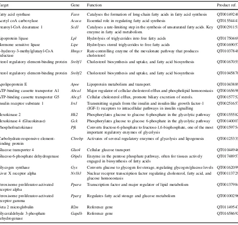
While a growing body of evidence points towards the presence of heterogeneity regarding insulin responsiveness and lipid homeostasis among different tissues [19], the mechan-isms by which testosterone may impart beneficial actions on insulin sensitivity and hence the development of MetS, T2D and cardiovascular risk remain unknown but are likely to be tissue dependent and involve multiple targets of lipid and carbohydrate metabolism. In the present exploratory study, we aim to investigate whether the metabolic protective effects of testosterone act via modulation of the expression of key targets involved in lipid and glucose metabolism in muscle, liver and adipose tissue of cholesterol-fed Tfm mice. Specifically, we investigate key regulatory enzymes of glycolysis, glycogen synthesis, pentose phosphate pathway, glucose transporters, fatty acid synthesis, fatty acid uptake, cholesterol synthesis and efflux, and master regulators of metabolism (see Table1).
Materials and methods
Animals
The Tfm mouse was used as a model of testosterone defi -ciency and androgen receptor (AR) dysfunction as pre-viously described [16–18]. The loss of 17α-hydroxylase, a
key enzyme necessary for testosterone synthesis, leads to serum levels of testosterone in the Tfm mouse that are severely (approximately 10-fold) reduced compared to normal XY littermate controls [20,21]. In addition, a nat-ural mutation in the gene encoding the AR leads to the formation of a truncated receptor protein which lacks both DNA-binding and steroid-binding domains, rendering it non-functional [22, 23]. This model therefore allows potential AR-dependent and independent effects to be investigated. All procedures were carried out under the jurisdiction of a UK Home Office project licence, governed by the UK Animals Scientific Procedures Act 1986. Mice were bred as previously described [20]. Animal numbers were calculated based on our previous investigation [16] for a significance level of 5 %, and a power of 90 % for the primary outcome measure of lipid deposition in the aortic root (see [18]). Where available, preliminary data was used for calculation of sample numbers of individual variables.
Experimental design and tissue collection
Eight-week-old Tfm and XY littermate mice were fed a high-fat, high-cholesterol diet, containing 42 % butterhigh-fat, 1.25 % cholesterol and 0.5 % cholate (Special Diet Services, Essex, UK) ad libitum for a period of 28 weeks. Separate 7-week-old Tfm mice werefirst randomly assigned to one of two groups: a placebo group receiving a once-fortnightly intramuscular injection of 10μL of saline (n=14), or testosterone group (n=14) receiving a once-fortnightly intramuscular injection of 10μL of 100 mg/mL testosterone esters (Sustanon100; tes-tosterone propionate 20 mg/mL, testes-tosterone phenylpropionate 40 mg/mL and testosterone isocaproate 40 mg/mL, Organon Laboratories Ltd, Cambridge, UK), providing a dose of 50 mg/kg, previously shown to replace circulating levels to those of wild-type littermate mice [16]. XY littermate mice (n=14) received placebo injections (10μL saline). Animals were caged under standard conditions in a temperature and humidity-controlled room on a 12 -h light:12-h darkness cycle. Water and food were unrestricted throughout the study.
Measurement of total testosterone and 17β-estradiol
Serum quantification of total testosterone (DRG Instruments GmBH, Marburg, Germany) and 17β-estradiol (Demeditec Diagnostics, Kiel, Germany) was measured in duplicate via ELISA (measurement range 0.2–16 ng/mL and 3–200 pg/mL, respectively).
Quantitative analysis of mRNA
Total RNA was extracted from approximately 100 mg of snap-frozen tissue, reverse transcribed and cDNA (2μL) used for qPCR, using commercial SYBR green reagents
[image:4.595.55.542.67.527.2](Qiagen) as described previously [17]. Primers were pur-chased pre-validated (QuantiTech primer assays; Qiagen), with specified amplification efficiencies of 100 % (±10 %) (see Table1). Primers for Β-2 microglobulin (B2m) were also included and served as an internal reference control, selected as the most stable gene from a panel of commonly used reference genes (Gapdh, beta-actin, ribosomal protein 13A). Each reaction was carried out in triplicate with cycling and detection offluorescent signal carried out using an Agilent Mx3000P QPCR System. Results were corrected for the expression of the house-keeping gene and normal-ised to the XY littermates as a control. Relative copy number was expressed as fold change 2-(ddCT).
Table 1 Qiagen qPCR primers
Target Gene Function Product ref.
Fatty acid synthase Fasn Catalyses the formation of long-chain fatty acids in fatty acid synthesis QT00149240 Acetyl coA carboxylase Acaca Essential role in regulating fatty acid synthesis QT01554441 Stearoyl-CoA desaturase 1 Scd1 Catalyses a rate-limiting step in the synthesis of unsaturated fatty acids. Key
enzyme in fatty acid metabolism.
QT00291151
Lipoprotein lipase Lpl Hydrolysis of triglycerides into free fatty acids QT01750469 Hormone sensitive lipase Lipe Hydrolyses stored triglycerides to free fatty acids QT00169057 3-hydroxy-3-methylglutaryl-CoA
reductase
Hmgcr Rate-controlling enzyme of the mevalonate pathway that produces cholesterol
QT01037848
Sterol regulatory element-binding protein 1
Srebf1 Cholesterol biosynthesis and uptake, and fatty acid biosynthesis QT00167055
Sterol regulatory element-binding protein 2
Srebf2 Cholesterol biosynthesis and uptake, and fatty acid biosynthesis QT01045870
Apolipoprotein E Apoe Lipoprotein metabolism and transport. QT01043889 ATP-binding cassette transporter A1 Abca1 Major regulator of cellular cholesterol efflux and phospholipid homoeostasis QT00165690 ATP-binding cassette transporter G5 Abcg5 Cellular cholesterol efflux, promote biliary excretion of sterols. QT00157752 Insulin receptor substrate 1 Irs1 Transmitting signals from the insulin and insulin-like growth factor-1
(IGF-1) receptors to intracellular pathways in insulin signalling
QT00251657
Hexokinase 2 Hk2 Phosphorylates glucose to glucose 6-phosphate in the glycolytic pathway QT00155582 Hexokinase 4 (Glucokinase) Gck Phosphorylates glucose to glucose 6-phosphate in the glycolytic pathway QT00140007 Phosphofructokinase Pfk Converts fructose-6-phosphate to fructose-1,6-bisphosphate, one of the most
important regulatory enzymes of glycolysis
QT00159754
Carbohydrate-responsive element-binding protein
Chrebp Activates of several regulatory enzymes of glycolysis and lipogenesis QT00125335
Glucose transporter 4 Glut4 Cellular glucose transport QT01044946 Glucose-6-phosphate dehydrogenase G6pdx Enzyme in the pentose phosphate pathway, often for tissues actively
engaged in biosynthesis of fatty acids
QT01748957
Glycogen synthase Gys Converts glucose to glycogen for storage, regulating glycogen/glucose levels QT00162099 Liver X receptor alpha Nr1h3 Nuclear receptor transcription factor regulating cholesterol, fatty acid, and
glucose homoeostasis
QT00113729
Peroxisome proliferator-activated receptor alpha
Ppara Transcription factor and major regulator of lipid metabolism QT00137984
Peroxisome proliferator-activated receptor gamma
Pparg Regulates fatty acid storage and glucose metabolism QT00100296
Beta 2 microglobulin B2m Reference gene QT01149547
Glyceraldehyde 3-phosphate dehydrogenase
Western immunoblotting
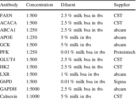
In this exploratory study we selected targets that were sig-nificantly altered at the gene expression level for analysis by western blotting. Due to low concentrations of protein ascertainable from limited availability of adipose tissue, western blotting was unable to be carried out on sub-cutaneous and visceral samples. Protein was extracted from 200 mg of mouse liver or muscle tissue as previously described [17]. In brief, 50µg of total isolated protein was separated by electrophoresis and transferred to nitrocellu-lose membranes (BioRad, Hertfordshire, UK). Membranes were blocked for 1 h in 5 % dried semi-skimmed milk diluted in tris/glycine (TG) buffer containing 0.05 % Tween 20 (TGT; BioRad, UK). Primary antibodies were incubated overnight at 4 °C diluted in either 5% bovine serum albu-min/TGT, 5 % milk/TGT or 2.5 % milk/BSA (see Table2). Immunoreactive proteins were detected using anti-rabbit IgG HRP-linked secondary antibody (1:500, Cell Signal-ling) for polyclonal antibody detection or anti-mouse IgG HRP-linked secondary antibody (1:500, Cell Signalling) followed by a chemiluminescence peroxidase substrate kit (Roche, Sussex, UK). Band intensities were quantified using Genetools software (Syngene, Cambridge, UK) rela-tive to the house-keeping protein GAPDH or Calnexin.
Statistical analysis
Results are presented as mean±SEM. Differences between groups with normally distributed data were compared using unpaired t tests without assuming consistent standard deviations of groups. Mann–Whitney U tests were used where data did not follow a normal distribution. Correc-tions for multiple comparisons were made using the
Sidak-Bonferroni post hoc test. Significance was accepted atp≤0.05.
Results
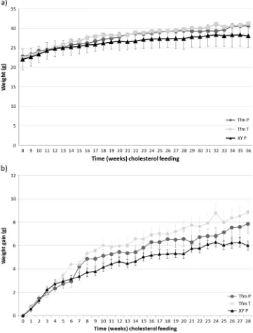
Serum testosterone levels were greatly reduced in Tfm mice (2.2±1.2 nM,p=0.03) compared to wild-type equivalents (16.5±4.3 nM).1 Testosterone treatment of Tfm mice increased serum levels of testosterone comparable to wild-type levels (14.7±5.2 nM, p=0.98). 17-β estradiol levels were similar between all groups, Tfm mice (94.2±15.5 pmol) compared to wild-type (106.0±33.9 pmol,p=0.17) and testosterone-treated Tfm mice (135.2±28.7 pmol,p= 0.99). Animal weights and weight gain did not significantly differ between groups over the duration of the 28 week feeding period but there was a trend towards Tfm mice gaining more weight compared to littermates by the end of the study period (p=0.066,n=14; Fig.1).
Carbohydrate metabolism
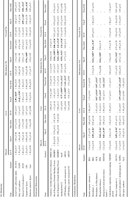
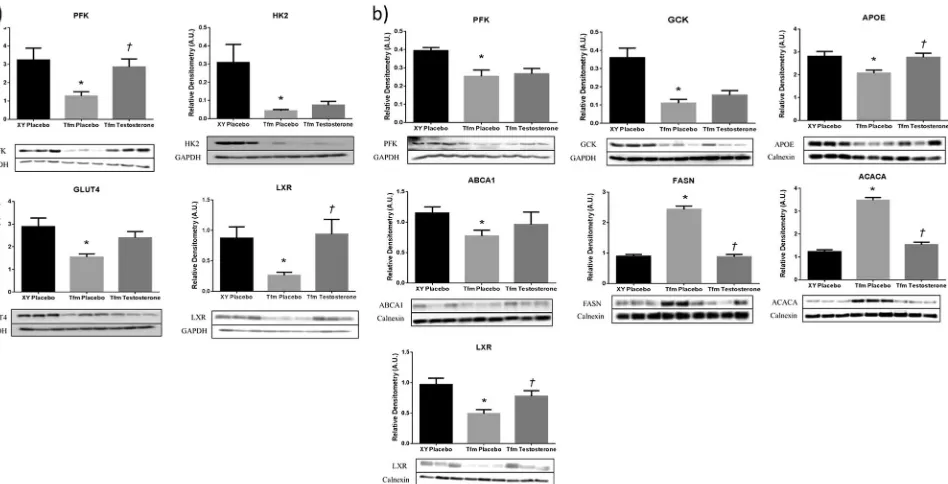
Gene expression of the glycolytic regulatory gateway enzymes hexokinase (Hk2,Gck) andPfkwas significantly lower in muscle (p=0.012, p=0.032), liver (p=0.002, p=0.04) and SAT (p=0.009,p=0.03) but not in VAT of Tfm-placebo mice compared to XY littermates (Table3). Testosterone administration increased Gck expression (p=0.015) in the liver of Tfm mice but these enzymes were not significantly altered in other tissues by treatment.Glut4 was similarly decreased in muscle (p=0.015) and SAT (p=0.014) of Tfm mice versus wild-type mice, with no effect of testosterone treatment. HepaticG6pdxwas elevated in Tfm mice compared to XY mice (p<0.001) and testosterone treatment showed a trend to reducing this expression in Tfm mice (p=0.056). All other gene targets were not altered between experimental groups in the tissues investigated.
[image:5.595.51.290.69.247.2]Protein expression of PFK in the liver of experimental animals matched gene expression data with reduced levels in Tfm placebo mice compared to wild-type (p=0.005) and no effect of treatment with testosterone (Fig.2). Muscle protein expression of PFK was reduced in Tfm mice (p=0.018) with a significant increase in expression fol-lowing treatment (p=0.01). Hepatic GCK protein was also reduced in Tfm mice receiving placebo (p=0.001) as demonstrated at the gene level; however, testosterone administration had no effect showing discrepancy between gene and protein expression. HK2 in muscle was also reduced at the protein level in Tfm mice (p=0.024), but there was no effect due to treatment. Muscle GLUT4 was decreased in Tfm mice compared to wildtype (p=0.037) Table 2 Antibody parameters
Antibody Concentration Diluent Supplier
FASN 1:500 2.5 % milk bsa in tbs CST ACACA 1:500 2.5 % milk bsa in tbs CST ABCA1 1:250 2.5 % milk bsa in tbs abcam APOE 1:250 5 % milk in tbs abcam GCK 1:500 5 % milk in tbs abcam PFK 1:250 0.01 % milk bsa in tbs Proteintech GLUT4 1:500 2.5 % milk bsa in tbs CST HK2 1:500 2.5 % milk bsa in tbs CST LXR 1:500 1 % milk bsa in tbs abcam G6PD 1:500 0.01 % milk bsa in tbs Sigma GAPDH 1:5000 2.5 % milk bsa in tbs abcam Calnexin 1:1000 5 % milk in tbs CST
bsabovine serum albumin,tbstris-buffered saline,CSTcell signalling technologies
and testosterone administration demonstrated a trend towards increasing this expression (p=0.053). We were unable to detect G6PD protein expression in the liver of experimental animals.
Lipid metabolism
Cholesterol metabolism
Expression of cholesterol transporters, Apoe and Abca1, were reduced in the liver of Tfm mice compared to litter-mates (p=0.009, p=0.002). Treatment with testosterone
significantly increased this expression (p=0.027,p=0.02), similar to wild-type levels (Table3). Similarly, Apoewas decreased in SAT of Tfm mice (p=0.01), an effect that was abolished by testosterone administration (p=0.015 versus Tfm P). Srebf1 and Srebf2 expression was significantly lower in the SAT of Tfm mice versus XY littermates (p= 0.002, p=0.003). Treatment with testosterone elevated these expression levels of Srebf1 (p=0.015) similar to those demonstrated in wild-type mice although not sig-nificantly so forSrebf2with only a trend towards increased expression observed (p=0.053). In support of gene expression data, ABCA1 protein was significantly reduced Fig. 1 Animal weights and
[image:6.595.182.543.60.535.2]in livers of Tfm mice compared to littermates and testos-terone treated Tfms (Fig.2). Hepatic APOE protein expression matched gene expression data with significantly lower levels in placebo-treated Tfm mice compared to XY littermates and testosterone-treated Tfm mice (p=0.011, p=0.007, respectively).
Fatty acid metabolism
Visceral adipose Scd1expression was significantly higher in Tfm mice receiving placebo than in XY littermates also receiving placebo injections (p=0.034, Table3). Testos-terone treatment of Tfm mice returned expression levels to those of XY mice with a significant reduction compared to placebo-treated Tfm mice (p=0.027). t test analysis similarly revealed an increase in hepaticScd1 expression in Tfm placebo mice although not statistically significant (p=0.08). DecreasedLplexpression was observed in SAT from Tfm mice compared to wildtype (p=0.016) although testosterone administration to Tfm animals had no effect on this. Hepatic gene expression of Fasn and Acaca, the key regulatory enzymes in de novo lipogenesis, were significantly increased in Tfm mice receiving placebo injections compared to wild-type littermates (p=0.049,p =0.042, respectively).2Testosterone treatment decreased this expression but not significantly. Gene expression of all other lipid metabolism targets in liver and adipose tissue were not significantly different between animal groups. Western blotting showed hepatic protein expres-sion of FASN and ACACA to be increased in Tfm mice confirming gene expression findings.2Testosterone treat-ment significantly reduced the protein expression of these enzymes versus placebo treated Tfm mice to similar levels as XY littermates.
No targets of fat metabolism and cholesterol homeostasis displayed altered gene expression in muscle tissue from the different experimental groups.
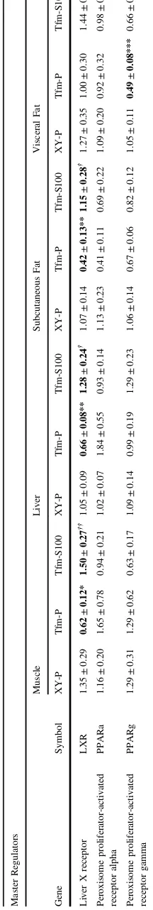
Master regulators
Gene expression of Lxr was significantly reduced in Tfm placebo mice in all tissues other than visceral adipose (muscle p=0.032, liver p<0.001, SAT p=0.003), and testosterone administration increased expression significantly and back to wild-type levels in these tissues (muscle p= 0.008, liver p=0.024, SAT p=0.03). Ppara and Pparg were significantly reduced in SAT of Tfm mice receiving placebo versus XY littermate controls (p=0.01, p=0.02, respectively). Pparg was also reduced in visceral adipose tissue of Tfm mice (p=0.001). Testosterone treatment had
Table 3 (continued) Master Regulators Muscle Liver Subcutaneous Fat Visceral Fat Gene Symbol XY-P Tfm-P Tfm-S100 XY-P Tfm-P Tfm-S100 XY-P Tfm-P Tfm-S100 XY-P Tfm-P Tfm-S100 Liver X receptor LXR 1.35 ± 0.29 0.62 ± 0.12* 1.50 ± 0.27 †† 1.05 ± 0.09 0.66 ± 0.08** 1.28 ± 0.24 † 1.07 ± 0.14 0.42 ± 0.13** 1.15 ± 0.28 † 1.27 ± 0.35 1.00 ± 0.30 1.44 ± 0.48 Peroxisome proliferator-activated receptor alpha PPARa 1.16 ± 0.20 1.65 ± 0.78 0.94 ± 0.21 1.02 ± 0.07 1.84 ± 0.55 0.93 ± 0.14 1.13 ± 0.23 0.41 ± 0.11 0.69 ± 0.22 1.09 ± 0.20 0.92 ± 0.32 0.98 ± 0.22 Peroxisome proliferator-activated receptor gamma PPARg 1.29 ± 0.31 1.29 ± 0.62 0.63 ± 0.17 1.09 ± 0.14 0.99 ± 0.19 1.29 ± 0.23 1.06 ± 0.14 0.67 ± 0.06 0.82 ± 0.12 1.05 ± 0.11 0.49 ± 0.08*** 0.66 ± 0.10 Relative tissue-speci fi c qPCR end-point analysis of selected genes of ( a ) fat metabolism, ( b ) cholesterol homeostasis, ( c ) carbohydrate metabolism and ( d ) master regulators between Tfm placebo-treated versus XY littermates placebo-treated, and Tfm placebo-treated versus Tfm testosterone-treated. N = 11. *p < 0.05, **p < 0.01, ***p < 0.001 versus XY placebo, † p < 0.05, †† p < 0.01 versus Tfm placebo, a p = 0.053, b p = 0.058, c p = 0.056.
[image:8.595.67.175.75.716.2]no effect on the altered expression ofPparswhen compared to placebo treated Tfm mice (see Table3).
LXR protein expression in liver and muscle demon-strated the same pattern indicated by gene expression ana-lysis with a reduction in Tfm placebo mice compared to wild-type littermates (p=0.001,p=0.01). Treatment with testosterone elevated LXR levels significantly in liver and muscle (p=0.024, p=0.022), to similar levels seen in placebo-treated Tfm mice (Fig.2).
Discussion
Exploratory evidence from this study suggests that testos-terone has tissue-specific metabolic effects in the regulation of gene targets which control glucose utilisation in liver, SAT and skeletal muscle, and lipid metabolism in liver and SAT. Some of these effects are, at least in part, androgen receptor-independent and may potentially explain some of the observed clinical benefit of testosterone in men with T2D and MetS.
Testosterone effects on expression of targets of glucose metabolism
GLUT4 expression is known to correlate positively with insulin responsiveness and defects in expression of GLUT4 have been observed in patients with T2D [24]. We have
shown that there is decreased expression of GLUT4 in muscle and SAT in the testosterone deficient Tfm mouse. Testosterone has previously been shown to increase the expression of GLUT4 in cultured skeletal muscle cells, hepatocytes and adipocytes [25–27] as well as augment-ing membrane translocation and promotaugment-ing glucose uptake in adipose and skeletal muscle tissue [27]. Key enzymes involved in glycolysis, PFK and HK, were sig-nificantly reduced in muscle, liver and SAT of Tfm mice. This supports previous studies which have demonstrated an increase in the activity of PFK and HK in cultured rat skeletal muscle cells and increased hexokinase activity in muscle tissue of castrated rats following testosterone treatment thus diminishing the raised blood levels of glucose seen in untreated control rats [27–29]. Improved glucose utilisation in muscle, liver and SAT by testos-terone may reduce the conversion of glucose to fat in times of excess and improve insulin sensitivity thus reducing lipid accumulation in these and other tissues. This clinically would be very important in muscle as this tissue accounts for approximately 75 % of whole-body insulin-stimulated glucose uptake [30,31].
We have also demonstrated in this study that the mRNA expression of Glucose-6-phosphate dehydrogenase (G6pd), the gateway enzyme in the pentose phosphate shunt path-way, is elevated in the liver of Tfm mice suggesting that glucose may also be utilised down this route during tes-tosterone deficiency. NADPH is produced by G6PD in the Fig. 2 Protein expression of selected targets of lipid and glucose
regulation in muscle and liver of Tfm mice. Semi quantitative western blot analysis in (a) muscle and (b) liver of Tfm mice receiving either placebo or testosterone and wild-type XY littermates receiving placebo
[image:9.595.65.539.56.298.2]pentose phosphate pathway supplying reducing power to contribute to fatty acid synthesis [32]. An aberrant increase of G6PD expression is present in obese and diabetic sub-jects, and overexpression of G6PD alters lipid metabolism, impairs insulin signalling and suppresses insulin-dependent glucose uptake in mouse adipocytes [32]. However, the exact role of hepatic G6PD in metabolic function is unknown.
Testosterone effects on expression of targets of lipid metabolism
In the present study we demonstrate that testosterone defi -ciency negatively alters the expression of targets of lipid metabolism primarily in liver and SAT but had little effect in VAT. DecreasedLplin Tfm mice with low testosterone may limit the hydrolysis of lipoproteins and the subsequent uptake of FFA into SAT. A previous study, however, has shown the expression of hormone sensitive lipase and LPL to be elevated in SAT of male mice with a selective adi-pocyte AR knockdown (fARKO) [33]. These mice were fed a normal chow diet and therefore LPL increase in the absence of testosterone activated AR signalling may reflect elevated subcutaneous lipid storage and decreased trigly-ceride usage as an energy source in other tissues in times of low fat intake. Treatment of hypogonadal men with TRT for 9 months resulted in a marked decrease in both LPL activity and triglyceride uptake in abdominal adipose tissue [34]. Following further investigation, although LPL expression or activity was not reported, the inhibition of lipid uptake after testosterone administration was apparent in visceral (omental plus mesenteric) and retroperitoneal but increased in abdominal SAT suggesting that inhibition of triglyceride assimilation may direct lipid to subcutaneous fat in TRT-treated men and may therefore involve altered lipase activity or expression in specific tissues [35], as suggested in the present study.
Human SCD1 is a critical control point of lipid parti-tioning with high SCD activity favouring fat storage and suppression of the enzyme activating metabolic pathways that promote the burning of fat and decrease lipid synthesis [36]. Mice with a targeted disruption of theScd1gene have very low levels of VLDL and impaired triglyceride and cholesterol ester biosynthesis, as well as markedly reduced adiposity and decreased hepatic steatosis on both lean and ob / ob background despite higher food intake [37,38]. In the present study we demonstrate significantly increased Scd1expression in VAT of Tfm mice and a trend towards increased expression in the liver. Beyond its role in fatty acid biosynthesis, SCD1 is an important factor in the pathogenesis of lipid-induced insulin resistance with SCD1 deficiency up-regulating insulin-signalling components and glycogen metabolism in insulin-sensitive tissues [38]. This
suggests that testosterone has the potential to improve both lipid and glucose metabolism via reducing Scd1 expression in VAT and the liver of Tfm mice.
Lower subcutaneous Apoeexpression in testosterone defi -cient Tfm mice may be indicative of decreased reverse cho-lesterol transport delivery of lipoproteins and chocho-lesterol from SAT to the liver for clearance. This difference was not apparent in VAT supporting an important depot-specific role of APOE in adipose tissue substrateflux and accumulation of triglyceride in these depots [39]. Additionally, in the present study we demonstrate that mRNA expression ofSrebf1 and Srebf2, key transcription factors and master regulators of lipogenesis [40], were significantly decreased in SAT of Tfm mice compared to testosterone treated animals and wild-type controls. Similarly, orchidectomy significantly reduced hepatic SREBP-1 expression in mice fed a high fat diet or normal chow, an effect that was ameliorated by testosterone treatment in high fat diet conditions [41]. As SREBPs are known to directly induce transcription of many genes needed for uptake and synthesis of cholesterol, fatty acids, triglycerides and phospholipids [42]; taken together, these data lead us to hypothesise that testosterone deficiency may diminish SAT metabolic function and reduce lipid storage capacity.
Increased liver fat in Tfm mice from the present study is considered partly due to increased de novo lipogenesis and the expression of FASN and ACACA [17], which supported earlier studies indicating that a lack of testosterone action results in hepatic lipid accumulation [41–43]. The present study additionally indicates that ABCA1 and APOE, involved in cholesterol and lipoprotein efflux, are reduced in the testosterone-deficient state in the liver of Tfm mice. The overexpression of hepatic Abca1 in transgenic mice results in a marked increase in HDL release, decreased LDL and significantly reduced atherosclerosis when compared with control mice [44]. Furthermore, increased hepatic cholesterol content was reported in these mice as the level of expression of the ABCA1 transporter decreased [45]. Indeed, Tfm mice from the present study have elevated total cholesterol and LDL compared to wild-type mice [18]. Therefore, the increased hepatic lipid accumulation in our Tfm mice may additionally result from absence of beneficial testosterone effects on lipid transport.
Testosterone effects on master regulators of lipid and glucose metabolism
its protective metabolic effects. LXRs are key transcriptional regulators of lipid and carbohydrate metabolism known to control molecular pathways including cholesterol efflux, glu-cose regulation, fatty acid synthesis and inflammation [46]. In parallel with testosterone-associated changes in LXR expres-sion in the present study, we saw alterations in known LXR target genes:Fasn, Apoe, Abca1, Lpl, Srebpf1. Rather than inducing hepatic steatosis as with many LXR agonists, tes-tosterone additionally protects against diet-induced hepatic lipid accumulation in this model [17]. Tfm mice also had reduced SAT and VAT expression of Pparg mRNA, indi-cating a potential mechanism by which testosterone deficiency may lead to metabolic dysregulation and adverse fat dis-tribution. Additionally, Tfm mice displayed lower SATPpara (a master regulator of fatty acid oxidation) expression, sug-gesting that testosterone deficiency may further inhibit lipid regulation.
The present study indicates that testosterone may sig-nal, at least in part, beyond its classical nuclear AR to modulate targets of lipid and glucose metabolism and that these actions are further differentially dependent on the target tissue. Whether the AR-independent effects in this study are via conversion to estradiol and subsequent activation of the oestrogen receptor (ER) was not addressed. We have previously shown, however, that testosterone has additional actions on hepatic and aortic lipid accumulation in Tfm mice even with aromatase inhibition and ER blockade [16,17]. Further investigation is required to elucidate the AR-independent signalling mechanisms of testosterone action.
Limitations
The present exploratory study is limited to target expression data, and while it indicates potential metabolic effects of tes-tosterone it does not directly assess metabolic function. Lack of tissue prevented protein analysis of SAT and VAT due to the reduced amounts of protein recoverable from available adipose tissue. In addition, the Tfm mouse is a model of global AR dysfunction and severely reduced testosterone levels from birth, therefore we cannot rule out any developmental effects of these factors on tissues which may influence the patho-genesis of metabolic disorders. Whilst the testosterone injec-tions produce levels within the normal range, diurnal patterns are absent and supraphysiological levels in thefirst few days are apparent with near-infraphysiologic levels towards the end of the interval [16]. Such administration may explain the influence of testosterone treatment on gene expression above and beyond that observed in wild-type controls. An additional orchidectomised XY littermate group receiving testosterone treatment would also allow us to control for pharmacological and dosing effects in animals with fully functional AR. These issues should be addressed in future studies.
Conclusion
We present exploratory evidence that suggests testosterone is a metabolic hormone that differentially regulates the expression of key targets of lipid and glucose metabolism in a tissue-specific manner to potentially reduce fat deposi-tion in pathologically relevant locadeposi-tions such as liver and the arterial tree. Indeed, as regional differences in the action of testosterone on subcutaneous and visceral adipose function are apparent, we hypothesise that low testosterone in the Tfm mouse leads to decreased lipid uptake and glucose utilisation in SAT resulting in its reduced capacity to act as a physio-logical ‘buffer’ in times of positive energy balance. This decreased ability to store excess lipid may then result in spillover into other tissues. Tfm mice have increased lipid accumulation in the aortic root and liver as early manifesta-tions of atherosclerosis and hepatic steatosis. These effects are significantly reduced by testosterone replacement [17]. While this study adds support to the literature implicating testosterone as a metabolic hormone, by combining expres-sional data from multiple metabolic tissues with pathological evidence that testosterone protects against the development of hepatic steatosis and atherosclerosis, we now suggest a system-wide androgenic action to offer new mechanistic insight to the observed clinical benefit of testosterone in men with T2D and MetS.
Acknowledgments The authors would like to thank Jonathan Brooke and David McLaren for their laboratory assistance.
Funding This research was supported by Barnsley Hospital Research Fund NHS Foundation Trust, the Cardiology Research Fund Sheffield NHS Foundation Trust, the Biomedical Research Centre, Sheffield Hallam University and Bayer Healthcare AG.
Compliance with ethical standards
Conflict of interest DMK has received funding to attend con-ferences from Bayer Pharma AG and Novo Nordisk. THJ has received research grants from Bayer Pharma AG and Besins Healthcare, con-sultancy fees from Clarus, Merck, honoraria for educational lectures and advisory boards, and travel grants from Bayer Pharma AG, Besin Healthcare and Prostrakan. SA, DJS, VM and KSC have no conflict of interest that could be perceived as prejudicing the impartiality of the research reported.
Ethical approval All applicable international, national, and/or institutional guidelines for the care and use of animals were followed.
Open Access This article is distributed under the terms of the Creative Commons Attribution 4.0 International License
(http://creativecommons.org/licenses/by/4.0/), which permits
References
1. T.H. Jones, Testosterone deficiency: a risk factor for cardiovas-cular disease? Trends Endocrinol. Metab.21, 496–503 (2010) 2. D.M. Kelly, T.H. Jones, Testosterone: a metabolic hormone in
health and disease. J. Endocrinol.217, R25–R45 (2013) 3. P. Mårin, S. Holmäng, L. Jönsson, L. Sjöström, H. Kvist,
G. Holm, G. Lindstedt, P. Björntorp, The effects of testosterone treatment on body composition and metabolism in middle-aged obese men. Int. J. Obes.16, 991–997 (1992)
4. N. Pitteloud, V.K. Mootha, A.A. Dwyer, M. Hardin, H. Lee, K.F. Eriksson, D. Tripathy, M. Yialamas, L. Groop, D. Elahi, F.J. Hayes, Relationship between testosterone levels, insulin sensitivity, and mitochondrial function in men.. Diabetes Care28, 1636–1642 (2005)
5. F. Saad, The emancipation of testosterone from niche hormone to multi-system player. Asian J. Androl.17, 58–60 (2015) 6. R.D. Stanworth, T.H. Jones, Testosterone for the aging male;
current evidence and recommended practice. Clin. Interv. Aging
3, 25–44 (2008)
7. C. Wang, G. Jackson, T.H. Jones, A.M. Matsumoto, A. Nehra, M.A. Perelman, R.S. Swerdloff, A. Traish, M. Zitzmann, G. Cunningham, Low testosterone associated with obesity and the metabolic syndrome contributes to sexual dysfunction and cardiovascular disease risk in men with type 2. Diabetes 34, 1669–1675 (2011)
8. T.H. Jones, Cardiovascular risk during androgen deprivation therapy for prostate cancer. Br. Med. J.342, d3105 (2011) 9. Levine, G.N., D’Amico, A.V., Berger, P., Clark, P.E., Eckel,
R.H., Keating, N.L., Milani, R.V., Sagalowsky, A.I., Smith, M.R., Zakai, N., American Heart Association Council on Clinical Car-diology and Council on Epidemiology and Prevention, the American Cancer Society, and the American Urological Asso-ciation: Androgen-deprivation therapy in prostate cancer and cardiovascular risk: a science advisory from the American Heart Association, American Cancer Society, and American Urological Association: endorsed by the American Society for Radiation Oncology121, 833–840 (2010)
10. C.J. Malkin, P.J. Pugh, R.D. Jones, D. Kapoor, K.S. Channer, T.H. Jones, The effect of testosterone replacement on endogenous inflammatory cytokines and lipid profiles in hypogonadal men. J. Clin. Endocrinol. Metab.89, 3313–3318 (2004)
11. D. Kapoor, E. Goodwin, K.S. Channer, T.H. Jones, Testosterone replacement therapy improves insulin resistance, glycaemic con-trol, visceral adiposity and hypercholesterolaemia in hypogonadal men with type 2 diabetes. Eur. J. Endocrinol. 154, 899–906 (2006)
12. S.Y. Kalinchenko, Y.A. Tishova, G.J. Mskhalaya, L.J. Gooren, E.J. Giltay, F. Saad, Effects of testosterone supplementation on markers of the metabolic syndrome and inflammation in hypo-gonadal men with the metabolic syndrome: the double-blinded placebo-controlled Moscow study. Clin. Endocrinol.73, 602–612 (2010)
13. T.H. Jones, S. Arver, H.M. Behre, J. Buvat, E. Meuleman, I. Moncada, A.M. Morales, M. Volterrani, A. Yellowlees, J.D. Howell, K.S. Channer, T. Investigators, Testosterone replacement in hypogonadal men with type 2 diabetes and/or metabolic syndrome (the TIMES2 study). Diabetes Care 34, 828–837 (2011)
14. D.M. Kelly, T.H. Jones, Testosterone: a vascular hormone in health and disease. J. Endocrinol.217, R47–R71 (2013) 15. S. Dhindsa, H. Ghanim, M. Batra, N.D. Kuhadiya, S. Abuaysheh,
S. Sandhu, K. Green, A. Makdissi, J. Hejna, A. Chaudhuri, M. Punyanitya, P. Dandona, Insulin resistance and inflammation in hypogonadotropic hypogonadism and their reduction after
testosterone replacement in men with type 2 diabetes. Diabetes Care.39, 82–91 (2016)
16. J.E. Nettleship, T.H. Jones, K.S. Channer, R.D. Jones, Physiolo-gical testosterone replacement therapy attenuates fatty streak for-mation and improves high-density lipoprotein cholesterol in the Tfm mouse: an effect that is independent of the classic androgen receptor. Circulation116, 2427–2434 (2007)
17. D.M. Kelly, J.E. Nettleship, S. Akhtar, V. Muraleedharan, D.J. Sellers, J.C. Brooke, D.S. McLaren, K.S. Channer, T.H. Jones, Tes-tosterone suppresses the expression of regulatory enzymes of fatty acid synthesis and protects against hepatic steatosis in cholesterol-fed androgen deficient mice. Life Sci.109, 95–103 (2014)
18. D.M. Kelly, D.J. Sellers, M.N. Woodroofe, T.H. Jones, K.S. Channer, Effect of testosterone on inflammatory markers in the development of early atherogenePlease provide the maintitle, volume number and page range in reference number 18.sis in the testicular-feminized mouse model. Endocr. Res. (2012)
19. C. Rask-Madsen, C.R. Kahn, Tissue-specific insulin signaling, metabolic syndrome, and cardiovascular disease. Arterioscler. Thromb. Vasc. Biol.32, 2052–2059 (2012)
20. R.D. Jones, P.J. Pugh, J. Hall, K.S. Channer, T.H. Jones, Altered circulating hormone levels, endothelial function and vascular reactivity in the testicular feminised mouse. Eur. J. Endocrinol.
148, 111–120 (2003)
21. L. Murphy, P.J. O’Shaughnessy, Testicular steroidogenesis in the testicular feminized (Tfm) mouse: loss of 17 alpha-hydroxylase activity. J. Endocrinol.131, 443–449 (1991)
22. N.J. Charest, Z.X. Zhou, D.B. Lubahn, K.L. Olsen, E.M. Wilson, F.S. French, A frameshift mutation destabilizes androgen receptor messenger RNA in the Tfm mouse. Mol. Endocrinol.5, 573–581 (1991)
23. W.W. He, M.V. Kumar, D.J. Tindall, A frame-shift mutation in the androgen receptor gene causes complete androgen insensi-tivity in the testicular-feminized mouse. J. Endocrinol. 19, 2373–2378 (1991)
24. J.E. Pessin, A.R. Saltiel, Signaling pathways in insulin action: molecular targets of insulin resistance. J. Clin. Invest. 106, 165–169 (2000)
25. X. Chen, X. Li, H.Y. Huang, J.F. Lin, [Effects of testosterone on insulin receptor substrate-1 and glucose transporter 4 expression in cells sensitive to insulin]. Zhonghua Yi Xue Za Zhi 86, 1474–1477 (2006). [in Chinese]
26. T. Muthusamy, P. Murugesan, K. Balasubramanian, Sex steroids deficiency impairs glucose transporter 4 expression and its trans-location through defective Akt phosphorylation in target tissues of adult male rat. Toxicology58, 1581–1592 (2009)
27. K. Sato, M. Iemitsu, K. Aizawa, R. Ajisaka, Testosterone and DHEA activate the glucose metabolism-related signaling pathway in skeletal muscle. Am. J. Physiol.294, E961–E968 (2008) 28. E. Bergamini, G. Bombara, C. Pellegrino, The effect of
testos-terone on glycogen metabolism in rat levator ani muscle. Biochim. Biophys. Acta177, 220–234 (1969)
29. A. Ramamani, M.M. Aruldhas, P. Govindarajulu, Differential response of rat skeletal muscle glycogen metabolism to testosterone and estradiol. Can. J. Physiol. Pharmacol.77, 300–304 (1999) 30. R.A. DeFronzo, E. Jacot, E. Jequier, E. Maeder, J. Wahren, J.P.
Felber, The effect of insulin on the disposal of intravenous glu-cose: results from indirect calorimetry and hepatic and femoral venous catheterization. Diabetes30, 1000–1007 (1981) 31. G.I. Shulman, D.L. Rothman, T. Jue, P. Stein, R.A. DeFronzo,
R.G. Shulman, Quantitation of muscle glycogen synthesis in normal subjects and subjects with non-insulin-dependent diabetes by 13C nuclear magnetic resonance spectroscopy. N. Engl. J. Med.322, 223–228 (1990)
associated with lipid dysregulation and insulin resistance in obe-sity. Mol. Cell Biol.25, 5146–5157 (2005)
33. K.J. McInnes, L.B. Smith, N.I. Hunger, P.T. Saunders, R. Andrew, B.R. Walker, Deletion of the androgen receptor in adipose tissue in male mice elevates retinol binding protein 4 and reveals independent effects on visceral fat mass and on glucose homeostasis. Diabetes61, 1072–1081 (2012)
34. P. Mårin, B. Odén, P. Björntorp, Assimilation and mobilization of triglycerides in subcutaneous abdominal and femoral adipose tis-sue in vivo in men: effects of androgens. J. Clin. Endocrinol. Metab.80, 239–243 (1995)
35. P. Mårin, L. Lönn, B. Andersson, B. Odén, L. Olbe, B.A. Bengtsson, P. Björntorp, Assimilation of triglycerides in sub-cutaneous and intraabdominal adipose tissues in vivo in men: effects of testosterone. J. Clin. Endocrinol. Metab.81, 1018–1022 (1996) 36. P. Dobrzyn, M. Jazurek, A. Dobrzyn, Stearoyl-CoA desaturase and insulin signaling–what is the molecular switch?. Biochem. Biophys. Acta1797, 1189–1194 (2010)
37. A.D. Attie, R.M. Krauss, M.P. Gray-Keller, A. Brownlie, M. Miyazaki, J.J. Kastelein, A.J. Lusis, A.F. Stalenhoef, J.P. Stoehr, M.R. Hayden, J.M. Ntambi, Relationship between stearoyl-CoA desaturase activity and plasma triglycerides in human and mouse hypertriglyceridemia. J. Lipid Res.43, 1899–1907 (2002) 38. P. Cohen, J.M. Ntambi, J.M. Friedman, Stearoyl-CoA
desaturase-1 and the metabolic syndrome. Curr. Drug Targets Immune Endocr. Metabol. Disord.3, 271–280 (2003)
39. Z.H. Huang, D.J. Espiritu, A. Uy, A.X. Holterman, J. Vitello, T. Mazzone, Adipose tissue depot-specific differences in adipo-cyte apolipoprotein E expression. Metabolism 60, 1692–1701 (2011)
40. Z. Xie, H. Li, K. Wang, J. Lin, Q. Wang, G. Zhao, W. Jia, Q. Zhang, Analysis of transcriptome and metabolome profiles
alterations in fatty liver induced by high-fat diet in rat. Metabolism
59, 554–560 (2010)
41. T. Senmaru, M. Fukui, H. Okada, Y. Mineoka, M. Yamazaki, M. Tsujikawa, G. Hasegawa, J. Kitawaki, H. Obayashi, N. Nakamura, Testosterone deficiency induces markedly decreased serum triglycerides, increased small dense LDL, and hepatic steatosis mediated by dysregulation of lipid assembly and secretion in mice fed a high-fat diet. Metabolism62, 851–860 (2013) 42. J.D. Horton, I. Shimomura, S. Ikemoto, Y. Bashmakov, R.E. Hammer, Overexpression of sterol regulatory element-binding protein-1a in mouse adipose tissue produces adipocyte hyper-trophy, increased fatty acid secretion, and fatty liver. J. Biol. Chem.278, 36652–36660 (2003)
43. H.Y. Lin, I.C. Yu, R.S. Wang, Y.T. Chen, N.C. Liu, S. Altuwaijri, C.L. Hsu, W.L. Ma, J. Jokinen, J.D. Sparks, S. Yeh, C. Chang, Increased hepatic steatosis and insulin resistance in mice lacking hepatic androgen receptor. Hepatology47, 1924–1935 (2008) 44. C.W. Joyce, M.J. Amar, G. Lambert, B.L. Vaisman, B. Paigen,
J. Najib-Fruchart, R.F. Hoyt, E.D. Neufeld, A.T. Remaley, D.S. Fredrickson, H.B. Brewer, S. Santamarina-Fojo, The ATP binding cassette transporter A1 (ABCA1) modulates the development of aortic atherosclerosis in C57BL/6 and apoE-knockout mice. Proc. Natl. Acad. Sci. USA 99, 407–412 (2002)