organic papers
Acta Cryst.(2005). E61, o567–o568 doi:10.1107/S1600536805001728 Noelia Sanchez Ballesteret al. C
12H15BN3
o567
Acta Crystallographica Section E Structure Reports Online
ISSN 1600-5368
Bis(2-pyridylmethyl)amine–borane
Noelia Sanchez Ballester,aJulia Barreira Fontecha,aJan
Wikairab* and Vickie McKeea
aChemistry Department, Loughborough University, Loughborough, Leics LE11 3TU, England, andbChemistry Department, Canterbury University, Christchurch, PB 4800, New Zealand
Correspondence e-mail: jan.wikaira@canterbury.ac.nz
Key indicators
Single-crystal X-ray study T= 150 K
Mean(C–C) = 0.002 A˚ Rfactor = 0.043 wRfactor = 0.116
Data-to-parameter ratio = 19.3
For details of how these key indicators were automatically derived from the article, see http://journals.iucr.org/e.
#2005 International Union of Crystallography Printed in Great Britain – all rights reserved
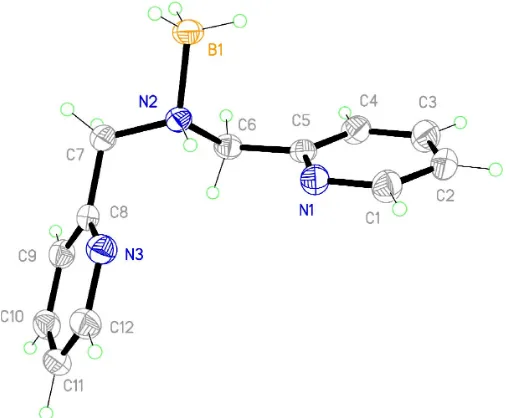
The title compound, C12H12N3BH3or C12H15BN3, contains a BH3 group and two picolyl groups attached to a central N atom. Both edge-to-face and face-to-face-stacking interac-tions are found.
Comment
The asymmetric unit of the title compound, (I), contains one molecule. The two planar pyridyl rings are twisted (Fig. 1) about the central N atom, with an interplanar angle of 110.9. The amine N atom is not involved in any hydrogen bonding but pyridyl atom N1 interacts with atom C3 in an adjacent ring (Table 1).
An edge-to-face interaction is found between the H atom on C2 and the plane of the pyridine ring containing atom N3 (Fig. 2). This H atom is 2.806 A˚ from the mean plane of the pyridine ring at (1
2+x, 1 2y,
1
2+z). The pyridine ring containing atom N3 is-stacked with its symmetry equivalent by inversion (symmetry code: 2x, y, 1z). The inter-planar and the centroid-to-centroid distances are 3.496 (2) and 3.971 A˚ respectively (Fig. 2).
[image:1.610.257.406.324.392.2] [image:1.610.206.459.518.727.2]Received 22 December 2004 Accepted 18 January 2005 Online 5 February 2005
Figure 1
Experimental
2-(Aminomethyl)pyridine (4.95 g, 44.77 mmol) and pyridine-2-carboxaldehyde (4.96 g, 46.31 mmol) were dissolved in methanol (150 ml) (Lambertet al., 1997). The solution was stirred for 2 h at room temperature (yellow–orange solution). After slow addition of an excess of sodium borohydride, stirring was continued for 1 h (pale-yellow solution). The solvent was removed by rotary evaporation to give bis(pyridin-2-ylmethyl)amine (6.43 g, 73%) as an orange oil. Colourless crystals of the borane adduct appeared as a minor product after the oil was stored in a freezer overnight.
Crystal data
C12H15BN3
Mr= 213.09 Monoclinic,P21=n
a= 5.3172 (4) A˚ b= 24.8494 (19) A˚ c= 9.3896 (7) A˚
= 102.938 (1)
V= 1209.14 (16) A˚3
Z= 4
Dx= 1.171 Mg m
3
MoKradiation Cell parameters from 3633
reflections
= 2.4–27.5
= 0.07 mm1
T= 150 (2) K Needle, colourless 0.550.170.09 mm
Data collection
Bruker SMART CCD area-detector diffractometer
’and!scans
Absorption correction: multi-scan (SADABS; Bruker, 1998) Tmin= 0.946,Tmax= 0.990
10293 measured reflections
2860 independent reflections 2156 reflections withI> 2(I) Rint= 0.023
max= 28.8
h=7!6 k=32!32 l=12!12
Refinement
Refinement onF2 R[F2> 2(F2)] = 0.043
wR(F2) = 0.116 S= 1.02 2860 reflections 148 parameters
H atoms treated by a mixture of independent and constrained refinement
w= 1/[2
(Fo2) + (0.0526P)2 + 0.3966P]
whereP= (Fo2+ 2Fc2)/3 (/)max< 0.001
max= 0.25 e A˚
3
min=0.20 e A˚
3
Table 1
Hydrogen-bonding geometry (A˚ ,).
D—H A D—H H A D A D—H A
C3—H3 N1i
0.95 2.66 3.5215 (19) 150
Symmetry code: (i)1 2þx;
1
2y;z
1 2.
H atoms bonded to C and B atoms were placed at calculated positions; the constrained C—H distances were 0.95, 0.98 and 0.99 A˚ for H atoms bonded to Csp2, Bsp3and methylene C atoms, respec-tively. They were refined using a riding model, with Uiso(H) =
1.2Ueq(B,C). The H atom bonded to the amine N atom was located in
a difference map and the coordinates freely refined with a fixedUiso
value of 0.03 A˚ .
Data collection:SMART(Bruker, 1998); cell refinement:SAINT
(Bruker, 1998); data reduction:SAINT; program(s) used to solve structure:SHELXS97(Sheldrick, 1997); program(s) used to refine structure: SHELXL97 (Sheldrick, 1997); molecular graphics:
SHELXTL (Bruker, 2001); software used to prepare material for publication:SHELXTL.
We are grateful to the Socrates Exchange Programme for support. JW thanks the Erskine fund at the University of Canterbury for support.
References
Bruker (1998).SMART, SAINTandSADABS.Bruker AXS Inc., Madison, Wisconsin, USA.
Bruker (2001). SHELXTL. Version 6.12. Bruker AXS Inc., Madison, Wisconsin, USA.
Lambert, E., Chabut, B., Chardon-Noblat, S., Deronzier, A., Chottard, G., Bousseksou, A., Tuchages, J.-P., Laugier, J., Bardet, M. & Latour, J.-M. (1997).J. Am. Chem. Soc.119, 9424–9437.
[image:2.610.315.566.72.175.2]Sheldrick, G. M. (1997). SHELXS97 and SHELXL97. University of Go¨ttingen, Germany.
Figure 2
View showing the C—H N bond (C3 and N1), the interaction between the H atom bonded to C2 and the pyridyl ring, and the-stacking of the N3-containing pyridine rings [symmetry codes: (i)1
2+x, 1 2y,z
1 2; (ii)
2x,y, 1z; (iii)1 2+x,
1 2y,
supporting information
sup-1
Acta Cryst. (2005). E61, o567–o568
supporting information
Acta Cryst. (2005). E61, o567–o568 [https://doi.org/10.1107/S1600536805001728]
Bis(2-pyridylmethyl)amine–borane
Noelia Sanchez Ballester, Julia Barreira Fontecha, Jan Wikaira and Vickie McKee
Bis(2-pyridylmethyl)amine–borane
Crystal data
C12H13N3·BH3
Mr = 213.09
Monoclinic, P21/n
a = 5.3172 (4) Å
b = 24.8494 (19) Å
c = 9.3896 (7) Å
β = 102.938 (1)°
V = 1209.14 (16) Å3
Z = 4
F(000) = 456
Dx = 1.171 Mg m−3
Mo Kα radiation, λ = 0.71073 Å Cell parameters from 3633 reflections
θ = 2.4–27.5°
µ = 0.07 mm−1
T = 150 K
Needles, colourless 0.55 × 0.17 × 0.09 mm
Data collection
Bruker SMART CCD area-detector diffractometer
Radiation source: normal-focus sealed tube Graphite monochromator
φ and ω scans
Absorption correction: multi-scan (SADABS; Bruker, 1998)
Tmin = 0.946, Tmax = 0.990
10293 measured reflections 2860 independent reflections 2156 reflections with I > 2σ(I)
Rint = 0.023
θmax = 28.8°, θmin = 2.4°
h = −7→6
k = −32→32
l = −12→12
Refinement
Refinement on F2
Least-squares matrix: full
R[F2 > 2σ(F2)] = 0.043
wR(F2) = 0.116
S = 1.02 2860 reflections 148 parameters 0 restraints
Primary atom site location: structure-invariant direct methods
Secondary atom site location: difference Fourier map
Hydrogen site location: inferred from neighbouring sites
H atoms treated by a mixture of independent and constrained refinement
w = 1/[σ2(F
o2) + (0.0526P)2 + 0.3966P]
where P = (Fo2 + 2Fc2)/3
(Δ/σ)max < 0.001
Δρmax = 0.25 e Å−3
Δρmin = −0.20 e Å−3
Special details
Refinement. Refinement of F2 against ALL reflections. The weighted R-factor wR and goodness of fit S are based on F2,
conventional R-factors R are based on F, with F set to zero for negative F2. The threshold expression of F2 > σ(F2) is used
only for calculating R-factors(gt) etc. and is not relevant to the choice of reflections for refinement. R-factors based on F2
are statistically about twice as large as those based on F, and R- factors based on ALL data will be even larger.
Fractional atomic coordinates and isotropic or equivalent isotropic displacement parameters (Å2)
x y z Uiso*/Ueq
N1 0.7734 (2) 0.21154 (5) 0.06397 (12) 0.0294 (3) C1 0.7038 (3) 0.26208 (6) 0.02464 (16) 0.0330 (3)
H1 0.5762 0.2787 0.0663 0.040*
C2 0.8076 (3) 0.29147 (6) −0.07349 (16) 0.0364 (3)
H2 0.7520 0.3273 −0.0985 0.044*
C3 0.9934 (3) 0.26772 (6) −0.13425 (17) 0.0391 (4)
H3 1.0690 0.2869 −0.2016 0.047*
C4 1.0676 (3) 0.21532 (6) −0.09506 (16) 0.0323 (3)
H4 1.1947 0.1979 −0.1354 0.039*
C5 0.9533 (3) 0.18883 (5) 0.00379 (14) 0.0249 (3) C6 1.0282 (3) 0.13226 (5) 0.05302 (14) 0.0268 (3)
H6A 1.1212 0.1153 −0.0157 0.032*
H6B 1.1461 0.1331 0.1509 0.032*
N2 0.7962 (2) 0.09966 (4) 0.05927 (12) 0.0247 (3) C7 0.8683 (3) 0.05115 (5) 0.15252 (14) 0.0315 (3)
H7A 1.0137 0.0326 0.1235 0.038*
H7B 0.7201 0.0260 0.1365 0.038*
C8 0.9453 (3) 0.06549 (5) 0.31264 (14) 0.0261 (3) C9 1.1746 (3) 0.04780 (5) 0.40128 (15) 0.0303 (3)
H9 1.2931 0.0271 0.3618 0.036*
C10 1.2282 (3) 0.06080 (6) 0.54864 (15) 0.0327 (3)
H10 1.3837 0.0491 0.6121 0.039*
C11 1.0516 (3) 0.09103 (6) 0.60112 (15) 0.0320 (3)
H11 1.0821 0.1003 0.7017 0.038*
C12 0.8290 (3) 0.10767 (6) 0.50462 (15) 0.0320 (3)
H12 0.7091 0.1288 0.5415 0.038*
N3 0.7732 (2) 0.09558 (5) 0.36190 (13) 0.0305 (3) B1 0.6297 (3) 0.08390 (7) −0.10096 (17) 0.0302 (3)
H13A 0.5839 0.1167 −0.1591 0.045*
H13B 0.4720 0.0652 −0.0916 0.045*
H13C 0.7321 0.0603 −0.1495 0.045*
H1N 0.700 (3) 0.1212 (6) 0.1050 (17) 0.030*
Atomic displacement parameters (Å2)
U11 U22 U33 U12 U13 U23
supporting information
sup-3
Acta Cryst. (2005). E61, o567–o568
C4 0.0362 (8) 0.0322 (7) 0.0319 (7) −0.0047 (6) 0.0148 (6) −0.0039 (6) C5 0.0260 (7) 0.0266 (7) 0.0217 (6) −0.0024 (5) 0.0044 (5) −0.0030 (5) C6 0.0249 (6) 0.0301 (7) 0.0251 (6) 0.0018 (5) 0.0048 (5) −0.0018 (5) N2 0.0294 (6) 0.0220 (5) 0.0227 (5) 0.0020 (4) 0.0057 (4) −0.0023 (4) C7 0.0448 (8) 0.0217 (6) 0.0259 (7) 0.0020 (6) 0.0035 (6) −0.0013 (5) C8 0.0339 (7) 0.0192 (6) 0.0249 (6) −0.0031 (5) 0.0059 (5) 0.0021 (5) C9 0.0334 (7) 0.0252 (7) 0.0325 (7) 0.0010 (6) 0.0077 (6) 0.0011 (5) C10 0.0324 (8) 0.0325 (8) 0.0302 (7) −0.0014 (6) 0.0005 (6) 0.0054 (6) C11 0.0399 (8) 0.0337 (7) 0.0222 (6) −0.0078 (6) 0.0066 (6) 0.0008 (5) C12 0.0346 (8) 0.0340 (8) 0.0291 (7) 0.0000 (6) 0.0108 (6) 0.0001 (6) N3 0.0315 (6) 0.0308 (6) 0.0283 (6) 0.0009 (5) 0.0051 (5) −0.0001 (5) B1 0.0334 (8) 0.0297 (8) 0.0248 (7) 0.0011 (6) 0.0012 (6) −0.0048 (6)
Geometric parameters (Å, º)
N1—C1 1.3375 (18) C7—C8 1.5097 (18)
N1—C5 1.3396 (17) C7—H7A 0.9900
C1—C2 1.384 (2) C7—H7B 0.9900
C1—H1 0.9500 C8—N3 1.3417 (17)
C2—C3 1.379 (2) C8—C9 1.385 (2)
C2—H2 0.9500 C9—C10 1.387 (2)
C3—C4 1.386 (2) C9—H9 0.9500
C3—H3 0.9500 C10—C11 1.377 (2)
C4—C5 1.3845 (19) C10—H10 0.9500
C4—H4 0.9500 C11—C12 1.383 (2)
C5—C6 1.5054 (18) C11—H11 0.9500
C6—N2 1.4881 (17) C12—N3 1.3402 (18)
C6—H6A 0.9900 C12—H12 0.9500
C6—H6B 0.9900 B1—H13A 0.9800
N2—C7 1.4887 (17) B1—H13B 0.9800
N2—B1 1.6138 (18) B1—H13C 0.9800
N2—H1N 0.910 (16)
C1—N1—C5 117.31 (12) N2—C7—C8 111.80 (11)
N1—C1—C2 123.53 (14) N2—C7—H7A 109.3
N1—C1—H1 118.2 C8—C7—H7A 109.3
C2—C1—H1 118.2 N2—C7—H7B 109.3
C3—C2—C1 118.61 (14) C8—C7—H7B 109.3
C3—C2—H2 120.7 H7A—C7—H7B 107.9
C1—C2—H2 120.7 N3—C8—C9 123.11 (12)
C2—C3—C4 118.67 (13) N3—C8—C7 114.89 (12)
C2—C3—H3 120.7 C9—C8—C7 121.98 (13)
C4—C3—H3 120.7 C8—C9—C10 118.83 (13)
C5—C4—C3 118.89 (13) C8—C9—H9 120.6
C5—C4—H4 120.6 C10—C9—H9 120.6
C3—C4—H4 120.6 C11—C10—C9 118.67 (13)
N1—C5—C4 122.98 (13) C11—C10—H10 120.7
C4—C5—C6 121.61 (12) C10—C11—C12 118.74 (13)
N2—C6—C5 110.83 (11) C10—C11—H11 120.6
N2—C6—H6A 109.5 C12—C11—H11 120.6
C5—C6—H6A 109.5 N3—C12—C11 123.60 (14)
N2—C6—H6B 109.5 N3—C12—H12 118.2
C5—C6—H6B 109.5 C11—C12—H12 118.2
H6A—C6—H6B 108.1 C12—N3—C8 117.02 (12)
C6—N2—C7 110.95 (11) N2—B1—H13A 109.5
C6—N2—B1 112.45 (10) N2—B1—H13B 109.5
C7—N2—B1 111.54 (10) H13A—B1—H13B 109.5
C6—N2—H1N 104.8 (10) N2—B1—H13C 109.5
C7—N2—H1N 107.2 (10) H13A—B1—H13C 109.5
B1—N2—H1N 109.5 (10) H13B—B1—H13C 109.5
Hydrogen-bond geometry (Å, º)
D—H···A D—H H···A D···A D—H···A
C3—H3···N1i 0.95 2.66 3.5215 (19) 150