www.wjpr.net Vol 8, Issue 10, 2019.
234
PATTERNS AND PRESENTATIONS OF LUNG CANCER IN IRAQI
PATIENTS
Dr. Ahmed Abdul Jabbar Jawad Alaskari* and Dr. Kudeir Jasim Sabeeh Al-Rawaq
Ministry of Higher Education, and Scientific Research, University of Baghdad, College of
Medicine, Clinical Oncologist, Kerbala, Iraq.
INTROUCTION
Lung cancer is the malignant transformation of lung tissue. The term
“lung cancer” is applied to tumors that develop from tracheal
parenchyma, bronchi, bronchioles or lung parenchyma.[1] Lung cancer
is the leading cause of cancer mortality for men and women, exceeding
the mortality of breast, colon, and prostate cancer combined.[18] The
major risk factor for lung cancer is undoubtedly cigarette smoking,
which probably accounts for about 90% of cases. There is a clear
relationship between risk and the age of smoking onset, the number of
years of smoking, and the number of cigarettes smoked per day,
smoking cessation at any age reduces the risk of lung cancer, which
drops to between 30 to50% of that of continuing smokers after 10
years. With chronic exposure to tobacco smoke, the ciliated epithelium
of the respiratory mucosa undergoes squamous metaplasia and
eventually a field change can occur with the development of carcinoma in situ before the start
of frankly invasive cancer.[3]
Approximately 10% of lung cancers are thought to be caused by carcinogens other than
cigarette smoking. Among non smokers, 30% of lung cancer deaths are attributed to radon
exposure.[18]
Increases in lung cancer risk also accompany exposure to carcinogens such as asbestos,
bis(chloromethyl) ether, polycyclic aromatic hydrocarbons, chromium, nickel and inorganic
arsenic compounds.[19]
Volume 8, Issue 10, 234-243. Research Article ISSN 2277– 7105
Article Received on 17 July 2019,
Revised on 06 August 2019, Accepted on 27 August 2019,
DOI: 10.20959/wjpr201910-15725
*Corresponding Author Dr. Ahmed Abdul Jabbar Jawad Alaskari
Ministry of Higher
Education, and Scientific
Research, University of
Baghdad, College of
Medicine, Clinical
Lung cancer is broadly separated into SCLC and NSCLC types, and NSCLC accounts for
approximately 80% to 85% of cases. Less than 50% of patients with NSCLC have resectable
disease on initial presentation, and 25% of patients present with locally advanced (regional
lymph node involvement without distant metastases) disease. Approximately 30% of patients
with SCLC have limited-stage disease on presentation.[6]
The Three Major Subtypes of NSCLC: Adenocarcinoma, Squamous cancer, Large cell
cancer.[1] adenocarcinoma (50%), squamous cell carcinoma (35%), and large cell lung cancer
(15%). Subtypes of adenocarcinoma include broncho-alveolar, acinar, and papillary; subtypes
of large cell lung cancer include giant cell and clear cell carcinomas, both of which carry
poor prognosis. Adenocarcinomas are the histologic type least associated with smoking
SCLC (oat cell cancer) contains dense neurosecretory granules containing neuroendocrine
hormones such as adrenocorticotropic hormone (ACTH) and vasopressin.[4]
Lung cancer can spread locally to the mediastinum, pleura (may produce effusion, especially
adenocarcinoma) chest wall and ribs, vertebral body or diaphragm. The disease can spread
via lymph nodes to the hilar nodes, the mediastinal nodes (pretracheal,
paratracheal,para-aortic, subaortic and subcarinal) or the supraclavicular fossa (N3). It can also spread via the
blood stream to the liver, adrenal gland, lung, brain,bone or skin.[3]
Although no set of signs or symptoms is pathognomonic for lung cancer, they may be divided
into three categories: (a) those due to local tumor growth and intrathoracic spread, (b) those
due to distant metastases, and (c) nonspecific systemic symptoms, or paraneoplastic
syndromes. Centrally located tumors produce cough, a localized wheeze, hemoptysis, and
symptoms and signs of airway obstruction and post obstructive pneumonitis such as dyspnea,
fever, and productive cough. Peripheral tumors are more likely to be asymptomatic when
they are small and confined within the lung; occasionally, cough and pleuritic chest pain may
be evident.[7]
Weight loss, which is usually but not always accompanied by anorexia, occurs in
approximately half of the patients, and generalized weakness occurs in one-third. Fever and
anemia occur less frequently, in fewer than 20% of patients. Fever is generally not considered
paraneoplastic in lung cancer patients; if present, it is usually associated with a documented
infection (e.g., postobstructive pneumonia) or with liver metastases. Paraneoplastic
from the primary tumor or its metastasis. The major categories of paraneoplastic syndromes
include endocrine, neurologic, cutaneous and musculoskeletal, and cardiovascular and
hematological manifestations.[7]
As with other medical conditions, the workup begins with a careful history and physical
examination Although sputum cytology reveals a diagnosis in only a small percentage of
patients, it is a simple and noninvasive evaluation that should be considered for patients with
respiratory symptoms and a suspicious lung mass. Traditionally, bronchoscopy has been
employed for biopsy of central lesions, whereas transthoracic needle biopsy is often the first
consideration for peripheral lesions. Endobronchial ultrasound is increasingly used to obtain
tissue from mediastinal lymph nodes or parenchymal lung lesions or both; esophageal
ultrasound may be used for biopsy of paraesophageal lymph nodes.[2]
Positron emission tomography combined with computed tomography
18F-fluorodeoxyglucose (18F-FDG-PET/CT) imaging is indicated in most patients with localized
NSCLC and SCLC and can replace the traditional workup that included CT imaging of the
chest and abdomen plus a bone scan. The addition 18F-FDG-PET imaging has been shown to
reduce the number of futile thoracotomies for patients with NSCLC and has a substantial
impact on the radiation treatment plan for NSCLC and SCLC, particularly when an approach
of involved field RT is used.[8]
Generally, biopsy of a suspected distant metastatic site should be considered in lieu of biopsy
of the primary lung lesion because this would confirm both histology and stage.
18F-FDG-PET imaging is typically not warranted in patients presenting with evidence of distant
metastases (on other imaging studies) because treatment would not likely be altered
according to the PET results. Magnetic resonance imaging (MRI) of the brain should be
obtained in patients with localized SCLC and patients with clinical stage III NSCLC, in
addition to patients presenting with neurologic symptoms.[2]
1 early-stage clinical node-negative NSCLC because CT and 18F-FDG-PET imaging have a
false-negative rate (in the mediastinum) ranging from 10% to greater than 25%.[9]
Combined anatomic and functional imaging is generally sufficient to assess mediastinal
Whether comprehensive mediastinal lymph node sampling with endobronchial ultrasound
can replace mediastinoscopy is an area of active investigation.[2]
In patients with NSCLC, the most important prognostic factor is tumor stage. This factor
largely determines treatment. Surgery is the standard mode of treatment of patients with stage
I and II tumors and for selective patients with stage III tumors. Neoadjuvant or adjuvant
therapy is recommended for many patients with stage II and III disease. Only about 20% of
all patients presenting with lung cancer are suitable candidates for curative surgery. The use
of combined-modality therapy including radiation and chemotherapy is recommended for
locally advanced stage III disease. Patients with stage IV disease are treated with
chemotherapy or palliative radiation therapy (RT) or with supportive therapy alone. Patients
with histologically documented unresectable or inoperable stage I-III NSCLC are evaluated
for definite radiotherapy with or without chemotherapy. If there are pressing symptomatic
needs for palliation, such as significant obstruction of a major airway, severe hemoptysis,
SVC obstruction, painful bony metastases in the weight-bearing areas, or symptomatic brain
metastases, the initial treatment is radiotherapy with or without chemotherapy. If a patient has
evidence of disseminated disease and there is no pressing need for radiotherapy, the approach
includes consideration of systemic chemotherapy, or supportive therapy alone if the patient's
general condition is not suitable for systemic chemotherapy. For limited-stage SCLC,
concurrent chemoradiotherapy should be considered. Radiotherapy should be delivered to 45
Gy, given 1.5 Gy per fraction and twice daily, with concurrent cisplatin and etoposide
chemotherapy. Patients should be encouraged to participate in research protocols using newer
chemotherapy and/or dose escalation/escalation radiotherapy. PCI should be considered for
complete clinical responders with a dose of 25 to 36 Gy.[7]
AIM OF THE STUDY
The aim of this study is to estimate the presentation of patients and types of lung cancers.
PATIENTS AND METHODS
A cross sectional study that included thirty patients already diagnosed with lung cancer who
attended the hospital of radiation oncology and nuclear medicine in Baghdad and Baghdad
Teaching Hospital, from January to July 2013 The patients included (20) males and (10)
females, their ages ranged from 50-86 years, with a mean age of 68 years. In this study we
evaluated factors including age, gender, cigarette smoking. Also we evaluated history of
voice or others symptoms including dyspnea, dysphagia, cough, hemoptysis and air way
obstruction.
The radiological investigations included, chest X-ray, chest CT scan, and if indicated
AbdominaL Ultra sound. (when there was suspicion of Liver metastasis).
All patients had histopathological report and sent for Bronchoscopic Biopsy or sputum
cytology or fine needle aspirate or diagnosed by supra clavicular lymph node biopsy. All
parameters estimated depended on history taken, physical examination, radiological
investigation and histopathological report information.
RESULTS
The median age for the lung cancer in our study was 68 years as 18 (60%) patients were > 50
year old and 12 (40%) were ≤ 50 year old, these results shown in table (1).
Table 1: shows age distribution of the lung cancer carcinoma in the studied group.
percentage No. patient
Age
40 % 21
≤50
60 % 21
>50
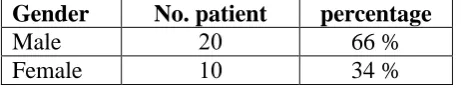
In our study thirty patients with lung cancer were evaluated for important risk factors
responsible for the tumour occurrence of them 20 patients were males (66%) and 10 were
females (34%), these results shown in table(2).
Table 2: shows the distribution of lung cancer according tothe gender in the studied
group.
percentage No. patient
Gender
66 % 12
Male
34 % 22
Female
In our study also thirty patients with lung cancer were evaluated for major risk factors which
is smoking responsible for the occurrence of lung cancer, of them 22 patients were smoker
(73.3%) and 8 were non smoker (26.6%), these results shown in table (3).
Table 3: shows the distribution of lung cancer according to smoking habit in the studied
group.
percentage No. patient
Smoking
73.3% 11
Smoker
26.6 % 1
[image:5.595.184.415.551.595.2]Two important pathological subtypes(NSCLC) and (SCLC) were found depending on the
histopathological reports performed on the primary of lung cancer, as shown in table (4).
Table 4: Shows the pathological subtypes of the primary lung cancer in the studied
group. percentage No. patient Histology 1- NSCLC 46 % 21
A- squamous cell ca
40 % 21 B- adenocarcinoma. 13 % 1 SCLC
In our study also thirty patients with lung cancer were evaluated for the main presenting sign
and symptom which include –shortness of breath, chest pain, hoarseness of voice, back pain,
vertigo, cough and haemoptysis.
Table 5: shows the main sign and symptom of the primary lung cancer in the studied
group. Cough and Hamoptysis Vertigo Back pain Hoarsness of voice Chest pain S.O.B. 15/30 1/30 1/30 1/30 5/30 7/30 50 % 3.3 % 3.3 % 3.3 % 16.6 % 23%
In our study also thirty patients with lung cancer which diagnosed with bronchoscopic
biopsy, sputum cytology, fine needle aspirate, supraclavicular lymph node biopsy.
Table 6: Shows the main methods for diagnosis of the primary lung cancer in the
studied group.
supraclavicular lymph node biopsy F.N.A Sputum Cytology Bronchoscopic Biopsy 2/30 3/30 5/30 20/30 6.7 % 10 % 16.6 % 66.6 %
The modality of treatment received by the patients was divided in our study as the use of
radiotherapy or chemotherapy or both modality for the treatment of the primary tumor so we
found that 18 patients (60%) were treated with both modality (radiotherapy and
chemotherapy) and 4 patient (13.34%) treated with radiotherapy while 8 patient(27%) treated
[image:6.595.132.466.540.597.2]Table 7: Shows the modalities of treatment used for the LUNG CANCER patients in the
studied group.
CHEMOTHEAPY. RADIOTHERAPY.
CHEMOTHEAPY. and RADIOTHERAPY.
8/30 4/30
18/30
27 % 13.34 %
60 %
In our study fifteen patient presented with stage four disease, seven person (46.67%) with
bone secondary and six person (40%) with liver and two person (13.34) with brain secondary.
Table 8: Shows the modalities of metastasis for LUNG CANCER patients in the studied
group.
Liver secondary Bone secondary
Brain secondary
6/15 7/15
2/15
40 % 46.67 %
13.34 %
Stage IV presentation no. patient 15/30.
DISCUSSION
In this study of the main risk factors for the lung cancer, The age of lung cancer patients was
which showed that the disease was more common between the ages 50-86 years, These
results were consistent with that of age, with the median age at diagnosis being 68 years, and
this result is consistent with that of the Beasley MB, Brambilla E, Travis.[11]
The median age of patients who present with lung cancer is 70 years. Among the patients of
this study 66% were found to be male (table2) and the remainder (34%) was female this
reflect that lung cancer are male predominant and this result is consistent with that of the
Elsayed I Salim, Abdul Rahman Jazieh, Malcolm A Moore; which they said that incidence
sharply increases particularly in males more than in female.[13]
Among the patients of this study 73.3% were found to be smokers (table 3); hence, smoking
was considered to be an important risk factor for developing lung cancer in this study also we
found that 26.6%was non smoker and this result is consistent with that of the Parkin DM.[10]
Among the patients of this study 87% were found to have non small cell histopathology and
13% have s mall cell histology (table 4); hence, n.s.c.l.c was considered to be more
predominant than s.c.l.c histology and this result is consistent with that of the In past decades,
squamous cell carcinoma was the most common type, but in recent years it has decreased in
United States. In some parts of the world, specifically Europe, squamous cell carcinoma is
apparently still the most common type.[7]
Among the patients of this study 23% presented with shortness of breath, 16.6% have chest
pain, 3.3% have hoarseness of voice, 3.3% presented with vertigoand 50% complain of cough
with haemoptysis and we found that cough with haemoptysis is the most common
presentation followed by shortness of breath and less with chest pain followed by back pain
and hoarseness of voice, vertigo this result is consistent with that of the G. Buccheri and D.
Ferrigno.
Among the patients of this study 66.6% of patient diagnosed with bronchscopic biopsy,
16.6%with sputum cytology, 10% with fine needle aspirateand6.7% with supra clavicular
lymph node biopsy in our study we found that bronchoscopic biopsy represent the most
dependable type of diagnosis followed by the sputum cytology and for less extent by fine
needle aspirate and at last the supra clavicular lymph node biopsy this result is consistent
with that of Toloza EM, Harpole L.[7]
Our patient in this study present either as locally advanced or metastatic stage and The
modality of treatment received by the patients was divided in our study as the use of
combination of both CHEMOTHEAPY and RADIOTHERAPY in18 patient (60%) and In
regard to the use of radiotherapy only 4 patients (13.3%) were in need for radiotherapy during
their course of the disease for palliative treatment; while 8 patients (27%) were in need for
chemotherapy as part of their management due to metastasis of disease, table(7) and this
result is consistent with that of Richard T. Hoppe, MD et al.[15]
The modality of presentation of stage four in our study present as 2 patient (13.34%) with
brain secondaryand7 patient (46.67%) with bone secondary and 6 patient (40%) with liver
secondary in our study patient with metastasis to bone represent the most common site of
lung secondary followed by liver secondary and then brain secondary table (8) and this result
is consistent with that of Gregory R. Mundy who said that; the most common human cancers
(lung, breast and prostate) have a great avidity for bone, leading to painful and untreatable
consequences.[17]
CONCLUSION AND RECOMMENDATIONS
1. Important risk factors for developing lung cancer are age, sex, smoking. lung cancer
could be suspected in elderly patients above 60 years of age who present with hemoptysis
and shortness of breath more than three weeks especially if associated with other risk
factors.
2. Smoking is the most important risk factor for developing lung cancer (73% of cases are
smokers). Smokers with high number of cigarette smoking and long duration of smoking
have higher risk than those with less amount and short period of smoking.
3. Hemoptysis and shortness of breath are the main presenting sign and symptom while
others depending on site of involvement and tumor extension.
4. Slightly more than half of cases presented with bone metastasis and for lesser extent with
liver secondary and then brain secondary.
5. Squamous cell carcinoma is the most histological type, followed by adenocarcinoma
while small cell type represent the least type.
6. Most common stage at time of presentation is advanced stage.
Recommendations
1. Giving particular care if more than one risk factor is present and activates and enhance
the role of primary care practitioners, as a part of a general evaluation, inquire about
hemoptysisin a patient from at risk population. If a positive response is obtained, the
patient can be referred to an respiratory specialist for appropriate evaluations.
2. Doing serial investigations especially chest x-ray and CT scan for those old and smoker
patients whom presented with cough and haemoptysis more than three weeks for early
detection of disease.
3. Cessation of smoking which is considered as the major risk factor.
4. Further studies regarding other parameters not mentioned in this study are recommended.
REFERENCES
1. Murat Beyzadeoglu, Gokhan Ozyigit, uneyt Ebruli. Basic Radiation Oncology, 2010.
2. Leonard L. Gunderson. Clinical Radiation Oncology, 2008.
3. Louise Hanna, Tom Crosby, Fergus Macbeth. Practical Radiation Oncology Practical,
2008.
4. L.W. Brady, H.-P. Heilmann, M. Molls, C. Nieder. Decision Making in Radiation
Oncology, 201.
5. Parkin D, Bray F, Ferlay. Global cancer statistics, journal of clinical oncology, 2005; 55:
6. Warde P, Payne D. Does thoracic irradiation improve survival and local control in
limited-stage small-cell carcinoma of the lung, journal of clinical oncology, 1992; 10:
890-895.
7. Edward C. Halper, Carlos A. Perez, Luther W. Brady. Principles and Practice of
Radiation Oncology, 2008.
8. Meyers BF, Downey RJ, Decker PA., Utility of positron emission tomography in staging
of potentially operable lung cancer, Journal of Thoracic Cardiovascular Surgery, 1982;
133: 738-745.
9. Friess GG, McCracken JD, Troxell ML. Effect of initial resection of small-cell carcinoma
of the lung, A review of Southwest Oncology Group Study 7628, journal of clinical
oncology, 1985; 964-968.
10.Parkin DM. Tobacco-attributable cancer burden in the UK in 2010, British Journal of
Cancer, 2011; 105(S2): S6-S13.
11.Beasley MB, Brambilla E, Travis WD. World Health Organization classification of lung
tumors, Seminar in Roentgenology, 2005; 40: 90-97.
12.Warde P, Payne D. Does thoracic irradiation improve survival and local control in
limited-stage small-cell carcinoma of the lung, Journal of Clinical Oncology, 1992; 10:
890-895.
13.Elsayed I. Salim, Abdul Rahman Jazieh, Malcolm A Moore, lung cancer, international Journal
of Radiation Oncology, 2007; 33: 57-65.
14.G. Buccheri, D. Ferrigno. Sign and symptoms of lung cancers, American Journal of
Clinical Oncology, 2010; 5: 47-78.
15.Toloza EM, Harpole L, Detterbeck F. Invasive staging of non-small cell lung cancer: a
review of the current evidence, 2003; 123: 157-166.
16.Richard T. Hoppe, Mack Roach, Theodore Locke Phillips. Leibel and Phillips Textbook
of Radiation Oncology, 2010.
17.Gregory R. Mundy. Metastasis to bone causes, consequences and therapeutic
opportunities, Nature Reviews of Cancer, 2002; 2: 584–593.
18.Jemal A. Cancer statistics, 2009. Cancer Journal for Clinicians, 2009; 59(4): 225-249.
19.Fraumeni Jr J, Blott W. Lung and pleura, Cancer epidemiology and prevention, 1982; 44:

![Dimethyl (2Z) 2 [4 ((1Z) 1 {2 [(2Z,5Z) 5 (2 methoxy 2 oxoethylidene) 4 oxo 3 phenyl 1,3 thiazolidin 2 ylidene]hydrazin 1 ylidene}ethyl)anilino]but 2 enedioate](data:image/gif;base64,R0lGODlhAQABAIAAAP///wAAACH5BAEAAAAALAAAAAABAAEAAAICRAEAOw==)