Original Article
MicroRNA-30b targets CBX3 and
regulates cell proliferation, apoptosis,
and migration in esophageal squamous cell
carcinoma via the JAK2/STAT3 signaling pathway
Lingxin Meng1, Fengsong Wang1, Shuyan Sun2, Yuxiu Zheng1, Zhaojun Ding1, Yanwei Sun1, Bingcheng Li1, Qin Meng1, Meiling Xu1
Departments of 1Oncology, 2Pathology, People’s Hospital of Rizhao, Rizhao 276826, Shandong, China
Received September 21, 2017; Accepted October 30, 2017; Epub December 1, 2017; Published December 15, 2017
Abstract: The present study aimed to investigate whether miR-30b plays a pivotal role in the progression of esopha-geal squamous cell carcinoma (ESCC) as well as to elucidate its possible regulatory mechanism. The expression levels of miR-30b in the ESCC tissues and cells were determined. The TE-1 cells were transfected with the miR-30b mimic, the mimic control, the miR-30b inhibitor, the inhibitor control, the pCDNA3.1-Chromobox 3 (pCDNA3.1-CBX3) and/or the blank vector, while the TE-2 cells were transfected with the miR-30b mimic and/or the mimic control. Cell proliferation, cell apoptosis, and cell migration of the different transfected groups were evaluated. The luciferase reporter assay was performed to detect the relationship between miR-30b, and CBX3. Furthermore, the
relation-ship between miR-30b, CBX3, and the JAK2/STAT3 signaling pathway was explored. Significant downregulation of
miR-30b was observed in the ESCC tissues and cells, while CBX3 was upregulated in the ESCC tissues and cells. The inhibition of miR-30b promoted cell proliferation, inhibited cell apoptosis, and enhanced cell migration in ESCC,
which was similar to the effects of CBX3 overexpression. In fact, CBX3 was confirmed to be a direct target of miR-30b. The overexpression of miR-30b decreased the expression levels of p-JAK2/JAK2 and p-STAT3/JAK3 signifi -cantly, which was obviously reversed after the simultaneous overexpression of CBX3. Our results revealed that the downregulation of miR-30b may increase cell proliferation, inhibit cell apoptosis, and promote cell migration in ESCC by targeting CBX3 and activating the JAK2/STAT3 signaling pathway. Thus, miR-30b may serve as a useful marker for predicting the progression of ESCC.
Keywords: Esophageal squamous cell carcinoma, miR-30b, Chromobox 3, JAK2/STAT3 pathway
Introduction
Esophageal cancer is one of the common upper gastrointestinal tract cancers with a high mor-tality across the world [1, 2]. Esophageal squa-mous cell carcinoma (ESCC) is the most pre-dominant type of esophageal cancer, accoun- ting for more than 90% of the esophageal can-cer cases [3, 4]. ESCC is characterized by a high rate of metastasis, limited therapeutic options, and poor prognosis [5]. Moreover, the molecular mechanisms, leading to the progres-sion and poor prognosis of ESCC remain to be elucidated [6]. Therefore, a better understand-ing of the key mechanisms will be helpful in designing a promising therapeutic approach for
lead to the progression of several types of cers, including gastric cancer, colorectal can-cer, and non-small cell lung cancer (NSCLC) [14-16]. Although a very recent study has reported that the low level of expression of miR-30b is correlated with lymph node metas-tasis, pathological stage, and poor overall sur-vival in patients with esophageal cancer, the roles and possible regulatory mechanisms of miR-30b in ESCC are largely unknown [17]. In the present study, the expression levels of miR-30b in the ESCC tissues and cells were determined. The expression of miR-30b in the ESCC cells was then altered in vitro, in order to elucidate further the effects of miR-30b on cell proliferation, cell apoptosis, and cell migration. In addition, the potential targets of miR-30b were identified and the relationship between miR-30b and the JAK2/STAT3 signaling path-way was explored. All these efforts were expect-ed to provide some theoretical basis for the better understanding of key mechanisms underlying the development of ESCC.
Materials and methods
Tissue sampling
A total of 20 ESCC patients, hospitalized in our hospital from June 2015 to January 2017 were included in this study. The diagnosis of ESCC was pathologically confirmed. Matched ESCC tissues and their adjacent normal tissues were collected from clinically diagnosed surgical specimens, snap-frozen in liquid nitrogen, and stored at -80°C. All the patients gave informed consent and this study was approved by the ethics committee of our hospital.
Cell culture
The normal esophageal cell line (NEEC) as well as the highly and poorly differentiated ESCC cell lines (TE-1 and TE-2, respectively) were pur-chased from the American Type Culture Collection (Manassas, VA, USA), cultured in RPMI 1640 medium (Gibco, Carlsbad, CA, USA) with 10% fetal bovine serum (FBS) (Gibco, Carlsbad, CA, USA), and incubated at 37°C in a humidified chamber with 5% CO2.
Cell transfection
The 30b mimic, the mimic control, the miR-30b inhibitor, the inhibitor control, the
overex-pressed pCDNA3.1-CBX3 vector, and the blank vector with no CBX3 sequence (used as a nega-tive control) were synthesized by RiboBio Corporation, China. The TE-1 cells were trans-fected with the miR-30b mimic, the mimic con-trol, the miR-30b inhibitor, the inhibitor concon-trol, the pCDNA3.1-CBX3 vector, and the blank vec-tor, using the Lipofectamine 2000 Reagent (Invitrogen, Carlsbad, CA, USA), according to the instructions of the manufacturer, while the TE-2 cells were transfected with the miR-30b mimic and the mimic control, using the same method. After 48 h of incubation, the transfec-tion efficiency was determined by quantitative real-time PCR (qRT-PCR).
The MTT assay
The viability of the transfected cells was mea-sured using the MTT assay. Briefly, the cells (5×10³ cells per well) were seeded in triplicate into 96-well microplates. Following different times of transfection, 20 μL of 10 mg/mL MTT solution was added and the plates were incu-bated at 37°C for 4 h. The culture medium was then removed and 150 μL of dimethyl sulfoxide (DMSO) was added to each well, in order to incubate the cells for 15 min. The absorbance (optical density) values at 490 nm were ana-lyzed with a microplate reader (BioTek, Winooski, VT, USA).
The colony formation assay
In order to detect cell proliferation, the colony formation assay was performed. Briefly, a total of 100 cells from each group, after 48 h of transfection were seeded in triplicate into 6-well dishes and incubated for an additional 14 days in RPMI 1640 medium with 10% FBS. Subsequently, the colonies were fixed in 10% formalin for 10 min and stained with 0.1% crys-tal violet (Sigma-Aldrich Chemical Company, St. Louis, MO, USA). Finally, the colonies were counted under an Olympus IX83 microscope (Olympus Inc., Tokyo, Japan).
Flow cytometry analysis
PI (propidium iodide) for 20 min at room tem-perature. The percentage of the apoptotic cells was then analyzed in a flow cytometer (FA- CSCalibur, Becton-Dickinson, San Jose, CA, USA), equipped with the CellQuest software (Becton-Dickinson, Franklin Lakes, NJ, USA). The transwell migration assay
The migration of the transfected cells was mea-sured by the transwell migration assay. Briefly, the cells in different groups were seeded into the upper layer of a transwell chamber of pore size 8 μm (Corning Inc., NY, USA), supplement-ed with serum-free RPMI 1640 msupplement-edium. The RPMI 1640 medium, containing 10% FBS was added to the lower layer of the chamber as a chemoattractant. Following 48 h of incubation, the transwell chamber was washed with PBS buffer, fixed in 4% paraformaldehyde, and stained with 0.1% crystal violet. Finally, the migrated cells were counted under an Olympus IX83 microscope (Olympus Inc., Tokyo, Japan). Each experiment was carried out in triplicate. The luciferase reporter assay
The CBX3 3’UTR sequence, containing putative binding sites for miR-30b (CBX3 3’UTR-wt) was amplified from human genomic DNA and insert-ed into the pGL3- Basic luciferase reporter plasmid, creating a luciferase reporter con-struct. The mutated CBX3 3’UTR sequence in the complementary site of miR-30b (CBX3 3’UTR-mut) was used as a negative control. The cells were then cotransfected with the miR-30b mimic and the luciferase reporter constructs, comprising either CBX3 3’UTR-wt or CBX3 3’UTR-mut. Subsequently, the transfected cells were serum shocked (20%) for 2 h and incubat-ed for 24 h with 2% mincubat-edia. Luciferase activity was then detected with the Dual Luciferase Assay kit (Promega Corporation, Madison, WI, USA), using a beta-counter or luminometer. The ratio of the raw firefly luciferase activity to the Renilla luciferase activity was calculated as the relative luciferase activity.
qRT-PCR analysis
Total RNA was isolated from the ESCC tissues and cells, using the TRIzol Reagent (Invitrogen, Carlsbad, CA, USA) and the quality of the extracted RNA was detected with a SMA4000 UV-VIS spectrophotometer (Merinton, Shanghai,
China). The synthesis of complementary DNA (cDNA) was then carried out from the extracted RNA, using the PrimeScript First Strand cDNA Synthesis Kit (Invitrogen, Carlsbad, CA, USA). In order to detect the relative expressions of miR-30b, RT-qPCR was performed using the SYBR ExScript RT-PCR kit (Takara, Shiga, Japan)in accordance with the protocols provided by the manufacturer. The sequences of the primers used in this study were as follow:miR-30b: sense, 5’-TGAAAGAGAGAACGATAAATGTT-3’ and antisense, 5’-ACTTCTGAATCAAAATATTGGTA-3’; U6: sense, 5’-CTGGTAGGGTGCTCGCTTCGGC- AG-3’ and antisense, 5’-CAACTGGTGTCGTGGA- GTCGGC-3’. The U6 gene was used as an endogenous control and the relative fold change in the expression of miR-30b was calcu-lated using the comparative 2-ΔΔCT method.
Each reaction was performed in triplicate. Western blot assay
The cells were lysed in radioimmunoprecipita-tion (RIPA) buffer (Sangon Biotech, Shanghai, China) and the resulting cell lysates were quan-titated using the bicinchoninic acid (BCA) assay (Pierce, Rochford, IL, USA). Equal amounts of the cell lysates were then subjected to a 10% SDS-PAGE, followed by blotting onto a Polyvinylidene fluoride (PVDF) membrane (Millipore, Bedford, MA, USA). The primary anti-bodies to the CBX3, BAX, BCL2, JAK2, p-JAK2, STAT3, p-STAT3, p-STAT5, and GAPDH (1:1000) genes were purchased from Cell Signaling Technology (Beverly, MA, USA) and were used for incubating the membranes overnight at 4°C, followed by incubation with a horseradish per-oxidase (HRP)-labeled secondary antibody (1:1000 dilution) at 37°C for 1 h. Subsequently, the protein bands were detected by an enhanced chemiluminescence (ECL) detection system (GE Healthcare, Piscataway, NJ, USA). The GAPDH gene was used as an internal control.
Statistical analysis
results were considered statistically significant at P<0.05.
Results
miR-30b is downregulated in the ESCC tissues and cells
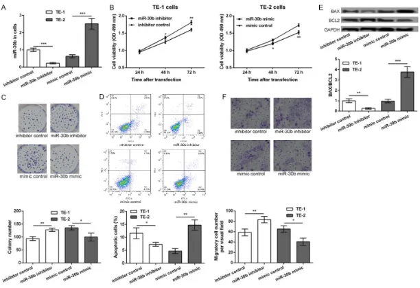
As shown in Figure 1, the qRT-PCR analysis was performed to detect the expression of miR-30b in the ESCC tissues and cells. The significant downregulation of miR-30b was observed in the ESCC tissues, compared to that in the adja-cent normal tissues (P<0.05, Figure 1A). Similarly, miR-30b was downregulated in the TE-1 and TE-2 cells, relative to that in the NEEC cells (P<0.05, Figure 1B). Moreover, the expres-sion of miR-30b in the TE-2 cells was signifi-cantly lower than that in the TE-1 cells (P<0.05, Figure 1B). Taken together, these data indicat-ed that the expression of miR-30b was associ-ated with the progression of ESCC.
The inhibition of miR-30b promotes cell prolif-eration, inhibits cell apoptosis, and enhances cell migration in ESCC
In order to detect the role of miR-30b in the development of ESCC, the TE-1 cells were trans-fected with the miR-30b inhibitor and the inhib-itor control, while the TE-2 cells were transfect-ed with the miR-30b mimic and the mimic control. After 48 h of transfection, the expres-sion of miR-30b was significantly decreased in the miR-30b inhibitor transfected TE-1 cells but was markedly increased in the miR-30b mimic transfected TE-2 cells, compared to their cor-responding controls (P<0.001, Figure 2A), indi-cating that the transfection efficiency was high and could be used for further analysis. After
tor significantly increased the number of colo-nies of the TE-1 cells, while transfection with the miR-30b mimic decreased the number of colonies of the TE-1 cells (P<0.05, Figure 2C). Furthermore, the results of the flow cytometry analysis showed that the apoptosis of the TE-1 cells was significantly inhibited after transfec-tion with the miR-30b inhibitor, while the apop-tosis of the TE-2 cells was markedly inhibited after transfection with the miR-30b mimic (P<0.05, Figure 2D). Similar changes in the expression levels of Bax/Bcl2 were obtained from the Western blot assay. The expression levels of Bax/Bcl2 were significantly decreased in the miR-30b inhibitor transfected TE-1 cells and were markedly increased in the miR-30b mimic transfected TE-2 cells, compared to their corresponding controls (P<0.05, Figure 2E). Besides, the results of the transwell migration assay showed that the number of the migratory TE-1 cells was significantly decreased after transfection with the miR-30b inhibitor but was increased after transfection with the miR-30b mimic (P<0.05, Figure 2E), indicating that the inhibition of miR-30b promoted cell migration. CBX3 is a target of miR-30b
According to the information provided by the TargetScan software, CBX3 was predicted to be a potential target of miR-30b (Figure 3A). The results of the luciferase reporter assay further confirmed that the miR-30b mimic significantly inhibited the luciferase reporter activity of the CBX3 3’UTR-wt construct (P<0.05), compared to the CBX3 3’UTR-mut construct (Figure 3B). The Western blot assay, performed to detect the expression of the CBX3 protein in the ESCC tissues and cells, showed that CBX3 was sig-nificantly upregulated in the ESCC tissues, rela-Figure 1. The expression levels of miR-30b in esophageal squamous cell
car-cinoma (ESCC) tissues and their adjacent normal tissues (A), as well as in ESCC cell lines (TE-1 and TE-2) and normal esophageal cell line NEEC (B). *P<0.05, ***P<0.001 compared with corresponding control.
tive to the adjacent normal tissues (P<0.05, Figure 3C). The expression of CBX3 was also upregulated in the TE-1 and TE-2 cells com-pared to the NEEC cells (P<0.05, Figure 3D). The expression of CBX3 in the TE-2 cells was significantly higher than that in the TE-1 cells (P<0.05, Figure 3D). Furthermore, the expres-sion of CBX3 was significantly increased in the miR-30b inhibitor transfected TE-1 cells but was decreased in the miR-30b mimic transfect-ed TE-2 cells, compartransfect-ed to their corresponding controls, following 48 h of transfection (P<0.05, Figure 3E). All these data indicated that CBX3 was upregulated in the ESCC cells and its expression was inhibited by miR-30b.
of the migratory TE-1 cells was significantly decreased after the overexpression of CBX3 (P<0.05, Figure 4F). Taken together, our data indicated that the overexpression of CBX3 pro-moted cell proliferation, inhibited cell apopto-sis, and strengthened cell migration in the TE-1 cells, which was similar to the effects of miR-30b inhibition.
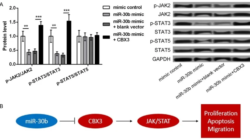
The JAK2/STAT3 pathway may be a key mechanism to mediate the role of miR-30b/ CBX3 in ESCC
As shown in Figure 5A, the expression of p-JAK2/JAK2 and p-STAT3/JAK3 was signifi-Figure 3. CBX3 as a target of miR-30b. A: The predicted sequence of CBX3
and miR-30b. B: Luciferase reporter assay showing the luciferase report ac-tivity of CBX3 3’UTR-wt and CBX3 3’UTR-mut after transfection with miR-30b mimic or mimic control. C: Western blot showing the protein expression level of CBX3 in ESCC tissues and their adjacent normal tissues. D: Western blot showing the protein expression level of CBX3 in ESCC cell lines (TE-1 and TE-2) and normal esophageal cell line NEEC. E: TE-1 cells transfected with miR-30b inhibitor and inhibitor control, while TE-2 cells transfected with miR-30b mimic and mimic control. The Western blot showing the protein expression level of CBX3 in different transfected groups. *P<0.05, **P<0.01 and ***P<0.001 compared with corresponding control.
The overexpression of CBX3 promotes cell proliferation, inhibits cell apoptosis, and strengthens cell migration in the TE-1 cells
cantly downregulated in the miR-30b mimic groups, compared to that in the mimic controls but was markedly reversed after the cells were cotransfected with the miR-30b mimic and the pCDNA3.1-CBX3 vector (P<0.05). However, there was no significant difference in the expression of p-STAT5/STAT5 among the differ-ent groups. Therefore, these data indicated that the JAK2/STAT3 signaling pathway might be a key mechanism to mediate the role of miR-30b/CBX3 in ESCC.
Discussion
The present study found the reverse expression of miR-30b and CBX3 in the ESCC tissues and cells. The study showed that miR-30b was downregulated and CBX3 was upregulated. In fact, CBX3 was confirmed to be a direct target of miR-30b. The inhibition of miR-30b
promot-ed cell proliferation, inhibitpromot-ed cell apoptosis, and enhanced cell migration in ESCC, which was similar to the effects of CBX3 overexpres-sion. In addition, the overexpression of miR-30b significantly decreased the expression lev-els of p-JAK2/JAK2 and p-STAT3/JAK3, which was markedly reversed after the simultaneous overexpression of CBX3. The mechanism of miR-30b in the development of ESCC is shown in Figure 5B, suggesting a regulatory role for miR-30b in the development of ESCC.
[image:8.612.93.519.162.398.2]In a previous study, miR-30b was found to be decreased in the esophageal cancer tissues, suppressing the growth and migration of tumor cells [17]. Similarly, in this study, we found that miR-30b was downregulated in the ESCC tis-sues and downregulation of miR-30b promoted the proliferation and migration of the ESCC cells. In addition, numerous studies have Figure 4. Overexpression of CBX3 promoted TE-1 cell proliferation, inhibited cell apoptosis, and strengthened cell migration. TE-1 cells were transfected with pc-CBX3 and blank control. A: The expression levels of CBX3 in different transfected cells. B: MTT assay showing the cell viability of different transfected cells. C: Colony formation assay showing the colony formation number of different transfected cells. D: Flow cytometry showing the apoptotic cells in different transfected groups. E: Western blot showing the expression of BAX/BCL2 in different transfected cells. F: Transwell assay showing the number of migratory cells in different transfected groups. Compared with correspond-ing control, *P<0.05, **P<0.01 and ***P<0.001.
exposed that miRNAs contribute to the devel-opment of esophageal cancer via the regula-tion of their target genes [18, 19]. In this study, CBX3 was confirmed to be a direct target of miR-30b. Accumulating evidence has con-firmed the overexpression of CBX3 in various cancer cells, thereby contributing to tumorigen-esis [20]. Saini et al. revealed that a higher expression of CBX3 was observed in spheres compared to monolayers, suggesting that CBX3 might be a marker for Tumor Stem Cell (TSC) enrichment in osteosarcoma [21]. Again, Fan et al. reported that CBX3 could promote cell cycle and cell proliferation in colon cancer [22]. Furthermore, the increased expression of CBX3 may be a promising marker for NSCLC [23]. In this study, we found that the overexpression of CBX3 promoted cell proliferation, inhibited cell apoptosis, and strengthened cell migration in the TE-1 cells, which was similar to the effects of miR-30b inhibition. Given the key roles of CBX3 in multiple cancers, we speculate that the downregulation of miR-30b may regulate cell proliferation, cell apoptosis, and cell migra-tion in ESCC by targeting CBX3.
In order to further elucidate the possible mech-anism of miR-30b in the development of ESCC, the relationship between miR-30b and the JAK2/STAT3 signaling pathway was investigat-ed. Several studies have confirmed that the activation of the JAK2/STAT3 signaling pathway widely occurs with high frequency in ESCC and disruption of this pathway can repress the tumorigenesis and progression of ESCC [24, 25]. Thus, blocking the JAK2/STAT3 signaling pathway can inhibit tumor growth in ESCC by regulating cell growth, cell cycle, and angiogen-esis [26]. In addition, the inactivation of the STAT3 signaling pathway promotes the inhibi-tory effect of metformin on the growth of tumor cells in ESCC [27]. Furthermore, the depletion of the transmembrane protein, B7-H4 can repress the secretion of the cytokine, IL-6 via the inactivation of the JAK2/STAT3 signaling pathway, thereby suppressing cell proliferation in ESCC [28]. In this study, the overexpression of miR-30b resulted in the decreased expres-sion of p-JAK2/JAK2 and p-STAT3/JAK3, which was reversed after the simultaneous overex-pression of CBX3. Taken together, these data indicate that the overexpression of miR-30b may inhibit the development of ESCC via the inactivation of the JAK2/STAT3 signaling
path-way. Thus, the miR-30b/CBX3/JAK2/STAT3 sig-naling pathway may play a key role in regulating the progression of ESCC.
In conclusion, our results reveal that the down-regulation of miR-30b may increase cell prolif-eration, inhibit cell apoptosis, and promote cell migration in ESCC by targeting CBX3 and acti-vating the JAK2/STAT3 signaling pathway. Thus, miR-30b may serve as a useful marker for pre-dicting the progression of ESCC.
Acknowledgements
This work was supported by Shandong Medical and Health Science and Technology Develop- ment program, Grant No: 2016WS0329.
Disclosure of conflict of interest
None.
Address correspondence to: Lingxin Meng, Depart- ment of Oncology, People’s Hospital of Rizhao, NO.126 Taian Road, Rizhao 276826, Shandong, China. E-mail: lingxinmeng191@sina.com
References
[1] Siegel RL, Ma J, Zou Z and Jemal A. Cancer sta-tistics, 2014. CA. CA Cancer J Clin 2014; 65. [2] Holmes RS and Vaughan TL. Epidemiology and
pathogenesis of esophageal cancer. Semin Radiat Oncol 2007; 17: 2-9.
[3] Enzinger PC and Mayer RJ. Esophageal cancer. N Engl J Med 2003; 349: 2241-2252.
[4] Jemal A, Bray F, Center MM, Ferlay J, Ward E and Forman D. Global cancer statistics. CA Cancer J Clin 2011; 61: 69-90.
[5] Lento A, Long A, Giroux V, Tang Q, Sammons M, Klein-Szanto A, et al. Investigating the role of mutant p53 in esophageal squamous cell car-cinoma. AACR 2017.
[6] Qin HD, Liao XY, Chen YB, Huang SY, Xue WQ, Li FF, Ge XS, Liu DQ, Cai Q and Long J. Genom-ic characterization of esophageal squamous cell carcinoma reveals critical genes underly-ing tumorigenesis and poor prognosis. The American Journal of Human Genetics 2016; 98: 709-727.
[7] Svoronos AA, Engelman DM and Slack FJ. On-comiR or tumor suppressor? The duplicity of microRNAs in cancer. Cancer Res 2016; 76: 3666-3670.
[8] Trang P, Weidhaas JB and Slack FJ. MicroRNAs and Cancer. In: editors. The molecular basis of human cancer. Springer; 2017. pp. 277-286. [9] Abba ML, Patil N, Leupold JH, Moniuszko M,
as novel targets and tools in cancer therapy. Cancer Lett 2017; 387: 84-94.
[10] Adams BD, Kasinski AL and Slack FJ. Aberrant regulation and function of microRNAs in can-cer. Curr Biol 2014; 24: R762-R776.
[11] Song Y, Li J, Zhu Y, Dai Y, Zeng T, Liu L, Li J, Wang H, Qin Y and Zeng M. MicroRNA-9 pro-motes tumor metastasis via repressing E-cad-herin in esophageal squamous cell carcinoma. Oncotarget 2014; 5: 11669.
[12] Ni Y, Meng L, Wang L, Dong W, Shen H, Wang G, Liu Q and Du J. MicroRNA-143 functions as a tumor suppressor in human esophageal squamous cell carcinoma. Gene 2013; 517: 197-204.
[13] Shafiee M, Aleyasin SA, Vasei M, Semnani SS
and Mowla SJ. Down-regulatory effects of miR–211 on long non-coding RNA SOX2OT and SOX2 genes in esophageal squamous cell car-cinoma. Cell Journal (Yakhteh) 2016; 17: 593. [14] Zhu ED, Li N, Li BS, Li W, Zhang WJ, Mao XH,
Guo G, Zou QM and Xiao B. miR-30b, down-regulated in gastric cancer, promotes apopto-sis and suppresses tumor growth by targeting plasminogen activator inhibitor-1. PLoS One 2014; 9: e106049.
[15] Zhao H, Xu Z, Qin H, Gao Z and Gao L. miR-30b regulates migration and invasion of human colorectal cancer via SIX1. Biochem J 2014; 460: 117-129.
[16] Zhong K, Chen K, Han L and Li B. MicroRNA-30b/c inhibits non-small cell lung cancer cell proliferation by targeting Rab18. BMC Cancer 2014; 14: 703.
[17] Li Q, Zhang X, Li N, Liu Q and Chen D. miR-30b inhibits cancer cell growth, migration, and in-vasion by targeting homeobox A1 in esopha-geal cancer. Biochem Biophys Res Commun 2017; 485: 506-512.
[18] Ren L, Chen W, Li S, He X, Zhang Z, Li M, Cao R, Hao B, Zhang H and Qiu H. MicroRNA-183 pro-motes proliferation and invasion in oesopha-geal squamous cell carcinoma by targeting programmed cell death 4. Br J Cancer 2014; 111: 2003.
[19] Meng H, Wang K, Chen X, Guan X, Hu L, Xiong G, Li J and Bai Y. MicroRNA-330-3p functions as an oncogene in human esophageal cancer by targeting programmed cell death 4. Am J Cancer Res 2015; 5: 1062.
[20] Takanashi M, Oikawa K, Fujita K, Kudo M, Kinoshita M and Kuroda M. Heterochromatin
protein 1γ epigenetically regulates cell differ -entiation and exhibits potential as a therapeu-tic target for various types of cancers. Am J Pathol 2009; 174: 309-316.
[21] Saini V, Hose CD, Monks A, Nagashima K, Han B, Newton DL, Millione A, Shah J, Hollingshead
MG and Hite KM. Identification of CBX3 and
ABCA5 as putative biomarkers for tumor stem cells in osteosarcoma. PLoS One 2012; 7: e41401.
[22] Fan Y, Li H, Liang X and Xiang Z. CBX3 pro-motes colon cancer cell proliferation by CDK6 kinase-independent function during cell cycle. Oncotarget 2017; 8: 19934.
[23] Han SS, Kim WJ, Hong Y, Hong SH, Lee SJ, Ryu DR, Lee W, Cho YH, Lee S and Ryu YJ. RNA
se-quencing identifies novel markers of non-small
cell lung cancer. Lung Cancer 2014; 84: 229-235.
[24] Zhang Y, Du XL, Wang CJ, Lin DC, Ruan X, Feng YB, Huo YQ, Peng H, Cui JL and Zhang TT. Re-ciprocal activation between PLK1 and Stat3 contributes to survival and proliferation of esophageal cancer cells. Gastroenterology 2012; 142: 521-530, e523.
[25] You Z, Xu D, Ji J, Guo W, Zhu W and He J. JAK/ STAT signal pathway activation promotes pro-gression and survival of human oesophageal squamous cell carcinoma. Clin Transl Oncol 2012; 14: 143-149.
[26] Fang J, Chu L, Li C, Chen Y, Hu F, Zhang X, Zhao H, Liu Z and Xu Q. JAK2 inhibitor blocks the
in-flammation and growth of esophageal squa -mous cell carcinoma in vitro through the JAK/ STAT3 pathway. Oncol Rep 2015; 33: 494-502. [27] Feng Y, Ke C, Tang Q, Dong H, Zheng X, Lin W,
Ke J, Huang J, Yeung SC and Zhang H. Metfor-min promotes autophagy and apoptosis in esophageal squamous cell carcinoma by downregulating Stat3 signaling. Cell Death Dis 2014; 5: e1088.