Int J Clin Exp Pathol 2017;10(2):2224-2230 www.ijcep.com /ISSN:1936-2625/IJCEP0033885
Case Report
Mucinous myoepithelial carcinoma of the parotid gland:
report of a new histological variant
Shaodong Yang1,2, Ming Zeng1, Jiangli Zhang2, Xinming Chen2
1The State Key Laboratory Breeding Base of Basic Science of Stomatology, (Hubei-MOST) & Key Laboratory of Oral
Biomedicine Ministry of Education, School and Hospital of Stomatology, Wuhan University, Wuhan, Hubei, China;
2Department of Pathology, School and Hospital of Stomatology, Wuhan University, Wuhan, Hubei, China
Received June 17 2016; Accepted July 12, 2016; Epub February 1, 2017; Published February 15, 2017
Abstract: Mucinous myoepithelial carcinoma (MMC) is a newly recognized histologic variant of myoepithelial carci-noma, and only 14 cases have been reported in the English literature. The authors herein report an additional case of MMC of the parotid gland. A 72-year-old man presented with a right-sided facial mass. Computed tomography demonstrated a 5.6×4.3×2.9 cm soft tissue mass in the right parotid, with peripheral enhancement and internal low-density. Total excision of the right parotid gland was performed. Histologically, the tumor was characterized by proliferation of predominantly mucin-containing signet-ring cells. Many polygonal to plasmacytoid cells with abun-dant eosinophilic, faintly granular cytoplasm were intermixed with the signet ring cells. Immunohistochemically, the tumor was diffuse positive for CK AE1/AE3, CK7, CK18, S100, and mammaglobin, but negative for CK14, CK20, vimentin, p63, p40, HHF-35, smooth muscle actin, calponin, DOG-1, Sox10, and Wilms’ tumor 1 gene protein. The patient’s postoperative course was uneventful and he was then lost to follow up.
Keywords: Myoepithelial carcinoma, myoepithelioma, mucinous, signet-ring cell adenocarcinoma, signet-ring cell, parotid gland
Introduction
Myoepithelial neoplasms are tumors composed almost exclusively of cells with myoepithelial differentiation. Most behave in a benign fash-ion and are designated myoepithelioma. Myo- epithelial carcinoma (MC), also known as malig-nant myoepithelioma, is the maligmalig-nant counter-part of benign myoepitheliom, and is distin-guished from benign myoepithelioma by its
infiltrative, destructive growth [1]. Myoepithelial
neoplasms account for about 1.5% of all sali-vary tumors, and MC is even rarer, representing
about 10% of myoepitheliomas [1-3]. However,
myoepitheliomas show a wide spectrum of morphologic and immunophenotypic variation,
resulting in difficulties in their diagnosis. They
remain underrecognized and might not be as rare as has been reported. The tumor cells of myoepithelioms can be quite diverse, including spindled, stellate, epithelioid, plasmacytoid,
basaloid, oncocytic, or clear cells [1-3]. In 2012,
Esteva et al. reported 2 cases of unrecognized
subtype of myoepithelial tumors that contained abundant intracellular mucin material, which
they termed mucinous myoepithelioma [4].
Bastaki et al. described the clinicopathologic, ultrastructural, immunophenotypic and molecu-lar features of four cases of primary signet ring cell adenocarcinoma, and designated them as
secretory MC [5, 6]. According to the review by
Gnepp, only 17 cases of mucinous myopithelio-mas have been reported in the English
litera-ture; Four have been classified as benign, and 13 as malignant neoplasms [7]. The authors
herein report an additional case of mucinous myoepithelial carcinoma (MMC) of the parotid gland in a 72-year-old man, and discuss its clini-copathologic and immunohistochemical fea- tures.
Case report
A 72-year-old man presented with a right-sided
facial mass. He had first noticed a mass in the
untreated, and the mass had gradually enl- arged. The lesion had recently increased in size and associated with pain for the last 3 months. The patient reported a 30-year history of smok-ing (2 packs per day) and alcohol consumption (250 ml per day). The patient’s past medical history included hypertension and hyperlipe-mia. His family history was unremarkable. Ph-
ysical examination revealed a 6-cm, firm, and
nodular mass in the right parotid region. The swelling was vaguely palpable perorally, with a normal overlying mucosa. No skin abnormali-ties were noted. There was no facial palsy or palpable lymphadenopathy. Routine hemato-logic and biochemical examination, chest radio-graph, and abdomen ultrasound appeared nor-mal. Computed tomography (CT) with contrast
demonstrated a 5.6×4.3×2.9 cm, ill-defined
soft tissue mass in the right parotid, with pe- ripheral enhancement and internal low-densi- ty (Figure 1). No abnormal lymphadenopathy was noted in the neck. Total excision of the right parotid gland was performed under a diag-nosis of a malignant parotid tumor, probably a carcinoma ex pleomorphic adenoma. Frozen section histopathological evaluation showed a malignant tumor with features suggestive of acinic cell carcinoma. The patient’s postopera-tive course was uneventful. He was then lost to follow up.
Gross examination of the surgical specimen
demonstrated an ill-defined mass involving the
entire parotid gland; the cut section was gray-ish white and gelatinous. Microscopic examina-tion revealed an unencapsulated neoplasm
with infiltrative margins. Variably sized mucin
lakes were divided into compartments by the
fibrous bands and accounted for approximately
two-thirds of the total tumor area, especially in the center (Figure 2A). Tumor cells nests and
single tumor cells were floating within the muci -nous material, resembling muci-nous (colloid) adenocarcinoma (Figure 2B). In other areas, the tumor was composed primarily of solid nests of pleomorphic cells separated by
inter-vening fibrous stroma, which often had a multi -nodular architecture with hypercellular periph-eral rims and myxoid and necrotic central zones (Figure 2C). Most of the tumor cells were
sig-net-ring cells, which demonstrated mucin-filled
cytoplasmic vacuoles and displaced indented nuclei (Figure 2D). Both intracellular and extra-cellular mucins were strongly positive for muci-carmine and periodic acid Schiff after diastase staining. In addition, many polygonal tumor cells with abundant eosinophilic, faintly granu-lar cytoplasm were intermixed with the signet ring cells (Figure 2E). Some of these tumor cells exhibited a plasmacytoid appearance, with ec- centric nuclei and abundant eosinophilic cyto-plasm. Tumor cells showed moderate to focally marked cellular pleomorphism with nuclear enlargement, vesicular or coarse chromatin, and variably prominent nucleoli (Figure 2F).
[image:2.612.90.527.71.270.2]Mucinous myoepithelial carcinoma of the parotid gland
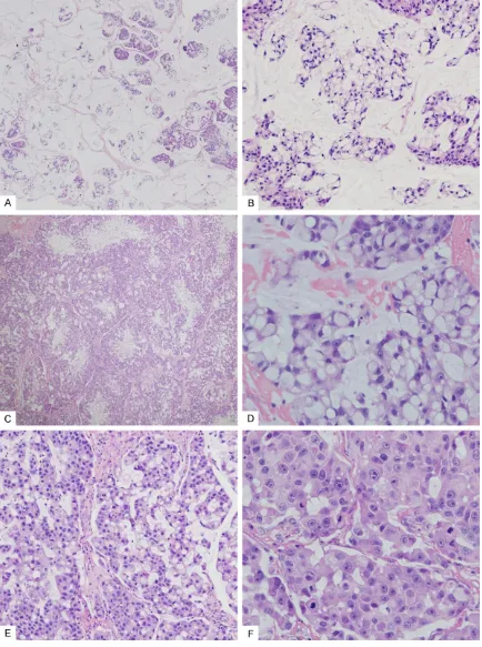
Figure 2. Histopathological findings. A. Mucinous area comprising pools of mucus separated into compartments by fibrous bands. Clusters of tumor cells appear to float within the mucus (hematoxylin-eosin, ×40); B. Cancer cells floated in the pools of mucin, a pattern that mimicked that of mucinous adenocarcinoma (colloid carcinoma). Most of the tumor cells exhibited signet ring morphology (hematoxylin-eosin, ×200); C. Low magnification shows a multi -nodular architecture with hypercellular peripheral rims and myxoid and necrotic central zones (hematoxylin-eosin,
×40); D. High magnification shows typical signet ring cells with intracytoplasmic mucin vacuoles and displaced
indented nuclei. (hematoxylin-eosin, ×400); E. Tumor cells with abundant eosinophilic cytoplasm often seen
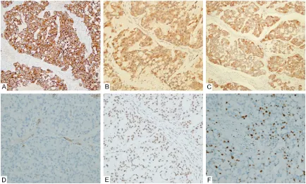
ses/10 high-power fields), and perineural inva -sion was observed. Focal tubule formation was noted. Immunohistochemically, the tumor cells showed diffuse reactivity for cytokeratin (CK) AE1/AE3, CK7 (Figure 3A), CK18, S100 (Figure 3B), and mammaglobin (Figure 3C), and focal
positivity for CK5/6 and CK 34βE12. CK14,
CK20, vimentin, p63, p40, HHF-35, smooth mu- scle actin (Figure 3D), calponin, DOG-1, Sox10, and Wilms’ tumor 1 gene (WT1) protein were all negative. The tumor cells showed intact expres-sion of SMARCB1/INI1 (Figure 3E). The prolif-erative index (Ki-67) was up to 20% in some tumor regions (Figure 3F).
Discussion
Signet-ring cells are characterized by round spaces, inclusions, or substance accumula-tions occupying the cytoplasms and compress-ing the nuclei toward the cellular borders. The signet-ring cell appearance can result from dif-ferent mechanisms, and the content of the vacuoles is variable (e.g. mucin, lipid, or
glyco-gen) [8]. Signet-ring cells are characteristically
found in carcinomas of high malignant
poten-tial. The gastrointestinal tract, breast, and lu- ngs are the most common primary sites. Alth- ough much less frequently, signet-ring cells have also been reported in single cases of pri-mary salivary gland neoplasms, such as onco-cytic cystadenoma, mucinous (colloid) carcino-ma, mucinous cystadenocarcinocarcino-ma, mucin-rich salivary duct carcinoma, mucoepidermoid
car-cinoma, and adenoid cystic carcinoma [8-11].
Moreover, salivary glands tumors described by name containing signet-ring cells have also rarely been reported. In 2004, Ghannoum and Freedman reported seven cases of a hitherto unknown salivary malignant neoplasm charac-terized by marked presence of mucin-contain-ing signet rmucin-contain-ing cells, which they termed signet ring cell (mucin producing) adenocarcinomas of
minor salivary glands [12]. Interestingly, a myo -epithelial phenotype was implied by immuno-histochemistry. Since then, a few similar cases, also including benign signet-ring cell tumors
have been reported [5, 13-15]. Recently, these
[image:4.612.89.523.71.333.2]tumors were considered to be a unique subtype of myopitheliomas, and the term mucinous myoepitheliomas and secretory MCs has been Figure 3. Immunohistochemical findings. A. The tumor cells were diffusely immunoreactivity for CK7 (×100). B. The
Mucinous myoepithelial carcinoma of the parotid gland
proposed by Esteva et al. [4] and Bastaki et al. [6], respectively.
To date only 15 cases of MMC, including the
present case, have been reported [6, 7, 12-14].
The male to female ratio is 7:8, and the mean age is 58.3 years ranging from 18 to 81 years. 11 cases arose in the minor salivary glands and 4 in the parotid. Tumors ranged in size from 1 to 5.6 cm. Most tumors were treated with sur-gical excision alone with negative margins. One patient presented with neck metastasis at the time of initial presentation. Seven patients available for follow-up had no evidence of dis-ease with an average follow-up of 33 months. Histologically, MMC is composed mainly of large mucin containing cells, arranged in solid nests, sheets, cords and trabeculae and em- bedded in a myxoid stroma. The mucin contain-ing cells often have a signet rcontain-ing cell appear-ance with abundant vacuolated clear cyto-plasm and displaced indented nuclei. The
intracellular mucin can be confirmed by muci -carmine, alcian blue and periodic acid schiff after diastase staining. A mixture of other cell types, including epithelioid or plasmacytoid
cells, have been identified [6, 7, 12-14]. The
epithelioid cells are polygonal, characterized by eosinophilic cytoplasm and ovoid nuclei. The plasmacytoid cells contain abundant brightly eosinophilic cytoplasm and eccentric nuclei. The tumor cells usually have minimal to moder-ate cytologic atypia. At present, the histological criteria for discriminating between benign and malignant mucinous myoepitheliomas are not
clearly defined. Gnepp proposed that the same
histological criteria should be used to evaluate for malignancy as any other myoepithelioma
[7].
Immunohistochemistry is almost essential to identify myoepithelial cells and reactivity for CKs and at least one of the other myoepithelial markers, including S100, vimentin, calponin,
p63, glial fibrillary acidic protein, CD10, smooth
muscle actin, and smooth muscle myosin heavy
chains, is required for diagnosis [1]. The immu -nophenotype of individual cases is highly vari-able. Therefore, a broad panel of markers
typi-cal of the myoepithelial immunoprofile should
be used. Of the 15 reported cases of MMC, all stained with cytokeratins (8/8 for pancytokera-tin, 11/11 for CAM5.2, and 6/6 for CK7). CK5/6 was focally positive in 3 of 5 tumors, and CK20
was negative for all 6 tumors that were exam-ined. 10 of 12 showed nuclear labeling for p63 (diffuse positive in 8 cases while sparse in 2 cases). 10 of 15 tumors showed positivty for S100 and 5 of 9 tumors showed positivty for
glial fibrillary acidic protein. 9 of 14 tumors
showed positivty for smooth muscle actin, 7 of 12 tumors showed positivty for HHF-35, and 3 of 12 tumors showed positivty for calponin. Of note, one case reported by Bastaki et al., was negative for all myoepithelial markers tested by immunohistochemistry, but it showed evidence of myoepithelial differentiation on
ultrastruc-tural analysis [6].
In terms of frequency, the finding of signet-ring
cells in a salivary neoplasm requires always to rule out metastatic adenocarcinoma. A full evaluation failed to identify any concomitant malignancy in our patient. The immunohisto-chemical study with CK7 and CK20 antibodies is sometimes advocated for the differential diagnosis. Salivary gland neoplasms often sh- ow the typical CK7+/CK20- immunophenotype,
whereas a conjointly CK7-/CK20+ profile may serve as a clue to an intestinal origin [16, 17].
Histopathological differential diagnosis of MMC also includes those primary mucin-rich adeno-carcinomas of salivary glands that may contain signet-ring cells, such as mucinous cystadeno-carcinoma, mucin-rich mucoepidermoid carci-noma, mucin-rich salivary duct carcinoma and
mucinous (colloid) adenocarcinoma [6, 7, 10, 17]. Mucinous cystadenocarcinoma has char -acteristic histological features with a
mucus-filled cystic space lined by malignant epithelial
cells. It is typically more cystic and papillary than MMC and do not stain with myoepithelial markers. Occasionally, mucoepidermoid carci-noma may be associated with prominent pools of acellular or paucicellular mucin and may also contain signet ring cells. However, mucoepider-moid carcinoma is characterized by the pres-ence of squamoid cells, mucinous cells, and intermediate cells. The mucin-rich salivary duct carcinoma is characterized by the presence of a mucinous component that comprises promi-nent mucinous lakes or pools divided into small
can-didate in the differential diagnosis of our case because approximately two-thirds of the tumor
consisted of a mucinous area with floating can -cer nests. However, the cells in mucinous (col-loid) adenocarcinoma are epithelial and pres-ent as single cells, cells forming ductlike str- uctures, or solid strands or clusters of cells. Moreover, mucinous (colloid) adenocarcinoma lacks the expression of S100 and other myoepi-thelial markers. Mammary analogue secretory carcinoma is a recently described new distinc-tive salivary gland tumor composed of uniform cells with bland-looking vesicular nuclei and eosinophilic vacuolated cytoplasm, arranged in tubular, microcystic and solid growth patterns. Immunohistochemically, mammary analogue secretory carcinomas show diffuse and strong expression of CK7, CK8, CK18, S-100,
vimen-tin, and mammaglobin [18]. The tumor in our
case contained abundant signet-ring cells, which are not seen in mammary analogue secretory carcinoma.
In summary, we presented a rare case of MMC of the parotid gland. For pathologists, it is im- portant to recognize this unusual variant of sali-vary gland-type neoplasm and to avoid confus-ing it with other primary and metastatic mucin-producing tumors, which require alternative management and have a correspondingly dif-ferent natural history.
Disclosure of conflict of interest
None.
Address correspondence to: Dr. Xinming Chen, De- partment of Pathology, School and Hospital of St- omatology, Wuhan University, 237 Luoyu Road, Wu- han 430079, Hubei, China. Tel: +86-27-87686229; Fax: +86-27-87686375; E-mail: xmchen3011@126. com
References
[1] Barnes L, Eveson JW, Reichart P. World Health
Organization classification of tumors: patholo -gy and genetics of the head and neck tumours. Lyon: IARC Press; 2005.
[2] Savera AT, Sloman A, Huvos AG and Klimstra DS. Myoepithelial carcinoma of the salivary glands: a clinicopathologic study of 25 pa-tients. Am J Surg Pathol 2000; 24: 761-774.
[3] Yang S, Li L, Zeng M, Zhu X, Zhang J and Chen X. Myoepithelial carcinoma of intraoral minor salivary glands: a clinicopathological study of
7 cases and review of the literature. Oral Surg Oral Med Oral Pathol Oral Radiol Endod 2010; 110: 85-93.
[4] Esteva CJ, Slater LJ and Gnepp DR. Mucinous Myoepithelioma, a Previously Unrecognized
Variant. Modern Pathology 2012; 25:
308A-308A.
[5] Bastaki JM, Purgina BM, Dacic S and Seethala RR. Primary Signet-Ring Cell (Mucin-Produc-ing) Adenocarcinoma of Minor Salivary Glands: A Clinicopathologic, Immunohistochemical and Molecular Survey. Modern Pathology 2012; 25: 304A-305A.
[6] Bastaki JM, Purgina BM, Dacic S and Seethala RR. Secretory myoepithelial carcinoma: a his-tologic and molecular survey and a proposed nomenclature for mucin producing signet ring tumors. Head Neck Pathol 2014; 8: 250-260.
[7] Gnepp DR. Mucinous myoepithelioma, a re-cently described new myoepithelioma variant. Head Neck Pathol 2013; 7 Suppl 1: S85-89.
[8] Altemani A, Costa AF, Montalli VA,
Mosqueda-Taylor A, Paes de Almeida O, Leon JE and Hermsen M. Signet-ring cell change in adenoid cystic carcinoma: a clinicopathological and im-munohistochemical study of four cases. Histo-pathology 2013; 62: 531-542.
[9] Michal M, Hrabal P and Skalova A. Oncocytic cystadenoma of the parotid gland with promi-nent signet-ring cell features. Pathol Int 1998; 48: 629-633.
[10] Yakirevich E, Sabo E, Klorin G, Alos L, Cardesa A, Ellis GL, Shumway BS and Gnepp DR. Pri-mary mucin-producing tumours of the salivary glands: a clinicopathological and morphomet-ric study. Histopathology 2013; 57: 395-409.
[11] Kusafuka K, Maeda M, Honda M and Nakaji-ma T. Mucin-rich salivary duct carcinoNakaji-ma with signet-ring cell feature ex pleomorphic adeno-ma of the subadeno-mandibular gland: a case report of an unusual histology with immunohisto-chemical analysis and review of the literature. Med Mol Morphol 2012; 45: 45-52.
[12] Ghannoum JE and Freedman PD. Signet-ring cell (mucin-producing) adenocarcinomas of minor salivary glands. Am J Surg Pathol 2004; 28: 89-93.
[13] Bastaki J and Summersgill K. Signet-ring cell (mucin-producing) adenocarcinoma of minor salivary glands: report of a case. Oral Surg Oral Med Oral Pathol Oral Radiol Endod 2010; 110: e33-36.
[14] Singh M, Khurana N, Wadhwa R and Gulati A. Signet ring carcinoma parotid gland: a case re-port. Head Neck 2011; 33: 1656-1659.
Mucinous myoepithelial carcinoma of the parotid gland
[16] Meer S and Altini M. CK7+/CK20-
immunoex-pression profile is typical of salivary gland neo -plasia. Histopathology 2007; 51: 26-32.
[17] Ide F, Mishima K, Tanaka A, Saito I and Kusa-ma K. Mucinous adenocarcinoKusa-ma of minor salivary glands: a high-grade malignancy prone
to lymph node metastasis. Virchows Arch
2009; 454: 55-60.