Original Article
GLUT-1 and its regulating factor HIF-1α expression
in epithelial ovarian tumors: GLUT-1 is associated with
molecular typing and grade of epithelial ovarian cancer
Xiu-Jie Yu, Jian-Chan Song, Jing Du, Yi-Quan Shi, Yi-Xin Liu, Yan Shen
Department of Pathology, Tianjin Central Hospital of Gynecology and Obstetrics, Tianjin, China
Received November 13, 2016; Accepted January 6, 2017; Epub April 1, 2017; Published April 15, 2017
Abstract: Objective: Hypoxia plays an important role in tumorigenesis and tumor progression. Under the hypoxia environment, glucose uptake is increased to support the growth of tumor. Here we analyzed the expression of GLUT-1 and hypoxia inducible factor-1α (HIF-1α) in epithelial ovarian tumors, investigated the correlation between their expression and the molecular typing (Kurman’s model) or histological grade of epithelial ovarian cancer. Then explore the possible significance of GLUT-1 in diagnosis and treatment of ovarian cancer. Materials and methods: 154 cases with primary epithelial ovarian tumors (malignant: 63 cases, borderline: 42 cases, benign: 49 cases) were studied. Immunohistochemistry was performed using GLUT-1 and HIF-1α antibody. GLUT-1 protein expression was quantified by Western blot analysis. GLUT-1 mRNA expression was analyzed by real time PCR (RT-PCR). Results: Both of the expressions of GLUT-1 and HIF-1α were increased gradually from benign to malignant ovarian tumors, they showed a sequence correlation. But, only GLUT-1 expression synchronously showed a statistical difference in molecular typing, histological grade and FIGO Stage of epithelial ovarian cancer (P<0.05). And there is a positive correlation between the expression of GLUT-1 and HIF-1α, the correlation coefficient was 0.232 (P<0.05). At the same time, the expression pattern showed that as the score of GLUT-1 staining increased the intensity and extent of HIF-1α expression was enhanced accordingly. Western blot and RT-PCR analysis were in accordance with the im-munohistochemical staining of GLUT-1. Conclusion: Our study suggests that the expression of GLUT-1 was closely related with molecular typing and histological grade of epithelial ovarian cancer. GLUT-1 could be a novel diagnosis biomarker and therapeutic target for epithelial ovarian cancer.
Keywords: GLUT-1, HIF-1α, epithelial ovarian cancer
Introduction
Epithelial ovarian tumor is the high incidence of gynecology tumor. According to the USA cancer statistics and analysis, in 2016, 1685210 new cancer cases and 595690 cancer deaths are projected to occur in the United States [1]. Therefore, ovarian cancer has become the most fatal gynecologic malignant tumor.
In recent ten years, the understanding of ovari-an covari-ancer has made great progress. Based on the distinctive clinicopathologic and molecular genetics feature, a dualistic model of ovarian carcinogenesis has been proposed by Kurman [2], which divided ovarian carcinomas into two groups: type I and type II. This new model reflects the finding that ovarian cancer
com-prises a group of heterogeneous tumors that develop and behave differently. At the same time, a two-tier system about ovarian serous adenocarcinoma has been put forward by Malpica [3], which classified ovarian serous adenocarcinoma into low grade and high grade. These concepts are closely related to assess-ment of prognosis, individualized treatassess-ment and increase of the survival rate.
needs a large amount of glucose as an energy source. Therefore, with the increase of glucose uptake, metabolism and glycolysis accelerated significantly. This process is mediated by spe-cific transmembranous glucose transporter proteins (GLUTs). GLUT family can be divided into three subfamilies, namely class I (the previ-ously known glucose transporters GLUT1-4), class II (the previously known fructose trans-porter GLUT5, the GLUT7, GLUT9 and GLUT11), and class III (GLUT6, 8, 10, 12, and the myoino-sitol transporter HMIT1) [4].
As a housekeeper of GLUTs, GLUT-1 is widely distributed in normal tissues such as erythro-cytes and endothelial cells at the blood-brain barrier. Overexpression is considered as self-adaptation in order to achieve rapid growth, proliferation, invasion and metastasis. GLUT-1 can be used as an intrinsic marker to detect
endogenous hypoxia status within the tumor [5]. Although the metabolic consequences of increased glucose transporter are not under-stood completely, the clinical importance of GLUT-1 expression has been demonstrated. GLUT-1 is primarily undetectable in normal epi-thelial tissues and benign epiepi-thelial tumors. However, GLUT-1 expression has been elevated in a significant proportion of human carcino-mas. Various studies have shown a close rela-tionship between GLUT-1 expression and carci-nogenesis, tumor development or the unfavor-able prognosis of various malignancies[6-8]. Hypoxia inducible factor-1 (HIF-1) is an upstream regulator of GLUT-1, also a key transcription factor to mediate the adaptive response to the hypoxic microenvironment. HIF-1 is an αβ het-erodimeric transcription factor, HIF-1β subunit is constitutively expressed, under normoxia the HIF-1α subunit is subject to prolyl hydroxylation, which targets this subunit for ubiquitinylation and subsequent proteasomal degradation [9]. Under hypoxia, however, HIF-1α degradation is inhibited leading to rapid induction of protein levels and formation of the functional heterodi-mer [9]. HIF-1α can regulate over 40 genes expression, for example GLUT-1, P53, VEGF (vascular endothelial growth factor), MDR-1 (multidrug resistance-1) etc. [9], and these tar-get genes are important in tumor energy sup-ply, apoptosis, angiogenesis, drug resistance and recurrence.
In this study we re-evaluate the typical expres-sion pattern of GLUT-1 and its regulator (HIF-1α) in epithelial ovarian tumors, focused on analyzing the correlation between GLUT-1 expression and Kurman’s model and grade. This would contribute to a better understanding of the role of GLUT-1 in ovarian epithelial tumor. Materials and methods
Patients
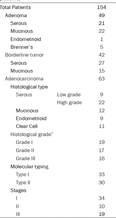
[image:2.612.93.286.92.454.2]A total of 154 patients with primary epithelial ovarian tumor who underwent surgical resec-tion in Tianjin Central Hospital of Gynecology and Obstetrics in 2006 were selected (Table 1). None of patients had received preoperative chemo- or radiotherapy. All patients were st- aged based on the International Federation of Gynecology and Obstetrics (FIGO). Histological type and grade were determined by two of the Table 1. Clinicopathologic characteristics of
patients
Total Patients 154
Adenoma 49
Serous 21 Mucinous 22 Endometrioid 1 Brenner’s 5 Borderline tumor 42 Serous 27 Mucinous 15 Adenocarcinoma 63 Histological type
Serous Low grade 9 High grade 22 Mucinous 12 Endometrioid 9 Clear Cell 11 Histological grade*
Grade I 19 Grade II 17 Grade III 16 Molecular typing
Type I 33 Type II 30 Stages
I 34 II 10 III 19
authors, using the World Health Organization (WHO) criteria. The concordance rate was 95% between the 2 pathologists. In case of dis-agreement, the slides were reviewed simulta-neously by the two pathologists seated at a multi headed microscope with a resolution of the difference in opinion. Molecular typing was assigned according to a dualistic model pro-posed by Kurman [2].
Immunohistochemical analysis
Formalin-fixed, paraffin-embedded tissue blo- cks were cut into 4 μm-thick sections for hema-toxylin and eosin (HE) staining and immunohis-tochemistry. The expression of GLUT-1 (poly-clonal rabbit antibody, diluted 1:200; DAKO, Carpinteria, CA, USA), HIF-1α (monoclonal mo- use antibody, ESEE122, diluted 1:1200; Novus Biologicals, Littleton, CO, USA) were investigat-ed using the standard horseradish peroxidase complex method. The paraffin sections were deparaffinized and immersed in 3% hydrogen peroxidase. Next, an antigen retrieval proce-dure was performed by microwaving in 10 mM citrate buffer (pH 6.0) at 500 W for 20 min. After washing in phosphate-buffered saline (PBS), the tissue sections were pre-blocked by 10% normal goat serum for 15 min. The proto-col for the DakoEnVisionTM+ kit was followed for
each section. The sections were incubated for 60 min with primary antibodies in a humidity chamber. The sections were rinsed with PBS for 15 min, and incubated for 30 min with horse-radish peroxidase complex using 3,3’-diamino-benzidine as a chromogen. Counterstaining was performed using hematoxylin. The Speci- ficity of the immunohistochemical reactions was checked by omitting the primary antibody. GLUT-1 and HIF-1α staining results were evalu-ated by semi-quantity method. GLUT-1 was assessed according to Airley’s [10] report, tumor sections were initially scanned at 40× magnification so that the distribution of stain-ing could be assessed, then analyzed field-by-field at 200× magnification. Membrane-predo- minant staining was regarded as positive. Each field was assigned a score of 1-4, representa-tive of the approximate area of immunohisto-chemical staining (0, 0%; 1, 0-5%; 2, 5-15%; 3, 15-30%; 4, >30%). To counteract the effect of variations in staining intensity, only areas of unequivocal staining were include, necrosis, stroma, normal epithelium and distinct edge effects were ignored. The overall scores used in
our study were derived from the average score for all fields. The final scoring scheme was neg-ative (0-1), weak 1+ (1-2), intermediate 2+ (2-3), marked 3+ (3-4). HIF-1α was assessed accord-ing to Wenyan X’s method [11], nuclear and/or cytoplasm staining was regarded as positive, the staining intensity and the percentage of stained cells were analyzed. The intensity was scored as 0 (negative), 1+ (weak), 2+ (medium), and 3+ (strong). The percentage of the positive cells was scored as 0 (<5%), 1+ (5%-25%), 2+ (25%-50%), 3+ (50%-75%), and 4+ (>75%). The final score was made by the above score multi-plying: negative (0-4), 1+ (5-8), 2+ (9-12).
Western blot analysis
To confirm the immumohistochemical staining result of GLUT-1, Western blot was performed in 20 cases. Using representative cases of ade-noma, borderline tumor and adenocarciade-noma, the total proteins and membrane protein were extracted and separated by electrophoresis on 10% SDS-polyacrylamide gels and transferred to immunobilon-NC membranes (Bio-Rad, Hercules, USA), respectively. GLUT-1 antibody (sc-7903, Santa Cruz Biotechnology, Inc., Texas, USA) was diluted 1:1000 in 5% nonfat milk in PBS-T. Immunoreactivity was visualized by horseradish peroxidase-conjugated anti-rabbit and chemiluminescence. β-actin was used as an internal control. The relative intensity of GLUT-1 is the ratio of GLUT-1 and β-actin gray value which digitalized by GIS1000 analysis software.
RNA extraction and RT-PCR
cycles of amplification at 95°C for 15 sec and 60°C for 1 min. The relative expression of each mRNA was calculated by the ΔCt method.
Statistical analysis
Statistical analysis were performed with SPSS 19.0. The Spearman’s rank correlation was used to evaluate the relationship between GLUT-1, HIF-1α expression and neoplastic nature (adenoma, borderline tumor and adeno-carcinoma) or clinicopathological characteris-tics (Molecular typing, grade, histological grade and FIGO stage) in ovarian tumors. The Spearman’s rank correlation coefficient was used to evaluate the relationship between GLUT-1 and HIF-1α expression. P<0.05 were considered statistically significant.
Results
Immunohistochemistry
GLUT-1: The expression was limited to
epitheli-al components, and the stroma was basicepitheli-ally negative except for erythrocytes. A positive reaction was seen in both cytoplasm and cell membrane. All of the epithelial ovarian tumors
showed various degrees of positive staining. The pattern of staining cells showed three forms: diffuse, focal or scattered, and usually expressed strongly in the area of papillary or stratifying structure. With the expression increased gradually and the cell membrane became more strongly stained. GLUT-1 staining showed a gradual enhancement with increas-ing distance from the vascular vessels. Interestingly, we observed giant tumor cells in stroma of ovarian adenocarcinoma which pos-sessed the characteristics of tumor stem-like cells[12] (round, lack branches, at least three times larger than the parental cancer cells) were positive.
HIF-1α: A positive reaction was most frequently
[image:4.612.97.523.72.326.2]observed in both cytoplasm and nuclei of tumor cells, and the staining pattern were focal or dif-fuse, similar to GLUT-1, HIF-1α expressed strongly in the area of papillary or stratifying structure and the area which far from vascular vessels. Positive cells in the stromal were small in number. In general, the staining extent paral-leled staining intensity and nuclear labeling. All of the epithelial ovarian tumors showed various degrees of positive staining.
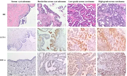
Here we showed H&E and immunohistochemi-cal staining of GLUT-1 and HIF-1α in ovarian serous tumors (Figure 1). GLUT-1 and HIF-1α expression are obviously different in benign, borderline and malignant tumor, the intensity is positive related with malignance and histologi-cal grade.
In most of the examined cases, HIF-1α expres-sion was more extensively and uniform than GLUT-1. However, GLUT-1 expression tended to be localized and generally contained in areas where positive for HIF-1α.
Statistically, a close examination of histomor-phological details was focused on analyzing the relationship between GLUT-1 and HIF-1α expression and neoplastic nature (adenoma, borderline tumor and adenocarcinoma) or clini-cal-pathological characteristics (Molecular typ-ing, grade, histological grade and FIGO stage) in
ovarian tumors. With the increase of malignan-cy of the tumor, the expression of GLUT-1 and HIF-1α increased gradually (P<0.05), they showed a sequence correlation (Table 2). Among them, only GLUT-1 expression synchro-nously showed a statistical difference between Type I and Type II, low grade and high grade serous carcinoma, different histological grade and FIGO Stage (P<0.05) (Table 3). At the time, there is a correlation between the expression of GLUT-1 and HIF-1α, the correlation coeffi-cients were 0.323 (P<0.05) (Table 4). As the score of GLUT-1 staining increased, the extent of HIF-1α enhanced accordingly.
Western blot
The electrophoretogram indicate that GLUT-1 protein content is different in ovarian tumors, adenocarcinoma showed a broad band around 45 kDa, but the intensity markedly decreased in borderline tumor and particularly in adeno-ma (Figure 2). There is a linear relationship from adenoma → borderline tumor → adeno-carcinoma (Figure 3). This result is in accor-dance with immunohistochemical staining.
Real-time PCR
Quantitative analysis demonstrated that the GLUT-1 mRNA levels are increased gradually from adenoma → borderline tumor → adenoca- Figure 2. Western blotting analysis of GLUT-1. Lane 1 and 2: adenoma, Lane 3: borderline tumor, Lane 4-6: adenocarcinoma. β-actin is a internal control. Adeno-carcinoma showed a broad band around 45 kDa, but the intensity markedly decreased in borderline tumor and particularly in adenoma.
Table 2. Immunohistochemical analysis of GLUT-1 and HIF-1α in epithelial ovarian tumors
GLUT-1 HIF-1α - + ++ +++ - + ++ Adenoma 43 1 4 1 28 16 5 Borderline tumor 29 10 2 1 8 27 7 Adeno-carcinoma 20 14 14 15 8 20 35
P value 1.69×10⁻⁸▲ 2.44×10⁻¹⁰▲
▲P<0.05.
Table 3. GLUT-1 expression in different clinico-pathologic characteristics
GLUT-1
P value
- + ++ +++ Molecular typing
Type I 17 7 7 2 4.92×10⁻⁴▲
Type II 3 7 7 13 Grade
Low Grade 2 3 3 1 0.040▲
High Grade 0 5 5 12 Histological grade
Grade I 8 4 6 1 0.015▲
Grade II 3 3 4 7 Grade III 0 7 3 6 FIGO Stage
I 14 6 9 6 0.002▲
II 0 6 3 0 III 3 4 3 9
▲P<0.05.
Table 4. Correlation between the expression of GLUT-1 and HIF-1α
GUTL-1 HIF-1α
- + ++
- 37 33 22
+ 4 14 7
++ 1 14 5
rcinoma, and the mRNA levels are positive cor-relation with protein levels (rs=0.454, P<0.05) (Figure 4). Also this result is in accordance with the immunohistochemical expression profiles. Discussion
In recent years, a lot of studies showed that GLUT-1 expression has been elevated in a sig-nificant proportion of human carcinomas. Various studies have shown a close relation-ship between GLUT-1 expression and carcino-genesis, tumor development or the unfavorable prognosis of various malignancies [6-8]. In this study, we analyzed the expression of GLUT-1 and hypoxia inducible factor-1α (HIF-1α) in epi-thelial ovarian tumors, investigated the correla-tion between their expression and the molecu-lar typing (Kurman’s model) or histological grade of epithelial ovarian cancer. Then explore the possible significance of GLUT-1 in diagnosis and treatment of ovarian cancer.
In our study, the immumohistochemistry results of GLUT-1 and its regulating factor HIF-1α
staining while GLUT-1 was heterogeneous, especially most of the GLUT-1 positive areas were contained within HIF-1α positive areas, suggested that GLUT-1 expression is consider-ably controlled by HIF-1α expression and GLUT-1 is more sensitive than HIF-α when microenvi-ronment change.
But the expression of GLUT-1 is not regulated by HIF-1α only, GLUT-1 is influenced by hypoxia, oncogene, antioncogene, glycogen’s storage, cell growth factor and hormones etc. The over-expression of GLUT-1 in tumor when microenvi-ronment hypoxia can be induced by two ways, one way is: the reduction of oxygen lead HIF-1 activation, then HIF-1 combined with DNA bind-ing site on GLUT-1 enhancer element to prompt the expression of GLUT-1 mRNA; another way is: hypoxia inhibit mitochondrial oxidative phos-phorylation, not only change the intrinsic activ-ity of GLUT-1 but also stimulate the transition of GLUT-1 from storage vacuoles to cell mem-brane. Mayer [14] discovered that although a robust induction of GLUT-1 by hypoxia has been demonstrated in vitro, this reaction is modulat-Figure 3. GLUT-1 protein content in ovarian epithelial tumors. GLUT-1 protein
[image:6.612.92.380.73.192.2]content of ovarian tumors is enhanced gradually from adenoma (A) → bor-derline tumor (B) → adenocarcinoma (AC).
Figure 4. GLUT-1 mRNA expression in ovarian epithelial tumors. GLUT-1 mRNA levels were increased gradually from adenoma (A) → borderline tumor (B) → adenocarcinoma (AC).
[image:6.612.91.377.253.376.2]ed both by confounding factors of the tumor microenvironment and intrinsic traits of malig-nant cells in vivo.
The immunochemistry staining result also shown that the expression of GLUT-1 and its regulator are obviously different in ovarian benign, borderline and malignant tumors, the intensity is positive related with the malignance and histological grade. And with the increase of GLUT-1 score, HIF-1α score increased. These results are accordance with Cantuaria et al [15], Ozcan et al [16]and Iica et al [17]’s, the overexpression of GLUT-1 and its regulators are related to malignant transformation of ovarian epithelial tumors, then accelerated the metab-olism of tumor cell when hypoxia, so GLUT-1 can be utilized as a molecular diagnosis marker of ovarian epithelial tumor in its early stage. To confirm the accuracy and significance of the immunohistochemical staining profiles, we per-formed Western blot and real time PCR to anal-ysis the protein and mRNA quantity of GLUT-1. We found that the protein of GLUT-1 is very low in benign tumor, increased in borderline tumor and highest in malignant tumor; the mRNA of GLUT-1 increased with the extent of malignan-cy, and positive correlation with protein. This result is in accordance with immunohistochem-istry staining result. And explain the phenome-non that with the level of malignancy increase, GLUT-1 expression in cell membrane is more obviously. So it is indicate that, under the action of regulator, GLUT-1 mRNA transcription and protein synthesis increase is the early event of malignant transformation of tumors.
Our study also found the expression of GLUT-1 and HIF-1α differed among the histological types of epithelial ovarian carcinoma, higher in serous carcinoma than mucinous adenocarci-noma, and the difference is statistically signifi-cant. This conclusion in accordance with Tsukioke et al [18] and Airley et al [19]’s study. These findings suggest that different histologi-cal types of epithelial ovarian carcinoma show different characteristic for glucose uptake, which may be related with the histological structure. In mucinous carcinoma, because glands often show back-to-back arrangement and lack papillary structure, at the same time, the stroma is rich relatively, so the extent of hypoxia is lower. So the positive rate of GLUT-1 shows decreased positive compared with se-
rous carcinoma. This phenomenon can also be seen in thyroid papillary carcinoma and follicu-lar carcinoma [20].
rate of GLUT-1 in low-grade (77.8% (7/9)) and high-grade (100% (22/22)) serous carcinoma showed no statistically significance. Because low-grade serous carcinoma is not sensitive to chemotherapy, so, conjugation of GLUT-1 to chemotherapeutics may be improving the che-motherapy effect of low-grade serous carcino-ma, this hypothesis is one of the aspects of GLUT-1 in treatment. If we can suppressing tumor growth by inhibit the transcription of GLUT-1, or kill tumor cells by restrain the activi-ty of GLUT-1 using antisense DNA, RNA may be a new way in therapy of ovarian carcinoma. Acknowledgements
This study has been financially supported by Sanitary Bureau of Tianjin, Research No. 2014KR20.
Disclosure of conflict of interest
None.
Address correspondence to: Dr. Yan Shen, Depart- ment of Pathology, Tianjin Central Hospital of Gyne- cology and Obstetrics, Tianjin 300100, China. Tel: 86-22-58287668; E-mail: serina_shen@163.com
References
[1] Siegel RL, Miller KD, Jemal A. Cancer statistics, 2016. CA Cancer J Clin 2016; 66: 7-30. [2] Shih IeM, Kurman RJ. Ovarian tumorigenesis:
a proposed model based on morphological and molecular genetic analysis. Am J Pathol 2004; 164: 1511-1518.
[3] Malpica A, Deavers MT, Lu K, Boduka DC, At-kinson EN, Gershenson DM, Silva EG. Grading ovarian serous carcinoma using a two-tier sys-tem. Am J Surg Pathol 2004; 28: 496-504. [4] Joost HG, Thorens B. The extended
GLUT-Fam-ily of sugar/polyol transport facilitators: no-menclature, sequence, characteristics and potential function of its novel members. Mol Membr Biol 2001; 18: 247-256.
[5] Cooper R, Sarioğlu S, Sökmen S, Füzün M, Küpelioğlu A, Valentine H, Görken IB, Airley R, West C. Glucose transpoter-1 (GLUT-1): a po-tential marker of prognosis in rectal carcino-ma? Br J Cancer 2003; 89: 870-876. [6] Cantuaria G, Fagotti A, Ferrandina G,
Magal-haes A, Nadji M, Angioli R, Penalver M, Man-cuso S, Scambia G. GLUT-1 expression in ovar-ian carcinoma: association with survival and response to chemotherapy. Cancer 2001; 92: 1144-1150.
[7] Kuwamura T, Kusakabe T, Sugino T, Watanabe K, Fukuda T, Nashimoto A, Honma K, Suzuki T. Expression of glucose transporter-1 in human gastric carcinoma: association with tumor ag-gressiveness, metastasis, and patient survival. Cancer 2001; 92: 634-641.
[8] Cho H, Lee YS, Kim J, Chung JY, Kim JH. Over-expression of glucose transpoter-1 (GLUT-1) predicts poor prognosis in epithelial ovarian cancer. Cancer Invest 2013; 31: 607-615. [9] Anderson CJ, Hoare SF, Ashcroft M, Bilsland
AE, Keith WN. Hypoxia regulation of telomer-ase gene expression by transcriptional and post-transcription mechanisms. Oncogene 2006; 25: 61-69.
[10] Airley RE, Loncaster J, Raleigh JA, Harris AL, Davidson SE, Hunter RD, West CM, Stratford IJ. GLUT-1 and CAIX as intrinsic markers of hypox-ia in carcinoma of the cervix: relationship to pimonidazole binding. Int J Cancer 2003; 104: 85-91.
[11] Xi W, Gao Y. The expression of HIF-1α and GLUT-1 in endometrial carcinoma and its clini-cal significance. Progress in Obstetrics and Gy-necology 2006; 15: 195-198.
[12] Zhang S, Mercado-Uribe I, Xing Z, Sun B, Kuang J, Liu J. Generation of cancer stem-like cells through the formation of polyploid giant cancer cells. Oncogene 2014; 33: 116-128.
[13] Yasuda M, Miyazawa M, Fujita M, Kajiwara H, Iida T, Hirasawa T, Muramatsu T, Murakami M, Mikami M, Saitoh K, Shimizu M, Takekoshi S, Osamura RY. Expression of hypoxia inducible factor-1 alpha (HIF-1alpha) and glucose trans-porter-1 (GLUT-1) in ovarian adenocarcinomas: difference in hypoxic status depending on his-tological character. Oncol Rep 2008; 19: 111-116.
[14] Mayer A, Höckel M, Vaupel P. Endogenous hy-poxia makers in locally advanced cancers of the uterine cervix: reality of wishful thinking? Strahlenther Onkol 2006; 182: 501-510. [15] Cantuaria G, Magalhaes A, Angioli R, Mendez
L, Mirhashemi R, Wang J, Wang P, Penalver M, Averette H, Braunschweiger P. Antitumor activ-ity of a novel glyco-nitric oxide conjugate in ovarian carcinoma. Cancer 2000; 88: 381-388.
[16] Ozcan A, Deveci MS, Oztas E, Dede M, Yenen MC, Korgun ET, Gunhan O. Prognostic value of GLUT-1 expression in ovarian surface epitheli-al tumors: a morphometric study. Anepitheli-al Quant Cytol Histol 2005; 27: 181-186.
HIF-1 alpha/GLUT-1 expression. Arch Gynecol Obstet 2008; 277: 539-546.
[18] Tsukioka M, Matsumoto Y, Noriyuki M, Yoshida C, Nobeyama H, Yoshida H, Yasui T, Sumi T, Honda K, Ishiko O. Expression of glucose trans-porters in epithelial ovarian carcinoma: corre-lation with clinical characteristics and tumor angiogenesis. Oncol Rep 2007; 18: 361-367. [19] Airley R, Evans A, Mobasheri A, Hewitt SM.
Glu-cose transporter Glut-1 is detectable in peri-necrotic regions in many human tumor types but not normal tissue: study using tissue mi-croarrays. Ann Anat 2010; 192: 133-138.
[20] Yasuda M, Ogane N, Hayashi H, Kameda Y, Mi-yagi Y, Iida T, Mori Y, Tsukinoki K, Minematsu T, Osamura Y. Glucose transporter-1 expression in the thyroid gland: clinicopathological signifi-cance for papillary carcinoma. Oncol Rep 2005; 14: 1499-1504.