Field-induced transformations in the biaxial order of non-tilted phases in a bent-core smectic
liquid crystal
This article has been downloaded from IOPscience. Please scroll down to see the full text article. 2010 EPL 92 26002
(http://iopscience.iop.org/0295-5075/92/2/26002)
Download details:
IP Address: 134.226.252.160
The article was downloaded on 02/03/2011 at 10:23
Please note that terms and conditions apply.
doi:10.1209/0295-5075/92/26002
Field-induced transformations in the biaxial order of
non-tilted phases in a bent-core smectic liquid crystal
Y. P. Panarin1,2, Mamatha Nagaraj1, J. K. Vij1(a), C. Keith3and C. Tschierske3
1Department of Electronic and Electrical Engineering, School of Engineering, Trinity College, University of Dublin
Dublin 2, Ireland, EU
2School of Electronic & Communication Engineering, Dublin Institute of Technology - Dublin, Ireland, EU 3Institute of Chemistry, Organic Chemistry, Martin-Luther-University Halle-Wittenberg - D-06120 Halle,
Germany, EU
received 11 August 2010; accepted in final form 27 September 2010 published online 5 November 2010
PACS 61.30.Gd– Orientational order of liquid crystals; electric and magnetic field effects on order
PACS 77.80.-e– Ferroelectricity and antiferroelectricity
PACS 78.20.Fm– Birefringence
Abstract – The structural and electro-optic investigations of an achiral bent-core molecule in SmAPA phase, in which the polar directors in the neighboring layers are arranged anti-ferroelectrically, show that it undergoes transformation from one biaxial to another biaxial structure via a quasi-stable uniaxial structure on the application of the electric field. The non-continuous change in biaxiality is explained by an intermediate state in which the secondary directors in the neighboring layers are perpendicular to each other.
Copyright cEPLA, 2010
Ferroelectricity in tilted smectic phases of chiral molecules predicted on symmetry considerations in 1975 was observed [1] and subsequently extensively investigated. In 1992, Brand et al. [2] suggested another model of ferroelectricity for an orthogonal smectic phase (CP) formed by non-chiral bent-core (or banana, bow, boomerang) molecules. Such a phase possesses an orthorhombic symmetry (C2v), in which one of the three mirror planes is absent and the spontaneous polarization is parallel to the 2-fold axis. The first experimental evidence of ferroelectricity in non-chiral bent-core smectic LCs was given in 1996 by Nioriet al.[3] through measurements of the polarization switching current and of the dielectric permittivity. An important feature of the bent-shaped molecules is their tendency to form tilted smectic phases with macroscopic chirality in the opposite directions [4–6]. Since the first works [1] on banana-shaped molecular systems, a wide variety of different tilted phases such as B1-B8, have been discovered and classified [7–10]. Among these B2 is the SmC-like structure which may form four different structures designated as SmCS/APF/A, where subscripts S/A refer to synclinic/anticlinic tilt and F/A refer to ferroelectric/antiferroelectric orderings in the neighboring
(a)E-mail:[email protected]
Y. P. Panarinet al.
CN
O O
O O
C12H25 C12H25
O
O O
O
Cr 37.8oC SmAPA111oC SmAPR157.5oC SmA 163.9oC I
δ
n=0.016
δ
n≈0
δ
n=0.016
∆n
I=0.112
∆n
L=0.104
(c)
(d)
(e)
(f)
(h)
(g)
(b)
∆n
H=0.12
δ
n=0.003
(a)
δ
n=0
P
A
E
P
A
R
R
50
µ
m
100
µ
m
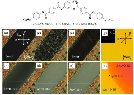
Fig. 1: (Colour on-line) Top: structural formula and the transition temperature obtained on cooling. Textural transformations in 8.7µm homeotropic (a)–(f) and 9.5µm planar cell (g), (h). (a) and (b) are in SmAPR phase at 113◦C, withoutE and with E= 1.4 V/µm respectively. (c)–(h) are in SmAPA phase at 95◦C, where (c) is withoutE, (inset: schlieren texture in a fresh cell), (d) at 0.35 V/µm (e) at 1.68 V/µm and (f) at 2.5 V/µm (g) in SmAPAphase at 95◦C for 0◦(inset) and 45◦angle of the rubbing direction with respect to the polarizer. (h) Three states of molecular alignment resulting in three birefringence values.
E is applied at 100 Hz.P, A,E, andRdenote polarizer, analyzer, electric field, and rubbing directions.
rotation around the long axis is faster by a factor of at least 100, there has thus been an increasing interest in the exploitation of biaxial nematics in fast displays as well their use in stabilizing the blue phases. In smectics, the switching in SmAPR and SmAPA phases is brought byE interacting with the dipole moment directed along the minor director. There is a considerable advantage to realizing switching in the smectic phases since the major director is already fixed by the smectic layers unlike in biaxial nematics where it needs to be anchored independently of the secondary director by external forces [16,17]. The switching speed in smectics may also be higher sinceE interacts with macroscopic polarization as opposed to dielectric biaxiality in biaxial nematics.
In this letter, we determine the biaxiality and electric-field–induced biaxialities in SmAPR and SmAPA phases for the first time. We find that in the SmAPA phase, the structure transforms from one biaxial into another biaxial via a quasi-stable uniaxial structure upon the application of electric field. We also observe a large second
[image:3.595.55.527.85.421.2]but its sign varies randomly from one neighboring layer to another, resulting in a random and macroscopically uniaxial structure [13].
The textural changes under crossed polarizers of this sample in both homeotropic and planar cells as functions of temperature and applied electric field are given in fig. 1. A polymer (JSR Co.) is used for homeotropic alignment. Two electrode stripes are formed by etching indium tin oxide (ITO) on the bottom glass plate. An in-planeE of frequency 100 Hz is applied between the ITO electrodes such thatE makes an angle of 45◦ to the polarizer. The distance between these electrodes is fixed to be∼200µm. Values of birefringence and biaxiality were measured using a tilting optical compensator. This technique allows for the measurements of an extremely small optical retardation. This also allows us to measure the birefringence of a partic-ular area (domain) in the cell even having a non-uniform texture such as the schlieren texture, etc. Strictly speaking the “birefringence” is applicable to the uniaxial structure such asne−no. For the biaxial structure there are three distinct eigenvaluesnX,nY andnZ. Therefore the smallest difference in refractive indices,nY −nX, is the “biaxial-ity” and the difference between the others two is the “bire-fringence”. Consequently, the term “birefringence” is used for planar cells while “biaxiality” is used for homeotropic ones. On cooling the sample in a homeotropic aligned cell in the absence ofE from the isotropic to SmAand then to SmAPR phases, the cell shows very large extinction and a change in the direction of the polarization of light does not affect this extinction. The smectic layers in this arrange-ment lie parallel to the substrate (fig. 1(a)). We note that the application of in-planeE produces no significant effect on the texture in the SmAphase, but induces biaxiality in SmAPR as shown in fig. 1(b) where the secondary direc-tors are directed parallel toE. This effect becomes stronger on approaching the phase transition to the SmAPAphase. The cell shows fast and reversible electro-optical switch-ing from the field-induced bright to the dark state after the field is removed. On a further cooling to 111◦C in the absence of E, the sample shows a phase transition to the SmAPA phase, characterized by the presence of a typical schlieren texture (fig. 1(c)), indicating the biaxial nature of this phase. On the application of E, textures show several, unexpected and interesting transformations (fig. 1(c)–(f)). Initially for a rather smallE(∼0.35 V/µm) the schlieren texture transforms into a uniform pattern (fig. 1(d)) with biaxiality of δn∼0.016 and the optical axis (i.e. secondary director m) is directed along the electric field. An increase in E causes an unexpected sharp drop in the macroscopic biaxiality to zero (δn= 0, fig. 1(e)) for a critical electric fieldEC(T) which increases on cooling. An additional small increase in E, causes a rapid change in the uniaxial texture to the texture with biaxiality as before δn= 0.016 (fig. 1(f)). The biaxiality remains saturated with a further increase inE. The initial (fig. 1(d)) and final (fig. 1(f)) textures have the same biaxiality and these are optically identical to each other.
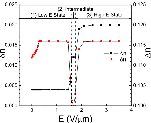
Fig. 2: (Colour on-line) Electric-field dependence of the biaxi-alityδn() and birefringence ∆n(•) at 95◦C measured in the SmAPA phase in 8.7µm homeotropic and 9.5µm planar cells, respectively; the frequency ofE is 100 Hz.
[image:4.595.303.548.82.282.2]Y. P. Panarinet al.
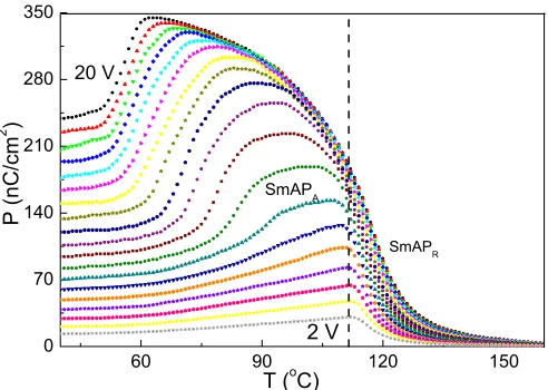
Fig. 3: (Colour on-line) Temperature dependence of the polar-ization measured in a 2µm planar cell for different applied volt-ages, from 2 V to 20 V, 100 Hz square wave with a step of 1 V.
are summarized in fig. 2. Three important features are being noted: i) a difference in the birefringence value (∆n) for high and low fields in a planar cell is equal to the biaxiality (δn) in a homeotropic cell; ii) for an intermediate E in the range (1.6–1.8 V/µm), the planar cell has an intermediate birefringence value, while biax-iality of the homeotropic cell drops to zero and iii) a difference in the birefringence values in the planar cell in between the intermediate and low/high field states is equal to one-half the value of the biaxiality in a homeotropic cell. The inter-relations between the measured birefringence values can be written as follows: ∆nH − ∆nL≈δnH≈
δnL≈0.016 and ∆nH − ∆nI≈∆nI − ∆nL≈δnL/2≈
0.008, where subscriptsL, H andI denote low, interme-diateand highelectric fields, respectively.
Before determining the physical mechanisms involved in such textural transformations, we present results of the net polarization,P (fig. 3). The important feature of this sample is the absence of P in the entire tempera-ture range when the field applied for its measurement is low. The application ofE above a threshold changes the distribution of the dipole moments (polar directors) and consequently gives rise to the net polarizationP. Figure 3 shows the temperature dependence of the measured polar-ization in a planar cell of thickness 2µm for both SmAPA and SmAPR phases. The polarization is measured using the integral current technique. In the higher temperature range of the SmAPR phase, we find that the induced polarization is linearly proportional to the applied field, which increases on cooling down to the SmAPAphase. It is observed thatP(T) increases on approaching the SmAPR -SmAPA phase transition. In the temperature range corre-sponding to the SmAPA phase, the net polarization is initially proportional to the applied field and then it jumps to a saturated value. The decrease in the polarization at lower temperatures is for the reason that much higher volt-age is needed to saturate it. It may be remarked that while
the field-induced polarization shows a smooth-like normal dependence onE, the biaxiality (fig. 2) shows a sharp and step-like behavior. This arises from the principal difference of the manner used in arriving at the results using the two techniques. The birefringence is measured for a localized small area in the cell, whereas the polarization is measured over the entire area of the cell. The latter may consist of three different domains shown in fig. 1(h) and hence the measured P corresponds to an integrated response over the entire area.
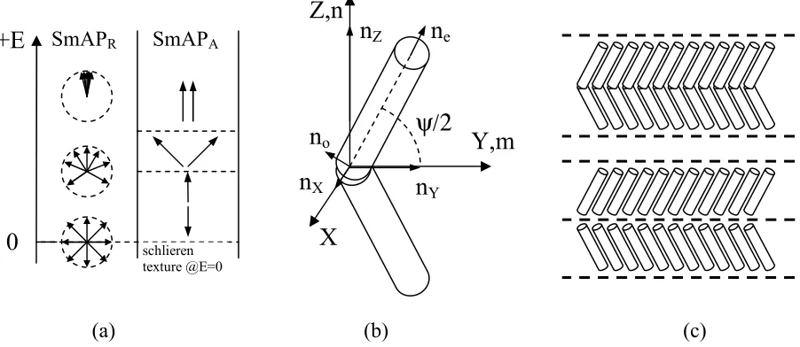
The suggested switching mechanisms in the sample under electric field are presented in fig. 4(a). In the SmAPR phase and in the absence of E across the cell, the dipoles and therefore the polar directors are randomly distributed as found in the literature [14,15]. On apply-ing an in-plane electric field, the dipoles gradually orient towards the field direction inducing macroscopic polar-ization and biaxiality; with the secondary optical axis of the polar directors in a homeotropic cell is directed along the applied field (fig. 1(b)). The distortion of the random distribution obeys the two-dimensional Langevin equation [15]. In the SmAPA phase, the short molecu-lar axes in single smectic layers are aligned simimolecu-larly but anti-parallelly in the neighboring layers thus forming an antiferroelectric structure. The response of this structure to the electric field is no longer continuous (Langevin-like) but shows three optically distinguishable states (fig. 1(h) and fig. 2). The application of a rather small electric field slightly distorts the initial antiferroelectric structure as shown in fig. 4(a) so that it gives rise to the measurable polarization. For higher fields, this initial antiferroelectric structure (fig. 1(d)) completely transforms into a ferro-electric (fig. 1(f)) structure. In the very narrow range of intermediate E, the homeotropic cell shows almost zero birefringence (fig. 1(e)) arising from zero biaxiality; this is only possible when the angle between the local polar-izations vectors (and therefore the polar directors) is 90◦ (fig. 4(a)). This state in the planar cell exhibits an inter-mediate birefringence value of 0.112, which lies exactly in between the birefringence values in the antiferroelectric and ferroelectric states (fig. 1(h)).
It is possible to estimate the birefringence as well as the biaxiality in different states for different alignment conditions. First, we introduce the biaxiality model of the orthogonal smectic layer formed by bent-shape mole-cules by two optically equivalent anticlinically tilted layers formed by rod-shaped molecules (i.e. fragments of bent-shaped molecules) with refractive indices ne (along the long fragment’s axis) and no, in each layer and with the tilt angle related to the opening angle (ψ) of the bent-shape molecules as shown in fig. 4(b), (c). This forms anisotropic bi-layered media with three distinct eigenvalues that can be labeled as refractive indices:
[image:5.595.42.288.83.258.2](a) (b)
(c)
Z,n
n
eψ/2
Y,m
X
n
Xn
Yn
Zn
o0
+E
SmAP
RSmAP
Aschlieren texture @E=0
Fig. 4: (a) Ordering and switching mechanisms in SmAPRand SmAPAphases with increase in fieldEin the vertical direction. The arrows represent the secondary molecular axes and local polarization vectors; (b) the biaxiality model for bent-core molecule. Z andY axes lie along the primary,n, and secondary,m, directors; (c) Model representation of one orthogonal bent-core smectic layer (upper diagram) by two optically equivalent tilted layers (lower diagram).
nX, nY and nZ can be estimated using the well-known
formula:n(θ) =none/[
n2
esin2(θ) +n2ocos2(θ)]. Here θis
an angle between the polarization of light and the opti-cal axis, ne and no are the refractive indices of the single layer, along and normal to the major axis. For the model proposed (fig. 4(b)): nZ=n(90−ψ/2), nY =
n(ψ/2),nX=no. Using this model, we find three different refractive indices values ∆nZX=nZ−nX; ∆nZY =nZ−
nY; ∆nYX=nY −nX; where ∆nZX−∆nZY = ∆nYX, the latter turns out to be the biaxiality (∆nYX=δn). In the homeotropic cell, both antiferroelectric and ferroelectric structures must have the same value of biaxiality, equal toδn= 0.016 (see fig. 1(d), (f) and fig. 2). For the planar cell in the absence or under lowE, the molecular planes are parallel to the surface and the birefringence must be equal to ∆nL= ∆nZY = 0.104. Under the high elec-tric field the molecular planes are perpendicular to the surface and the macroscopic birefringence must equal to ∆nH= ∆nZX= 0.12. According to the model proposed here, all three refractive indices depend on only two inde-pendent parameters:ne−no, ψ. By fitting this equation to the experimental data, these are found to be:ne−no= 0.14 andψ= 138◦. The opening angle,ψ= 138◦, obtained from fitting the data lies in the range 135–140◦ [19] and this agrees with that found by NMR experiments for systems similar to CK64. Also note that for intermedi-ateE, the macroscopic biaxiality in the homeotropic cell is observed to be zero and in the planar cell the birefrin-gence is found to be 0.112 (fig. 2). Finally, let us examine the possible application based on the electro-optic effect in a homeotropic cell to displays. The feasibility of the electro-optic effect in SmAPRwas studied in [14] showing several advantages over existing LCD modes, such as the
fast response (100µs), high contrast ratio (3000 : 1), wide viewing angle and continuous gray scale. Nevertheless, it requires a rather large fieldE (up to 10 V/µm) which for the homeotropic sample may reach 100 V or more. This voltage is also strongly temperature dependent (fig. 3). In contrast to SmAPR, the electro-optic effect in SmAPA requires much smaller applied voltage. The high contrast electro-optic switching can be achieved between the inter-mediate dark (fig. 1(e)) and bright high/low-voltage states (fig. 1(d)/(f)). The feasibility study of this effect is in progress.
In conclusion, the structural and electro-optical phenomena are investigated in an achiral biaxial bent-core molecular compound with SmAPA and SmAPR phases. The homeotropic cell in the SmAPA phase shows an unusual transition from an initially biaxial texture to a quasi-stable uniaxial one and then again to a biaxial structure upon the application of electric field. The last state (high E) has the same biaxiality as the initial one (low E) with high and low fields, respectively. These transformations are explained with successive transitions from antiferroelectric to ferroelectric states. In the inter-mediate state, a uniaxial structure is formed where the secondary directors in the neighboring smectic layers are perpendicular to each other. The biaxiality/birefringence calculated with the model for the bent-core molecules shows a good quantitative agreement with those obtained experimentally.
∗ ∗ ∗
[image:6.595.71.522.88.284.2]Y. P. Panarinet al.
REFERENCES
[1] Meyer R. B., Liebert L., Strzelecki L.andKeller P.,J. Phys.(Paris),36(1975) L69.
[2] Brand H. R., Cladis P. E. and Pleiner H.,
Macro-molecules,25(1992) 7223.
[3] Niori T., Sekine T., Watanabe J., Furukawa T.and Takezoe H.,J. Mater. Chem.,6(1996) 1231.
[4] Link D. R., Natale G., Shao R., Maclennan J. E., Clark N. A., Korblova E.andWalba D. M.,Science, 278(1997) 1924.
[5] Kruerke D. and Heppke G., presented at the Gordon
Conference on Liquid Crystals, 1997, New Hampshire; Banana-shaped Liquid Crystal Workshop, 1997, Berlin, Germany.
[6] Sekine T., Niori T., Watanabe J., Furukawa T., Choi S. W.andTakezoe H.,J. Mater. Chem.,7(1997) 1307; Sekine T., Niori T., Sone M., Watanabe J., Choi S. W., Takanishi Y. and Takezoe H., Jpn. J.
Appl. Phys.,36(1997) 6455.
[7] Pelzl G., Diele S. and Weissflog W., Adv. Mater., 11(1999) 707.
[8] Reddy A. R.andTschierske C.,J. Mater. Chem.,16 (2006) 907961.
[9] Ros M. B., Serrano J. L., de la Fuente M. R. and Folcia C. L.,J. Mater. Chem.,15(2005) 5093.
[10] Takezoe H.andTakanishi Y.,Jpn. J. Appl. Phys.,45 (2006) 597.
[11] Eremin A., Diele S., Pelzl G., Nadasi H., Weissflog W., Salfetnikova J.andKresse H.,Phys. Rev. E,64 (2001) 051707.
[12] Schroder M. W., Diele S., Pancenko N., Weissflog W.andPelzl G.,J. Mater. Chem.,12(2002) 1331.
[13] Pociecha D., Cepic M., Gorecka E. and
Mieczkowski J.,Phys. Rev. Lett.,91(2003) 185501. [14] Shimbo Y., Takanishi Y., Ishikawa K., Gorecka
E., Pociecha D., Mieczkowski J., Gomola K. and Takezoe H.,Jpn. J. Appl. Phys.,45(2006) L282. [15] Shimbo Y., Gorecka E., Pociecha D., Araoka F.,
Goto M., Takanishi Y., Ishikawa K., Mieczkowski J., Gomola K. and Takezoe H.,Phys. Rev. Lett., 97 (2006) 113901.
[16] Acharya B. R., Primak A. andKumar S.,Phys. Rev.
Lett.,92(2004) 145506.
[17] Nagaraj M., Panarin Y. P., Manna U., Vij J. K., Keith C. and Tschierske C., Appl. Phys. Lett., 96 (2010) 011106.
[18] Keith C., Prehm M., Panarin Y. P., Vij J. K. and Tschierske C.,Chem. Commun.,46(2010) 3702. [19] Wirth I., Diele S., Eremin A., Pelzl G., Grande
S., Kovalenko L., Panfenko N. andWeissflog W.,