Original Article
Significance of TLR4/MyD88 expression
in breast cancer
Xiangjin Chen1, Feng Zhao2, Huihao Zhang1, Youzhi Zhu1, Kunlin Wu1, Guozheng Tan2
1Department of Thyroid and Breast Surgery, First Affiliated Hospital of Fujian Medical University, China; 2Fujian Medical University Graduate School, China
Received April 17, 2015; Accepted May 29, 2015; Epub June 1, 2015; Published June 15, 2015
Abstract: Objective: To investigate the expression of TLR4/MyD88 in breast cancer, and explore the relationship be-tween their expression and breast cancer tumor growth and invasion. Methods: We examined the protein expression of TLR4 and MyD88 in 60 cases of histologically confirmed breast cancer. The relationship of their protein expres -sions with clinical features including age at diagnosis, tumor size and stage, lymph node metastasis and distant metastasis were analyzed. Results: The IHC results showed that TLR4 and MyD88 were expressed in 63.3% (38/60) and 58.3% (35/60) of malignant breast tumors respectively. TLR4 expression in breast cancer were significantly higher than in fibroadenoma (n = 4, 20.0%) and adjacent normal tissues (n = 2, 10.0%) (P < 0.001). MyD88 expres-sion in breast cancer were also significantly higher than in fibroadenoma (n = 4, 20.0%) and adjacent normal tissue (n = 3, 15.0%) (P < 0.001). The gene expressions of TLR4 and MyD88 were significantly higher in breast cancer than in fibroadenoma and adjacent normal tissues (P < 0.05). The protein expressions of TLR4 and MyD88 were also significantly associated with poor clinical features (P < 0.05). Conclusion: TLR4 and MyD88 expression might be associated with breast cancer growth and regional and distant metastases.
Keywords: Breast cancer, MyD88, TLR4
Introduction
Breast cancer is a heterogeneous malignant and life-threatening disease amongst females. Its incidence rate is increasing worldwide. According to the latest report of “GLOBOCAN 2008” by American Cancer Society [1], breast cancer is the most frequent cancer among women with an estimated 1.38 million new cases and 458,400 cases died of the disease. Most of breast cancer patients have prolonged disease-free survival and overall survival or even been cured after receiving systemic thera-py. Nevertheless, there are still some patients died of the disease due to development of che-moresistance, lack of therapeutic targets such as estrogen and HER-2 receptors in tumors. It is therefore important to study pathogenesis and biologic behavior of the malignant disease to help develop new treatment strategy and subsequently prolong survival and improve quality of life of patients.
Toll-like receptor 4 (TLR4) and myeloid
differen-tiation factor 88 (MyD88) specific binding plays
an important biological function in pathogene-sis by mediating tumor invasion and migration, escaping from immunosurveillence, promoting tumor proliferation, inhibiting apoptosis and developing chemoresistance in colorectal can-cer [2], ovarian cancan-cer [3], and prostate cancan-cer [4]. In breast cancer, lipopolysaccharide acting on downstream signaling molecules TLR4 and MyD88 could regulate the growth rate of tumor cells by reducing the expression of TLR4 or MyD88 molecules [5]. Downregulation of MyD88 expression or formation of MyD88 homodimerization with inhibitory peptitde could effectively reduce lung metastasis in breast cancer mouse model as well as decreased CCL2 and CCL5 expression. Yang et al [6] found that the expression of TLR4 was the highest among other toll-like receptors TLR1-TRL10 in human breast cancer cell line MDA-MB-231 and a dramatic reduction of breast cancer cell viability and subsequently decreased IL-6 and IL-8 levels were observed after knockdown of TLR4 gene in the cell line. At present, the
expression and significance of TLR4 and MyD88
action are not widely studied. In this study, we aimed to explore the potential prognostic val-ues of TLR4 and MyD88, and their involvement in malignant biological behavior in breast cancer.
Materials and methods
Patients and human tissues
A total 60 cases of histologically confirmed
breast cancer with 20 cases of matched
nor-mal adjacent tissues and 20 cases of fibroade
-noma were obtained from The First Affiliated
Hospital of Fujian Medical University, China between January 2008 and September 2012. Female patients diagnosed primary breast can-cer at AJCC stage I to III and underwent surgical resection of tumor were selected. Patients who were given neoadjuvant therapy and proven distant metastasis at presentation were exclud-ed. The study was approved by local ethics committee and informed consents were ob- tained for collection of tissue samples.
Immunohistochemistry
An immunohistochemical streptavidin-peroxi-dase (SP) method was used in this study. Mouse anti-human monoclonal antibodies TLR4 and MyD88 were purchased from Zhongshan Jinqiao Biotechnology Co., Ltd. (Beijing, China). All specimens were routinely
fixed with 10% formalin and embedded in par
-affin, and sectioned at a thickness of 2.5 μm.
The slides were incubated at 65°C overnight
and the tissue sections were deparaffinized
and hydrated. The antigens were recovered by natural cooling to room temperature. After rinsed with PBS, the specimen was treated with 3% hydrogen peroxide and incubated at room temperature for 10 mins. After rinsing samples with PBS for three times three minutes each, the slides were incubated for 1 hour with pri-mary antibodies (anti-TLR4 at 1:300 dilution and anti-MyD88 at 1:200 dilution). The slides were then rinsed with PBS for three times three minutes each followed by incubation with pri-mary reagent for another 30 minutes. Further rinsed with PBS for three times three minutes each, the slides were incubated with secondary reagent for another 20 minutes. The DAB stain-ing agent was added to samples stained for 3 minutes after rinsed with PBS for three times three minutes each and the sections were
counterstained with hematoxylin, dehydrated, transparent and mounted for storage. Known TLR4-positive sample obtained from mouse spleen and MyD88-positive sample obtained from papillary carcinoma of lung were used as positive control and the use of PBS without pri-mary antibody was used as negative control. RNA preparation and RT-PCR
RNA was isolated using Trizol (Beijing ComWin Biotech Co., Ltd., China) as per the instruction. The purity and concentration of the total extracted RNA was determined using UV spec-trophotometer (Nano Photometer, Germany) and the absorbance ratio of each sample at OD 260/280 was between 1.8 and 2.0. We applied Prime Premier 5.0 software to design the sequences of primers for TLR4, MyD88 and actin as internal control. The primers were syn-thesized by Sangon Biotech (Shanghai) Co., Ltd as follow: TLR4, 5’-GACCTGTCCCTGAACCCTAT- GA-3’ (upstream), 5’-CTTCTAAACCAGCCAGACC-
TTGA-3’ (downstream), amplified fragment size
of 139 bp; MyD88, 5’-CGGTCTCCTCCACATC- CTCCCTTCC-3’ (upstream), 5’-CTGCCAGTGGG-
GTCCGCTTGTGTCT-3’ (downstream), amplified
fragment size of 181 bp; actin, 5’-ACTTAGTT- GCGTTACACCCTT-3’ (upstream), 5’-GTCACCTT-
CACCGTTCCA-3’ (downstream), amplified frag
-ment size of 156 bp. The following conditions
were adopted for the PCR amplification:
pre-denaturation for 3 minutes at 94°C, denatur-ation for 30 seconds at 94°C, annealing for 30 seconds at 60°C and extension for 1 minute at 72°C. These steps were repeated for 35 cycles followed by a step of extension for 5 minutes at 72°C. The PCR products were separated by 1.5% agarose gel eletrophoresis, stained with GoldView nucleic acid dye and visualized by a gel HR camera (EC3 300, USA). The experiment was repeated for three times.
Immunohistochemistry analysis
The number of positively stained cells was eval-uated using a numeric score ranging from 0 to 4, representing the percentage of positively stained cells as follows: 0, less than 5%; 1, 5% to less than 25%; 2, 25% to less than 50%; 3, 50% to less than 75%; 4, greater than 75%. The immunostaining intensity was evaluated using
a numeric score ranging from 0 to 3, reflecting
the intensity as follows: 0, no staining; 1, weak staining (light yellow); 2, moderate staining
(yel-tics 19.0 (Chinese version). Chi-square test and Fisher’s exact test were used to compare the differences in expression of TLR4 and MyD88 and explore the relationship between their expressions and clinicopathological features of breast cancer. P-values of less than 0.05 were
considered statistically significant.
Results
Clinicopathological characteristics
Of 60 female primary breast cancer patients
aged 31-86 years (mean = 51.3 years, median = 50 years), all were diagnosed stage I to III
without distant metastasis at presentation. All patients underwent surgery including radical
mastectomy (n = 3) and modified radical mas
-tectomy (n = 57) followed by adjuvant therapy. Ninety-five percent of patients did not have
family history of malignant disease and 35 (58.3%) patients were premenopausal whereas 25 (41.7%) patients were postmenopausal.
Primary tumor sizes classified as T1, T2, T3 and
T4 were observed in 31 (26.7%), 66 (56.9%), 16 (13.8%) and 3 (2.6%) patients respectively. According to the AJCC TNM staging for breast cancer (version 7) [7], 41 (68.3%) and 19 (31.7%) patients were diagnosed stage I/II and stage III diseases respectively. Histological grading of tumor was based on Chinese Breast Cancer Diagnostic and Treatment Practices (2011 edition) [8] and 32 (53.3%) and 28
(46.7%) cases were classified as histological
grade I/II and grade III respectively. Figure 1. Overall survival of 60 breast cancer patients according to TLR4
expression.
low); and 3, intense staining (brownish yellow). The total score was obtained by multi-plying the score of percentage of positively stained cells and the intensity score. The total score of less than 6 repre-sented low level expression and that of greater or equal to 6 represented high level of expression.
Statistical analysis
All clinicopathological data, treatment history of individual patients, and results of sur-vival follow-up were compiled into a statistical database analyzed by IBM SPSS
[image:3.612.89.375.69.261.2] [image:3.612.91.289.311.535.2]Survival follow-up
All patients were followed up after surgery until December 2012 with a median follow-up dura-tion of 21 months. No patient defaulted follow-up and the survival data was retrieved at last follow-up visit. Of 60 breast cancer patients, 7 patients had distant metastasis among which lung metastasis was observed in 5 cases and bone metastasis was observed in 2 cases. One patient died of tumor progression. Survival
analysis stratified according to the expression
of TLR4 has shown that patients with low TLR4 expression have longer survival than that with high TLR expression in tumors (Figure 1).
Expression of TLR4 and MyD88 in breast can
-cer, adjacent normal tissue and fibroadenoma
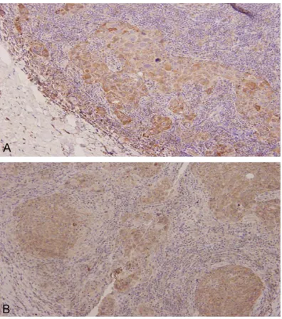
Protein expression of TLR4 detected by IHC: Positive IHC staining of TLR4 was mainly located at cytoplasm and some at the nuclei showing brownish yellow granules (Figure 2A). TLR4 expression was observed in 38 (63.3%), 2 (10.0%) and 4 (20.0%) cases of breast cancer,
adjacent normal tissue and fibroadenoma
respectively. The expression of TLR4 in ma-
lignant tumor was significantly higher than
adjacent normal tissues and benign tumor (P < 0.001) (Table 1).
Protein expression of MyD88 detected by IHC: Positive IHC staining of MyD88 was also located at cytoplasm showing brownish yellow granules (Figure 2B). MyD88 expression was observed in 35 (58.3%), 3 (15.0%) and 4 (20.0%) cases of breast cancer, adjacent normal tissue and
fibroadenoma respectively. Its expression was also significantly higher in malignant tumor
than adjacent normal tissues and benign tumor (P < 0.001) (Table 1).
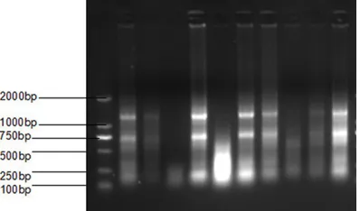
Gene expression of TLR4 and MyD88 detected by RT-PCR: The expression of TLR4 and MyD88 genes were comparatively stronger in breast cancer tissues than adjacent normal tissues and benign tumor (Figure 3).
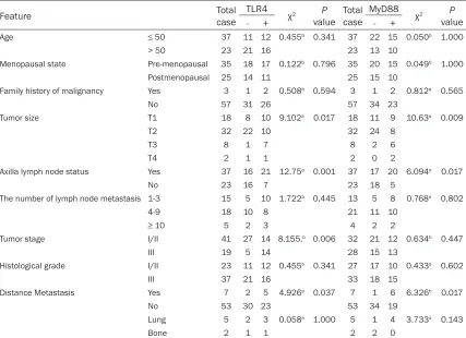
Association between TLR4 and MyD88 protein expressions and clinical parameters in breast
cancer: High expression of TLR4 was signi-
ficantly associated with axillary lymph node metastasis (P = 0.006), tumor size (P = 0.017), tumor staging (P = 0.013) and distant me-tastasis (P = 0.038), and high expression of MyD88 was also significantly associated with tumor size (P = 0.009), tumor staging (P = 0.03), axillary lymph node metastasis (P = 0.006) and distant metastasis (P = 0.004)
(Table 2).
Discussion
Toll-like receptors (TLRs) belong to a class of innate immune receptors commonly found in mammals and form a family of TLRs because of their similar homology. TLRs have emerged as a family of pattern recognition receptors (PRRs) able to recognize a variety of pathogen-associ-ated molecular patterns and interact with other families of PRRs which triggers a series of sig-nal transduction leading to the release of
inflammatory mediators as part of important
innate immunity and ultimately activation of the acquired immune system. To date, there are at least 12 different TLRs which possess different ligands and among which TLR4 is the major receptor mediated by lipopolysaccharide [9].
TLR-4 was first discovered by Medzhitov et al
[10] on extracellular domain of human cells which were mostly immune cells to identify pathogens, endogenous ligands and drug and involve in innate and adaptive immune respons-es. In recent years, TLR4 expression was also
observed in different human tumors. The first identified effector actively involved in TLR4 acti -vation and subsequent signal transduction was TIR-containing adaptor MyD88.
[image:4.612.91.290.73.190.2]Our study demonstrated that TLR4 and MyD88 signaling factors were highly active and strongly expressed in breast cancer as seen with high Figure 3. Agarose gel electrophoresis of total RNA
protein and gene expressions. However, their expressions were both lower in adjacent nor-mal tissues and benign breast tumors. Despite the unclear causal relationship between the expression of TLR4 and MyD88 and the tumor progression at this stage, we at least recog-nized a strong association between the signals and tumor growth. In our study, high expression of TLR4 was found in patients with positive axil-lary lymph node metastasis, which is similar to a study by Ehsan et al [11] that high tumoral
expression of TLR4 was significantly associat
-ed with local metastasis. The possible prognos-tic indication was also observed with poor sur-vival in patients with high expression of TLR4 expression. However, its correlation with
sur-vival was not observed in another study by Petricevic et al [12]. Our study provided us with preliminary results on the possible involvement of TLR4 and MyD88 signaling factors in patho-genesis of breast cancer. The upstream signal-ing factors might activate the downstream sig-naling factors via TLR4 and MyD88 to activate other signaling pathways. However, further investigation is required to explore the role of these signaling factors in tumor formation, development and progression.
Conclusion
[image:5.612.92.522.85.152.2]It is now more evident that TLR4 and MyD88 are one of the major signaling factors involved
Table 1. Expression of TLR4 and MyD88 in breast cancer, adjacent normal tissue and fibroadenoma
Group No. of cases TLR4 χ2 P value MyD88 χ2 P value
Positive Negative Positive Negative
Breast cancer 60 28 32 89.62 < 0.001 25 35 84.71 < 0.001
Adjacent normal tissue 20 2 18 3 17
Fibroadenoma 20 4 16 4 16
Table 2.Expression of TLR4 and MyD88 in breast cancer and its relationship with clinicopathological characteristics
Feature Total case TLR4 χ2 P
value Total case MyD88
χ2 P
value
- + - +
Age ≤ 50 37 11 12 0.455b 0.341 37 22 15 0.050b 1.000
> 50 23 21 16 23 13 10
Menopausal state Pre-menopausal 35 18 17 0.122b 0.796 35 20 15 0.049b 1.000
Postmenopausal 25 14 11 25 15 10
Family history of malignancy Yes 3 1 2 0.508b 0.594 3 1 2 0.812a 0.565
No 57 31 26 57 34 23
Tumor size T1 18 8 10 9.102a 0.017 18 11 9 10.63a 0.009
T2 32 22 10 32 24 8
T3 8 1 7 8 2 6
T4 2 1 1 2 0 2
Axilla lymph node status Yes 37 16 21 12.75a 0.001 37 17 20 6.094a 0.017
No 23 16 7 23 18 5
The number of lymph node metastasis 1-3 15 5 10 1.722a 0.445 13 5 8 0.768a 0.802
4-9 18 10 8 21 11 10
≥ 10 5 2 3 4 2 2
Tumor stage I/II 41 27 14 8.155.b 0.006 32 21 12 0.634b 0.447
III 19 5 14 28 15 13
Histological grade I/II 23 11 12 0.455b 0.341 27 17 10 0.433a 0.602
III 37 21 16 33 18 15
Distance Metastasis Yes 7 2 5 4.926a 0.037 7 1 6 6.326b 0.017
No 53 30 23 53 34 19
Lung 5 2 3 0.058a 1.000 5 1 4 3.733a 0.143
Bone 2 1 1 2 2 0
[image:5.612.93.520.199.509.2]in carcinogenesis and pathogenesis of various types of cancer as well as breast cancers. TLR4 and MyD88 expressions might be associated with breast tumor growth and progression. Further investigation on their involvement in breast cancer pathogenesis is warranted.
Acknowledgements
This study was funded by the young teacher education research projects in Fujian (JB13392), Key Clinical Specialty Discipline Construction Program of Fujian, People’s Republic of China and Fujian medical universi-ty professor development fund (JS14021).
Disclosure of conflict of interest
None.
Address correspondence to: Dr. Xiangjin Chen, Department of Thyroid and Breast Surgery, First Affiliated Hospital of Fujian Medical University, 20 Cha Zhong Road, Tai Jiang District, Fu Zhou 350005, Fujian Province, China. Tel: +86-13600865958; E- mail: xiangjinchen2014@yeah.net
References
[1] Jemal A, Bray F, Center MM, Ferlay J, Ward E, Forman D. Global cancer statistics. CA Cancer J Clin 2011; 61: 69-90.
[2] Wang EL, Qian ZR, Nakasono M, Tanahashi T, Yoshimoto K, Bando Y, Kudo E, Shimada M, Sano T. High expression of Toll-like receptor 4/ myeloid differentiation factor 88 signals corre-lates with poor prognosis in colorectal cancer. Br J Cancer 2010; 102: 908-915.
[3] Wang AC, Su QB, Wu FX, Zhang XL, Liu PS. Role of TLR4 for paclitaxel chemotherapy in human epithelial ovarian cancer cells. Eur J Clin Invest 2009; 39: 157-164.
[4] Hua D, Liu MY, Cheng ZD, Qin XJ, Zhang HM, Chen Y, Qin GJ, Liang G, Li JN, Han XF, Liu DX.
Small interfering RNA-directed targeting of Toll-like receptor 4 inhibits human prostate cancer cell invasion, survival, and tumorigenicity. Mol Immunol 2009; 46: 2876-2884.
[5] Egunsola AT, Zawislak CL, Akuffo AA, Chalmers SA, Ewer JC, Vail CM, Lombardo JC, Perez DN, Kurt RA. Growth, metastasis, and expression of CCL2 and CCL5 by murine mammary carci-nomas are dependent upon Myd88. Cell Immunol 2012; 272: 220-229.
[6] Yang H, Zhou H, Feng P, Zhou X, Wen H, Xie X, Shen H, Zhu X. Reduced expression of Toll-like receptor 4 inhibits human breast cancer cells proliferation and inflammatory cytokines se -cretion. J Exp Clin Cancer Res 2010; 29: 92. [7] Edge SB, Compton CC. The American Joint
Committee on Cancer: the 7th edition of the AJCC cancer staging manual and the future of TNM. Ann Surg Oncol 2010; 17: 1471-1474. [8] Breast cancer diagnostic and treatment
prac-tices (2011 edition). Chinese Journal of Practical Surgery 2011; 10: 902-907.
[9] Jun LB, Zhang HY, W XH. TLR4 protein expres-sion in breast cancer tissue and its relation-ship with apoptosis and proliferation. Gener- al Surgery (Electronic Version) 2011; 05: 211-213.
[10] Medzhitov R, Preston-Hurlburt P, Janeway CA Jr. A human homologue of the Drosophila Toll protein signals activation of adaptive immuni-ty. Nature 1997; 388: 394-397.
[11] Ehsan N, Murad S, Ashiq T, Mansoor MU, Gul S, Khalid S, Younas M. Significant correlation of TLR4 expression with the clinicopathological features of invasive ductal carcinoma of the breast. Tumour Biol 2013; 34: 1053-1059. [12] Petricevic B, Vrbanec D, Jakic-Razumovic J,