Original Article
Clinicopathological characteristics and outcomes of
patients with ANCA-positive lupus nephritis: a large
cohort study from a single Chinese center
Xiaojiang Zhan,Qinghua Liu, Li Fan, Lingyao Hong, Qian Zhou, Wei Chen, Xueqing Yu
Department of Nephrology, The First Affiliated Hospital of Sun Yat-sen University, Key Laboratory of Nephrology, Ministry of Health, Guangzhou, China
Received October 21, 2015; Accepted December 22, 2015;Epub February 1, 2016; Published February 15, 2016
Abstract: Very limited clinicopathological and clinical outcome data are available regarding lupus nephritis (LN) patients with antineutrophil cytoplasmic antibodies (ANCAs). The present study therefore investigated such data in a large cohort of LN patients in China. 478 patients with renal biopsy-proven lupus nephritis diagnosed in The First Affiliated Hospital of Sun Yat-sen University from January 1998 to December 2011 were enrolled. The primary endpoint was the composite of a doubling of baseline serum creatinine, end-stage renal disease, or death. The prevalence of ANCA seropositivity in this cohort was 14.0% (67/478). At the time of biopsy,ANCA-positive patients had higher CRP levels, higher ESR values, and were more frequently positive for anti-cardiolipin antibodies. How-ever, they had lower eGFR values, and lower levels of HDL-C, LDL-C, and hemoglobin when compared with ANCA-negative patients. ANCA-positive patients had a higher prevalence of 2003 ISN/RPS class IV LN, and higher scores for karyorrhexis/fibrinoid necrosis, cellular glomerular crescents, interstitial inflammation, and tubular atrophy. At a median follow-up time of 56 months (range, 3-162 months), the event-free survival rate for a composite outcome was significantly lower among patients with ANCAs than patients without (P = 0.015). Multivariate Cox regression analysis revealed that initial serum creatinine and CI score were the most important risk factors significantly af-fecting the primary outcome. Notably, ANCA was not independently associated with the primary outcome. Patients with ANCA-positive LN have more severe kidney injury and a worse long-term outcome compared with patients with ANCA-negative LN.
Keywords: Antineutrophil cytoplasmic antibodies, lupus nephritis, clinical outcomes
Introduction
Renal involvement significantly contributes to the morbidity and mortality among patients with systemic lupus erythematosus (SLE) [1, 2]. Moreover, the clinical manifestations of SLE and various histopathological characteristics of its associated renal lesions are closely related to the therapeutic responses and clinical out-comes achieved when treating patients with lupus nephritis (LN) [3], which is the most prev-alent pathologic type (54.3%) of secondary glo-merulonephritis diagnosed in China [4].
Antineutrophil cytoplasmic antibodies (ANCAs) constitute a class of autoantibodies directed against the cytoplasmic constituents of primary granules often found in the lysosomes of neu-trophils and monocytes [5]. Davies et al. [6] first described ANCAs while conducting studies
large cohort of ANCA-positive LN patients in China.
Patients and methods
The clinical data, pathological features, and clinical outcomes of 478 patients with renal biopsy-proven lupus nephritis diagnosed at The First Affiliated Hospital of Sun Yat-sen University between January 1998 and December 2011 were retrospectively analyzed for this study. Additionally, renal histopathology data were re- viewed and reclassified according to the In- ternational Society of Nephrology and Renal Pathology Society (ISN/RPS) 2003 classifica-tion [18].
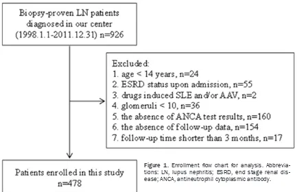
Each study patient satisfied the revised crite- ria for SLE developed in 1997 by the American College of Rheumatology [19]. The study exclu-sion criteria were as follows: (1) age < 14 years; (2) dialysis dependence upon admission; (3) dr- ugs (such as hydralazine, propylthiouracil or th- ioridazine etc.) induced SLE and/or AAV; (4) the absence of ANCAs test results; (5) glomeruli < 10; (6) the absence of up data or follow-up time shorter than 3 months. The flow chart of this study is shown in Figure 1. The conduct of this study complied with ethical principles outlined in the Helsinki Declaration, and the
Human Ethics Committees of Sun Yat-sen Uni- versity reviewed and approved the study proto-col. All study participants provided a signed written Informed Consent prior to enrollment. Clinical data and laboratory tests
Baseline demographic and clinical data for each patient were collected immediately prior to biopsy. Each patient’s estimated glomerular filtration rate (eGFR) was calculated using the CKD-EPI equation [20]. Disease activity was classified according to guidelines in the Sy- stemic Lupus Erythematosus Disease Activity Index (SLEDAI) [21].
[image:2.612.90.523.70.350.2]ANCAs testing were conducted with an indire- ct immunofluorescence (IIF) assay and/or an enzyme-linked immunosorbent assay (ELISA). Standard IIF assays were performed in accor-dance with instructions provided by the manu-facturer of the assay kit (EUROIMMUN; Lübeck, Germany). The human neutrophil ANCAs anti-gens proteinase 3 (PR3) and myeloperoxidase (MPO) were purified as previously reported [13], and used as solid-phase ligands in the antigen-specific ELISAs. A subset of the patients en- rolled in this study had been tested for ANCAs only with either an IIF assay or ELISA rather than with both assays. Therefore, patients who
tested positive for ANCAs with either type of assay were classified ANCA positive. Further- more, it has been reported that ~5% of serum samples are positive by ELISA only [5].
Renal histopathology
Each renal biopsy specimen was examined by both light microscopy and direct immunofluo- rescence.
Light microscopy examination
Renal biopsy specimens for light microscopy were fixed in 4% buffered formaldehyde, serial-ly cut into consecutive 2 µm sections, and th- en stained with either hematoxylin and eosin (H&E), periodic acid-Schiff, silver methenami- ne, or Masson’s trichrome. Next, renal patholo-gists evaluated the stained sections for various pathological parameters including activity indi-ces (AIs) and chronicity indiindi-ces (CIs); after whi- ch, specific biopsy features were semi-quanti-tatively scored as described in previous studies [13, 22, 23].
Direct immunofluorescence examination The intensity of immunofluorescence indicating immunoglobulin deposition (IgG, IgA, IgM, C3 or C1q) or the presence of fibrin deposits was semi-quantitatively graded on a scale of 0 to 4+ [24].
Patient follow-up and clinical outcomes
All enrolled patients participated in follow-up visits until one of the following events occurred: (1) doubling of baseline serum creatinine (D- SCr), (2) progression to ESRD, (3) all-cause de- ath or (4) the date of December 31, 2012. If patients were lost to follow-up, they were fol-lowed until the last recorded visit. The primary end point was the composite of D-SCr, the onset of ESRD, or patient mortality. ESRD was defined as the initiation of maintenance dialy-sis or renal transplantation [25, 26]. Survival time was defined as the time elapsed between a patient’s enrollment in the study and the start of D-SCr, permanent dialysis, a renal transplan-tation, the occurrence of patient death or the date of the last follow-up; whichever occurred first.
Statistical analysis
All study data were analyzed using SPSS for Windows, Version 13.0. Chicago, IL: SPSS Inc.
Continuous variables are expressed as the mean ± standard deviation (SD) or a median value with the interquartile range. Categorical data are presented as the frequency and per-centage. Clinical, laboratory, and pathology da- ta were compared using the Student’s t-test, non-parametric Mann-Whitney test or chi-sq- uare test.
A Kaplan-Meier survival curve was created and used to calculate the cumulative event-free sur-vival rates, and the log-rank test was used to compare survival differences. Risk factors wh- ich might affect clinical outcomes were evalu-ated using univariate analysis followed by a multivariate Cox proportional hazards regres-sion model. The following variables were exam-ined in the multivariate analysis: ANCA, sex, age, SLEDAI, systolic blood pressure, 24-h uri- ne protein, hemoglobin, creatinine, HDL-C, C-re- active protein, AI score, CI score, and immuno-suppressive therapy. The results of these re- gression analyses are expressed as hazard ratios (HRs) with 95% confidence intervals (CIs). Two-tailed P-values < 0.05 were considered statistically significant.
Results
Baseline patient demographic and clinical characteristics
Each patient in the ANCA-positive group was identified by an ANCA positive ELISA or IIF test result. Nearly all ANCA-positive patients (64/ 67) had been tested for ANCAs with an IIF assay, and the results showed that 60.9% (39/64) were p-ANCA positive, 12.5% (8/64) were c-ANCA positive, 4.7% (3/64) were bo- th p-ANCA and c-ANCA positive, and 21.9% (14/64) were IIF negative. Among patients in the ANCA-positive cohort, 79.1% (53/67) had been tested by ELISA, and those results showed that 37.7% (20/53) were MPO-ANCA positive, 15.1% (8/53) were PR3-ANCA positive, 24.5% (13/53) were both MPO-ANCA and PR3-ANCA positive, and 22.7% (12/53) were ELISA nega-
Comparison of baseline characteristics of pa-tients with ANCA-negative and ANCA-positive LN
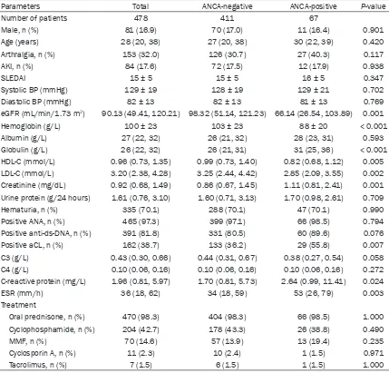
Table 1 shows clinical characteristics of the 478 patients with LN after they were divided into two groups consisting of ANCA-negative and ANCA-positive patients. At the time of renal biopsy, patients in the ANCA-positive groups had higher levels of serum creatinine, globulin, and C-reactive protein (CRP), higher ESR val-ues, and were more likely to test positive for anticardiolipin (aCL) antibodies. However, the ANCA-positive patients had lower eGFR values, and lower levels of high-density lipoprotein cho-Table 1. Comparison of baseline clinical characteristics of patients with ANCA-negative or ANCA-posi-tive lupus nephritis
Parameters Total ANCA-negative ANCA-positive P-value
Number of patients 478 411 67
Male, n (%) 81 (16.9) 70 (17.0) 11 (16.4) 0.901
Age (years) 28 (20, 38) 27 (20, 38) 30 (22, 39) 0.420
Arthralgia, n (%) 153 (32.0) 126 (30.7) 27 (40.3) 0.117
AKI, n (%) 84 (17.6) 72 (17.5) 12 (17.9) 0.938
SLEDAI 15 ± 5 15 ± 5 16 ± 5 0.347
Systolic BP (mmHg) 129 ± 19 128 ± 19 129 ± 21 0.702
Diastolic BP (mmHg) 82 ± 13 82 ± 13 81 ± 13 0.769
eGFR (mL/min/1.73 m2) 90.13 (49.41, 120.21) 98.32 (51.14, 121.23) 66.14 (26.54, 103.89) 0.001
Hemoglobin (g/L) 100 ± 23 103 ± 23 88 ± 20 < 0.001
Albumin (g/L) 27 (22, 32) 26 (21, 32) 28 (23, 31) 0.593
Globulin (g/L) 26 (22, 32) 26 (21, 31) 31 (25, 36) < 0.001
HDL-C (mmol/L) 0.96 (0.73, 1.35) 0.99 (0.73, 1.40) 0.82 (0.68, 1.12) 0.005
LDL-C (mmol/L) 3.20 (2.38, 4.28) 3.25 (2.44, 4.42) 2.85 (2.09, 3.55) 0.002
Creatinine (mg/dL) 0.92 (0.68, 1.49) 0.86 (0.67, 1.45) 1.11 (0.81, 2.41) 0.001
Urine protein (g/24 hours) 1.61 (0.76, 3.10) 1.60 (0.71, 3.13) 1.70 (0.98, 2.61) 0.709
Hematuria, n (%) 335 (70.1) 288 (70.1) 47 (70.1) 0.990
Positive ANA, n (%) 465 (97.3) 399 (97.1) 66 (98.5) 0.794
Positive anti-ds-DNA, n (%) 391 (81.8) 331 (80.5) 60 (89.6) 0.076
Positive aCL, n (%) 162 (38.7) 133 (36.2) 29 (55.8) 0.007
C3 (g/L) 0.43 (0.30, 0.66) 0.44 (0.31, 0.67) 0.38 (0.27, 0.54) 0.058
C4 (g/L) 0.10 (0.06, 0.16) 0.10 (0.06, 0.16) 0.10 (0.06, 0.16) 0.272
C-reactive protein (mg/L) 1.96 (0.81, 5.97) 1.70 (0.81, 5.73) 2.64 (0.99, 11.41) 0.024
ESR (mm/h) 36 (18, 62) 34 (18, 59) 53 (26, 79) 0.003
Treatment
Oral prednisone, n (%) 470 (98.3) 404 (98.3) 66 (98.5) 1.000
Cyclophosphamide, n (%) 204 (42.7) 178 (43.3) 26 (38.8) 0.490
MMF, n (%) 70 (14.6) 57 (13.9) 13 (19.4) 0.235
Cyclosporin A, n (%) 11 (2.3) 10 (2.4) 1 (1.5) 0.971
Tacrolimus, n (%) 7 (1.5) 6 (1.5) 1 (1.5) 1.000
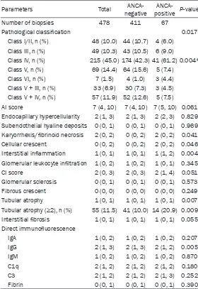
[image:4.612.92.521.97.508.2]terol (LDL-C), and hemoglobin when compared with the ANCA-negative LN patients. Additi- onally, ANCA-positive patients were more likely to present with Class IV lupus nephritis (42.3% versus 61.2%, P = 0.004), and had higher scores for karyorrhexis/fibrinoid necrosis, cel-lular glomerular crescents, interstitial inflam-mation, and tubular atrophy (P = 0.041, P = 0.046, P = 0.004, and P = 0.007, respectively). The two groups showed no significant differ-ences regarding other pathological features as determined by light microscopic examinations. Additionally, with the exception of slightly less intense IgG staining in the ANCA-positive group (P = 0.005), there was no significant difference
ANCA-positive group, 10 patients died; among whom, 4 died of CVD events, and 6 died of unknown reasons; while 6 reached ESRD and 2 reached D-SCr.
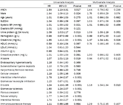
[image:5.612.89.369.96.503.2]Univariate survival analysis was used to evalu-ate factors that may affect composite outcome in all the patients with lupus nephritis. We found that ANCA was a risk factor of reaching the composite outcomes in lupus nephritis (HR, 1.93; 95% CI, 1.13-3.31; P = 0.017), other uni-variate risk factors included male gender (HR, 1.84; 95% CI, 1.09-3.13; P = 0.024), systolic blood pressure (HR, 1.01; 95% CI, 1.00-1.02; P = 0.021), 24-h urine protein (HR, 1.09; 95% CI, Table 2. Comparison of renal pathological data from groups of
patients with ANCA-negative or ANCA-positive lupus nephritis
Parameters Total negativeANCA- positiveANCA- P-value
Number of biopsies 478 411 67
Pathological classification 0.017
Class I/II, n (%) 48 (10.0) 44 (10.7) 4 (6.0)
Class III, n (%) 49 (10.3) 43 (10.5) 6 (9.0)
Class IV, n (%) 215 (45.0) 174 (42.3) 41 (61.2) 0.004*
Class V, n (%) 69 (14.4) 64 (15.6) 5 (7.4)
Class VI, n (%) 7 (1.5) 4 (1.0) 3 (4.4)
Class V + III, n (%) 33 (6.9) 30 (7.3) 3 (4.5) Class V + IV, n (%) 57 (11.9) 52 (12.6) 5 (7.5)
AI score 7 (4, 10) 7 (4, 10) 7 (5, 10) 0.061
Endocapillary hypercellularity 2 (1, 3) 2 (1, 3) 2 (2, 3) 0.829 Subendothelial hyaline deposits 0 (0, 1) 0 (0, 1) 0 (0, 1) 0.969 Karyorrhexis/fibrinoid necrosis 2 (0, 2) 0 (0, 2) 2 (0, 2) 0.041
Cellular crescent 0 (0, 2) 0 (0, 2) 2 (0, 2) 0.046
Interstitial inflammation 1 (0, 1) 1 (0, 1) 1 (1, 2) 0.004 Glomerular leukocyte infiltration 1 (0, 2) 1 (0, 2) 1 (0, 1) 0.345
CI score 2 (0, 3) 2 (0, 3) 2 (1, 4) 0.051
Glomerular sclerosis 0 (0, 1) 0 (0, 1) 0 (0, 1) 0.573
Fibrous crescent 0 (0, 0) 0 (0, 0) 0 (0, 0) 0.249
Tubular atrophy 1 (0, 1) 1 (0, 1) 1 (0, 1) 0.007
Tubular atrophy (≥2), n (%) 55 (11.5) 41 (10.0) 14 (20.9) 0.009
Interstitial fibrosis 1 (0, 1) 1 (0, 1) 1 (0, 1) 0.055
Direct immunofluorescence
IgA 1 (0, 2) 1 (0, 2) 1 (0, 2) 0.207
IgG 2 (1, 3) 2 (1, 3) 2 (1, 2) 0.005
IgM 1 (0, 2) 1 (0, 2) 1 (0, 2) 0.870
C1q 2 (1, 2) 2 (1, 2) 2 (1, 2) 0.180
C3 2 (1, 2) 2 (1, 2) 2 (1, 3) 0.252
Fibrin 0 (0, 1) 0 (0, 1) 0 (0, 1) 0.390
Notes: Data are presented as number (%) and/or median (25th, 75th); *compari-son of Class IV; AI, activity indices; CI, chronicity indices.
between the two groups re- garding the majority of their immune deposits. The renal hi- stopathological characteristics of ANCA positive and negative patients are shown in Table 2. Treatment and clinical out-comes
1.02-1.17; P = 0.013), baseline serum creati-nine concentration (HR, 1.65; 95% CI, 1.41-1.93; P < 0.001), AI score (HR, 1.07; 95% CI, 1.01-1.13; P = 0.019), cellular crescent (HR, 1.19; 95% CI, 1.05-1.36; P = 0.006), interstitial inflammation (HR, 1.78; 95% CI, 1.34-2.37; P < 0.001), CI score (HR, 1.24; 95% CI, 1.13-1.36; P < 0.001), glomerular sclerosis (HR, 1.60; 95% CI, 1.24-2.07; P < 0.001), tubular atrophy (HR, 1.77; 95% CI, 1.34-2.35; P < 0.001), and inter-stitial fibrosis (HR, 1.74; 95% CI, 1.33-2.27; P < 0.001). Higher hemoglobin was a protective factor (HR, 0.98; 95% CI, 0.97-0.99; P < 0.001). However, on multivariate regression, only base-line serum creatinine concentration (HR, 1.37; 95% CI, 1.04-1.81; P = 0.028) and CI score (HR, 1.18; 95% CI, 1.04-1.34; P = 0.010) were inde-pendently associated with primary outcome events. ANCA was not found to be a risk factor for composite outcome on multivariate Cox regression (Table 3).
Discussion
In this present study, we reported the preva-lence of ANCA-positivity in one Chinese LN cohort and identified certain characteristics in LN patients that were associated with ANCA seropositivity. Furthermore, the ANCA-positive LN patients were shown to have a poor progno-sis; e.g., ~60.1% of ANCA seropositive patients
positivity might result from the following fac-tors: (1) we excluded patients who were treated by chronic dialysis upon their first admission; (2) we excluded patients who did not have any follow-up data; (3) the different sample sizes and patient populations in the studies may have affected their results. Various studies have provided conflicting ideas concerning the clinical implications of ANCAs being detected in SLE patients. While the results of some investi-gations have suggested that ANCAs, disease activity, and organ involvement are all associ-ated [11, 15, 27, 28], other studies have failed to show such associations [16, 29]. Manolova et al. [27]found a positive correlation between the presence of BPI-, LF-, and PR3-ANCAs and disease activity, as well as an association between PR3-ANCAs and arthritis. A study by Chin et al. [15]of 59 patients with SLE in South Korea revealed an association between ANCA seropositivity and the presence of both nephri-tis and anti-ds-DNA antibodies. Additionally, a study by Lee et al. [28]of 79 SLE patients in Hong Kong found that LF-ANCA positivity was correlated with disease activity, lymphadenop-athy, and crescentic glomerulonephritis. Me- anwhile, Galeazzi et al. [11] studied a large cohort of SLE patients from 11 European cen-ters and found a positive correlation between the presence of ANCAs detected by IIF and vari-ous disorders including serositis, live do
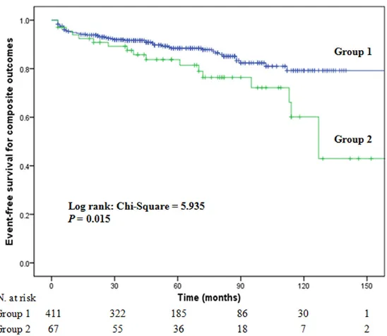
reticu-Figure 2. Kaplan-Meier plots for composite outcomes of LN patients with (group 2) and without (group 1) ANCAs.
[image:6.612.96.370.70.308.2]ANCA-laris, venous thrombosis, and arthritis. How- ever, other autoantibodies such as aCL and anti-SSA/Ro showed stronger correlations than ANCAs with the same clinical disorders. Mo- reover, other studies have failed to identify any correlation between ANCAs and either disease activity or organ involvement [16, 29]. In our current study, we failed to identify a significant difference between positive and ANCA-negative LN patients regarding the majority of their clinical manifestations and laboratory test results. However, we did find that ANCA-positive LN patients had higher rates of seropositivity for aCL antibodies, as well as higher ESR values and levels of CRP; however, they had lower val-ues for eGFR and lower levels of hemoglobin. These findings suggest that the presence of ANCAs might be associated with the active
phases of lupus nephritis and higher degrees of kidney damage. A previous study showed an association between ANCAs and the presence of diffuse proliferative LN (WHO class IV) [15], as well as crescent formation in patients with WHO class IV glomerulonephritis [28]. In agree-ment with those results, our current study also found a significantly higher proportion of 2003 ISN/RPS class IV LN in an ANCA-positive group, as well as higher scores for karyorrhexis/fib- rinoid necrosis, cellular crescents, interstitial inflammation, and fewer IgG deposits in that group. These findings suggest that ANCA sero-positivity in LN patients may represent more than the coincidence. Nasr et al. [30] proposed that either one of the two conditions (lupus nephritis or ANCA seropositivity) may facilitate development of the second. They also pro-Table 3. Results of Cox proportional analyses for composite outcomes of patients with lupus nephritis
Univariate Analysis Multivariate Analysis
HR 95% CI P-value HR 95% CI P-value
ANCA 1.93 1.13-3.31 0.017 1.44 0.79-2.62 0.238
Male 1.84 1.09-3.13 0.024 1.76 0.95-3.29 0.074
Age (years) 1.01 0.99-1.03 0.275 1.01 0.98-1.03 0.682
SLEDAI 1.04 0.99-1.09 0.097 1.03 0.97-1.09 0.329
Systolic BP (mmHg) 1.01 1.00-1.02 0.021 1.01 0.99-1.02 0.456
Diastolic BP (mmHg) 1.01 1.00-1.03 0.121
Urine protein (g/24 hours) 1.09 1.02-1.17 0.013 1.09 1.00-1.19 0.051
Hemoglobin (g/L) 0.98 0.97-0.99 < 0.001 0.99 0.97-1.00 0.110
Creatinine (mg/dL) 1.65 1.41-1.93 < 0.001 1.37 1.04-1.81 0.028
HDL-C (mmol/L) 0.59 0.33-1.04 0.067 0.74 0.38-1.46 0.391
LDL-C (mmol/L) 1.04 0.91-1.20 0.544
Albumin (g/L) 0.98 0.94-1.01 0.158
C-reactive protein (mg/L) 1.01 1.00-1.03 0.091 1.00 0.99-1.02 0.805
AI score 1.07 1.01-1.13 0.019 0.94 0.87-1.02 0.112
Endocapillary hypercellularity 1.16 0.84-1.60 0.368
Subendothelial hyaline deposits 1.10 0.78-1.55 0.580
Karyorrhexis/fibrinoid necrosis 1.03 0.88-1.20 0.736
Cellular crescent 1.19 1.05-1.36 0.006
Interstitial inflammation 1.78 1.34-2.37 < 0.001
Glomerular leukocyte infiltration 1.15 0.87-1.51 0.329
CI score 1.24 1.13-1.36 < 0.001 1.18 1.04-1.34 0.010
Glomerular sclerosis 1.60 1.24-2.07 < 0.001
Fibrous crescent 1.09 0.59-2.02 0.776
Tubular atrophy 1.77 1.34-2.35 < 0.001
Interstitial fibrosis 1.74 1.33-2.27 < 0.001
Immunosuppressive therapy 1.11 0.66-1.86 0.694 1.29 0.71-2.35 0.407
[image:7.612.91.522.84.452.2]posed that ANCA-associated necrotizing and crescentic glomerulonephritis may occur super-imposed on lupus nephritis [30].
While there are no established guidelines for treating ANCA-positive lupus nephritis, the cur-rent regimen used for treating AAV is similar to that used for treating proliferative lupus nep- hritis, and has components of both initial and maintenance therapy [31]. In cases with dis-ease relapse, maintenance immunosuppres-sive therapy may be extended to ≥ 18-24 mo- nths [32, 33]. At our center, similar therapeutic regimens are used for treating LN patients with and without ANCAs. There is minimal informa-tion available concerning the long-term outco- mes of ANCA-positive LN patients. A Korean study of 51 LN patients found that patients with ANCAs, and especially p-ANCAs, more frequent-ly showed deterioration of renal function than LN patients without ANCAs after 32.5 months (range, 0.2-178 months) of follow-up [15]. Ho- wever, due to the small sample size and the fact that only a few patients had renal deterio-ration, the investigator did not assess the asso-ciation of ANCA and the clinical outcomes. Our current results obtained from a large cohort study showed that LN patients who were ANCA-positive had significantly worse long-term clini-cal outcomes compared with ANCA-negative patients. Nevertheless, the presence of ANCAs was not independently associated with occur-rence of the primary outcome, and a patient’s initial serum creatinine concentration was the only independent risk factor. This finding is con-sistent with previous reports indicating that serum creatinine concentrations are highly pre-dictive of long-term outcomes for patients with lupus nephritis or AAV [34, 35].
Strength of this study is that to our best knowl-edge, it is the largest study ever conducted to investigate long-term outcomes in patients with ANCA-positive lupus nephritis. Additionally, the studied patients displayed had a wide range of clinical manifestations and were followed up for a median time of 56 months (range, 3-162 months). However, this study also has some limitations that should be mentioned. First, although it was the largest LN cohort study con-ducted to date, it had a retrospective rather than a prospective design. Second, not all pa- tients were tested for ANCAs by both an IIF assay and ELISA. Some patients were tested only by IIF and others only by ELISA. Third, there
was only limited data available concerning patient response to treatment and causes of death.
In conclusion, the prevalence of ANCA sero- positivity among LN patients at our center was 14.0%. Patients with ANCA-positive lupus ne- phritis had worse renal function and more active renal pathological changes when com-pared to ANCA-negative patients. Lupus nephri-tis patients with ANCAs had less favorable cli- nical outcomes compared to lupus nephritis patients without ANCAs. The initial serum cre-atinine concentration and CI score were the strongest predictors of primary outcome events for a lupus nephritis patient; whereas ANCAs were not independently associated with those events. In this regard, our data will be of impor-tance to physicians developing strategies for treating lupus nephritis patients.
Acknowledgements
This work was supported by grants from the National Key Technology Research and Deve- lopment Program of the Ministry of Science and Technology of China (No. 2011BAI10B05) the 5010 Clinical Research Program of Sun Yat-sen University (No. 2007007) to Dr Xueqing Yu, National Natural Science Foundation of China (No. 81470952), Scientific Research Founda- tion for the Returned Overseas Chinese Scholars, State Education Ministry, Outstand- ing Young Scholars Foundation of The First Affiliated Hospital, Sun Yat-sen University to Dr Wei Chen.
Disclosure of conflict of interest
None.
Address correspondence to: Dr. Wei Chen, De- partment of Nephrology, The First Affiliated Hospital of SunYat-sen University, 58Zhongshan Road II, Guangzhou 510080, China. Tel: +86-20-87766335; Fax: +86-20-87769673; E-mail: vvchen66qq@sina. com
References
[1] Schwartz N, Goilav B, Putterman C. The patho-genesis, diagnosis and treatment of lupus ne-phritis. Curr Opin Rheumatol 2014; 26: 502-509.
man-agement of lupus nephritis? Clin J Am Soc Nephrol 2013; 8: 138-145.
[3] Wu LH, Yu F, Tan Y, Qu Z, Chen MH, Wang SX, Liu G, Zhao MH. Inclusion of renal vascular le-sions in the 2003 ISN/RPS system for classify-ing lupus nephritis improves renal outcome predictions. Kidney Int 2013; 83: 715-723. [4] Li LS, Liu ZH. Epidemiologic data of renal
dis-eases from a single unit in China: Analysis based on 13,519 renal biopsies. Kidney Int 2004; 66: 920-923.
[5] Bosch X, Guilabert A, Font J. Antineutrophil cy-toplasmic antibodies. Lancet 2006; 368: 404-418.
[6] Davies DJ, Moran JE, Niall JF, Ryan GB. Seg-mental necrotising glomerulonephritis with an-tineutrophil antibody: Possible arbovirus aeti-ology? Br Med J (Clin Res Ed) 1982; 285: 606. [7] Flores-Suarez LF, Cabiedes J, Villa AR, van der
Woude FJ, Alcocer-Varela J. Prevalence of anti-neutrophil cytoplasmic autoantibodies in pa-tients with tuberculosis. Rheumatology (Ox-ford) 2003; 42: 223-229.
[8] Wu YY, Hsu TC, Chen TY, Liu TC, Liu GY, Lee YJ, Tsay GJ. Proteinase 3 and dihydrolipoamide dehydrogenase (E3) are major autoantigens in hepatitis C virus (HCV) infection. Clin Exp Im-munol 2002; 128: 347-352.
[9] Ruffatti A, Sinico RA, Radice A, Ossi E, Cozzi F, Tonello M, Grypiotis P, Todesco S. Autoantibod-ies to proteinase 3 and myeloperoxidase in systemic sclerosis. J Rheumatol 2002; 29: 918-923.
[10] Savige J, Dimech W, Fritzler M, Goeken J, Ha-gen EC, Jennette JC, McEvoy R, Pusey C, Pol-lock W, Trevisin M, Wiik A, Wong R. Addendum to the International Consensus Statement on testing and reporting of antineutrophil cyto-plasmic antibodies. Quality control guidelines, comments, and recommendations for testing in other autoimmune diseases. Am J Clin Pathol 2003; 120: 312-318.
[11] Galeazzi M, Morozzi G, Sebastiani GD, Bellisai F, Marcolongo R, Cervera R, De Ramon GE, Fernandez-Nebro A, Houssiau F, Jedryka-Goral A, Mathieu A, Papasteriades C, Piette JC, Scor-za R, Smolen J. Anti-neutrophil cytoplasmic an-tibodies in 566 European patients with sys-temic lupus erythematosus: Prevalence, clinical associations and correlation with other autoantibodies. European Concerted Action on the Immunogenetics of SLE. Clin Exp Rheuma-tol 1998; 16: 541-546.
[12] Sen D, Isenberg DA. Antineutrophil cytoplas-mic autoantibodies in systecytoplas-mic lupus erythe-matosus. Lupus 2003; 12: 651-658.
[13] Yu F, Tan Y, Liu G, Wang SX, Zou WZ, Zhao MH. Clinicopathological characteristics and out-comes of patients with crescentic lupus ne-phritis. Kidney Int 2009; 76: 307-317.
[14] Zhao MH, Liu N, Zhang YK, Wang HY. Antineu-trophil cytoplasmic autoantibodies (ANCA) and their target antigens in Chinese patients with lupus nephritis. Nephrol Dial Transplant 1998; 13: 2821-2824.
[15] Chin HJ, Ahn C, Lim CS, Chung HK, Lee JG, Song YW, Lee HS, Han JS, Kim S, Lee JS. Clini-cal implications of antineutrophil cytoplasmic antibody test in lupus nephritis. Am J Nephrol 2000; 20: 57-63.
[16] Pauzner R, Urowitz M, Gladman D, Gough J. An-tineutrophil cytoplasmic antibodies in systemic lupus erythematosus. J Rheumatol 1994; 21: 1670-1673.
[17] Nishiya K, Chikazawa H, Nishimura S, Hisaka-wa N, Hashimoto K. Anti-neutrophil cytoplas-mic antibody in patients with systecytoplas-mic lupus erythematosus is unrelated to clinical features. Clin Rheumatol 1997; 16: 70-75.
[18] Weening JJ, D’Agati VD, Schwartz MM, Seshan SV, Alpers CE, Appel GB, Balow JE, Bruijn JA, Cook T, Ferrario F, Fogo AB, Ginzler EM, Hebert L, Hill G, Hill P, Jennette JC, Kong NC, Lesavre P, Lockshin M, Looi LM, Makino H, Moura LA, Nagata M. The classification of glomerulone-phritis in systemic lupus erythematosus revis-ited. Kidney Int 2004; 65: 521-530.
[19] Hochberg MC. Updating the American College of Rheumatology revised criteria for the classi-fication of systemic lupus erythematosus. Ar-thritis Rheum 1997; 40: 1725.
[20] Levey AS, Stevens LA, Schmid CH, Zhang YL, Castro AR, Feldman HI, Kusek JW, Eggers P, Van Lente F, Greene T, Coresh J. A new equa-tion to estimate glomerular filtraequa-tion rate. Ann Intern Med 2009; 150: 604-612.
[21] Gladman DD, Ibanez D, Urowitz MB. Systemic lupus erythematosus disease activity index 2000. J Rheumatol 2002; 29: 288-291. [22] Austin HR, Boumpas DT, Vaughan EM, Balow
JE. Predicting renal outcomes in severe lupus nephritis: Contributions of clinical and histo-logic data. Kidney Int 1994; 45: 544-550. [23] Austin HR, Muenz LR, Joyce KM, Antonovych
TT, Balow JE. Diffuse proliferative lupus ne- phritis: Identification of specific pathologic features affecting renal outcome. Kidney Int 1984; 25: 689-695.
[24] Hill GS, Delahousse M, Nochy D, Tomkiewicz E, Remy P, Mignon F, Mery JP. A new morphologic index for the evaluation of renal biopsies in lupus nephritis. Kidney Int 2000; 58: 1160-1173.
[26] Porter A, Fischer MJ, Wang X, Brooks D, Bruce M, Charleston J, Cleveland WH, Dowie D, Fa- ulkner M, Gassman J, Hiremath L, Kendrick C, Kusek JW, Norris KC, Thornley-Brown D, Gr- eene T, Lash JP. Quality of life and outcomes in African Americans with CKD. J Am Soc Nephrol 2014; 25: 1849-1855.
[27] Manolova I, Dancheva M, Halacheva K. Anti-neutrophil cytoplasmic antibodies in patients with systemic lupus erythematosus: Prevale- nce, antigen specificity, and clinical associa-tions. Rheumatol Int 2001; 20: 197-204. [28] Lee SS, Lawton JW, Chan CE, Li CS, Kwan TH,
Chau KF. Antilactoferrin antibody in systemic lupus erythematosus. Br J Rheumatol 1992; 31: 669-673.
[29] Spronk PE, Bootsma H, Horst G, Huitema MG, Limburg PC, Tervaert JW, Kallenberg CG. Anti-neutrophil cytoplasmic antibodies in systemic lupus erythematosus. Br J Rheumatol 1996; 35: 625-631.
[30] Nasr SH, D’Agati VD, Park HR, Sterman PL, Goyzueta JD, Dressler RM, Hazlett SM, Pursell RN, Caputo C, Markowitz GS. Necrotizing and crescentic lupus nephritis with antineutrophil cytoplasmic antibody seropositivity. Clin J Am Soc Nephrol 2008; 3: 682-690.
[31] Schonermarck U, Gross WL, de Groot K. Treat-ment of ANCA-associated vasculitis. Nat Rev Nephrol 2014; 10: 25-36.
[32] Ntatsaki E, Carruthers D, Chakravarty K, D’Cr- uz D, Harper L, Jayne D, Luqmani R, Mills J, Mooney J, Venning M, Watts RA. BSR and BHPR guideline for the management of adults with ANCA-associated vasculitis. Rheumatolo-gy (Oxford) 2014; 53: 2306-9.
[33] Mukhtyar C, Guillevin L, Cid MC, Dasgupta B, de Groot K, Gross W, Hauser T, Hellmich B, Jayne D, Kallenberg CG, Merkel PA, Raspe H, Salvarani C, Scott DG, Stegeman C, Watts R, Westman K, Witter J, Yazici H, Luqmani R. EU-LAR recommendations for the management of primary small and medium vessel vasculitis. Ann Rheum Dis 2009; 68: 310-317.
[34] Zheng ZH, Zhang LJ, Liu WX, Lei YS, Xing GL, Zhang JJ, Quan SX, Liu D, Hu DS, Li LL, Liu ZS. Predictors of survival in Chinese patients with lupus nephritis. Lupus 2012; 21: 1049-1056. [35] de Joode AA, Sanders JS, Stegeman CA. Renal