Original Article
SFRP5 as a prognostic biomarker for patients with
pancreatic ductal adenocarcinoma
Wence Zhou, Ming Tian, Jinjing Hu, Lihong Li, Yong He
Second Department of The General Surgery, The First Hospital of Lanzhou University, Lanzhou University, Lanzhou, China
Received November 19, 2015; Accepted January 23, 2016; Epub March 1, 2016; Published March 15, 2016
Abstract: Secreted frizzled-related protein 5 (SFRP5) is a member of the SFRP family and plays an important role in Wnt (wingless-type) signaling pathway. Aberrant expression of SFRP5 has recently been reported in several human cancers, but studies on human pancreatic ductal adenocarcinoma (PDAC) are lacking. Therefore, the aim of this study was to measure the expression of SFRP5 in pancreatic cancer patients and its correlation with prognosis. Semiquantitative reverse transcriptase-polymerase chain reaction (qRT-PCR) and Western blotting (WB) were used to explore the expression of SFRP5 in human PDAC tissues. Using immunohistochemistry to determine the expres-sion of SFRP5 in 71 patients with PDAC. The results of qRT-PCR and WB showed that the expresexpres-sion of SFRP5 was lower in PDAC tissues than that of precancerous tissues. The expression of SFRP5 was correlated with distant me-tastasis (absent vs. present; P<0.001), TNM Stage (I-II vs. III-IV; P=0.008), first symptom (pain vs. others; P=0.023). Low expression of SFRP5 in PDAC was a poor prognostic factor for human PDAC. In conclusion, our data suggest that low expression of SFRP5 severs as a prognostic biomarker for PDAC therapy.
Keywords: SFRP5, PDAC, prognosis, biomarker
Introduction
Pancreatic cancer is one of the most common gastrointestinal tumors with the characteristics of hidden onset, high malignant degree, poor prognosis, low survival, and the lack of clinical typical presentation, the early diagnosis and treatment are more difficult [1]. Despite improvements in the treatment of pancreatic cancer, the 5-year survival of pancreatic cancer is still under 5% [2]. Given lack of early diagno-sis is the primary reason, therefore, specific biomarkers are important for predicting prog-nosis for pancreatic cancer. SFRP5 acts as a soluble modulator of Wnt signaling pathway that contains a cysteine-rich domain homolo-gous to the putative Wnt-binding site of Frizzled proteins [3]. With a 300 amino acids in length, SFRP5 has a frizzled-like cysteine-rich domain (CRD). The CRD could compete with the Frizzled receptors to bind Wnt ligands. Then, it will increase the free β-catenin levels [4]. The tumor cell cycle progression and proliferation can also be affected by SFRP5. It has been reported
that several cancers has the low expression of SFRP5, such as prostate cancer, breast cancer, ovarian cancer, bladder cancer and gastric can-cer [5]. However, the expression pattern and function of SFRP5 are still unknown in PDAC. In the present study, we examined 71 cases of pancreatic cancer from 2010 to 2013, we also analyzed the association between the SFRP5 expression and clinicopathological factors as well as prognosis. Then, immunohistochemistry is used to investigate the relation between SFRP5 expression and prognosis.
Materials and methods
Patients and tissue samples
Quantitative real-time PCR (qRT-PCR) analysis
Total RNA was extracted from tumor specimens using Trizol reagent (Invitrogen, Carlsbad, CA) according to the manufacturer’s protocol. qRT-PCR was done by a SYBR PrimeScript RT-qRT-PCR kit (Takara Bio, Shiga, Japan) according to the manufacturer’s instructions. GAPDH was used as an internal control. The primers were as follows: SFRP5-Forward: 5’-ACGGTATTGGGGA- GTATATC-3’, SFRP5-Reverse: 5’-CCAATAAAAAA- TAATCCGA-3’, GAPDH-Forward: 5’-CGCATCCT- GGGCTACACTGA-3’, GAPDH-Reverse: 5’-GTG- GTCGTTGAGGGCAATG-3’. The relative expres-sion of SFRP5 was compared to GAPDH by using the equation: 2-ΔCt [ΔCt = Ct (SFRP5) -
Ct (GAPDH)]. All experiments were done in triplicate.
were as follows: (1) All the patients were treat-ed by pancreat-oduodenectomy. (2) None of these patients received anticancer therapy before pancreat-oduodenectomy. (3) Abundant and accurate clinical details and follow-up data could achieve. The clinicopathological charac-teristics were carefully reviewed from patholo-gy records. The study was approved by the Human Research Ethics Committee, the first hospital of Lanzhou University.
Immunohistochemical analysis
Four micron-thick sections were deparaffinized by xylene and then subjected to antigen retriev-al (citrate buffer, PH=6.0) for twenty minutes. Block endogenous peroxidase by hydrogenper-oxide incubating 30 minutes. Sections were
incubated for 60 minutes with mouse anti-human SFRP5 antibody (1:200 dilution; Abcam, catalog number: ab198206). Reaction products were visualized with 3,3’-diaminobenzidine tetrahydrochloride and counterstained with hematoxylin showed a relatively homogenous staining across each paracancerous section. We measured the staining intensity of SFRP5 as: 0, negative; 1, weak; 2, moderate; 3, strong; The job was assessed by three independent investigators who were blinded to patient character-istics, and comparisons were made between tumor and precancerous tissues.
Western blot analysis
[image:2.612.91.340.97.492.2]Briefly, total tissue protein were sepa-rated and loaded in the 10% sodium dodecyl sulfate-polyacrylamide gel electrophoresis (SDS-PAGE), followed by transfer to polyvinylidenedifluoride (PVDF) membranes. After the mem-branes were washed and blocked, incubated with the primary antibody mouse anti-human SFRP5 antibody (1:1000 dilution; Abcam) and Hor- seradish peroxidase (HRP)-conju- gated secondary antibodies. Antibody binding was detected by enhanced chemiluminescence (ECL) assays. Be- taactin (1:2000 dilution, Sigma-Aldrich) was used as the loading control.
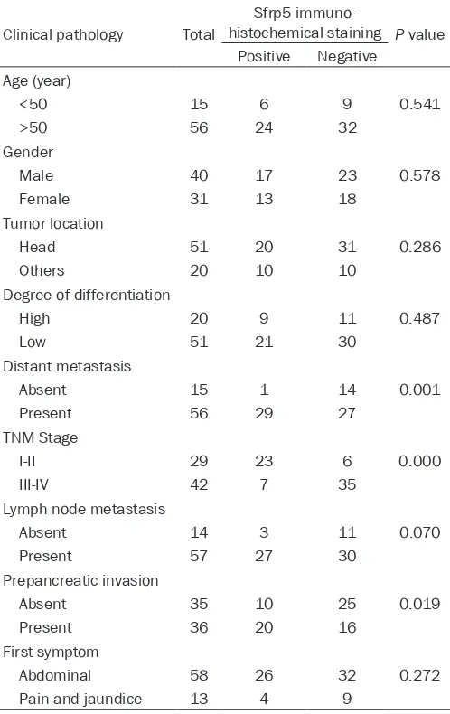
Table 1. Correlations between Sfrp5 expression and clini-copathological data
Clinical pathology Total
Sfrp5 immuno-
histochemical staining P value Positive Negative
Age (year)
<50 15 6 9 0.541
>50 56 24 32
Gender
Male 40 17 23 0.578
Female 31 13 18
Tumor location
Head 51 20 31 0.286
Others 20 10 10
Degree of differentiation
High 20 9 11 0.487
Low 51 21 30
Distant metastasis
Absent 15 1 14 0.001
Present 56 29 27
TNM Stage
I-II 29 23 6 0.000
III-IV 42 7 35
Lymph node metastasis
Absent 14 3 11 0.070
Present 57 27 30
Prepancreatic invasion
Absent 35 10 25 0.019
Present 36 20 16
First symptom
Abdominal 58 26 32 0.272
Statistical analysis
The X2 and Fisher’s exact test was used to
ana-lyze the SFRP5 expression and clinical charac-teristics. The prognostic value of SFRP5 was analyzed by Kaplan-Meier’s method. Univariate and multivariate Cox regression models were used to calculate hazard ratios. Then indepen-dent prognostic factors were iindepen-dentified from 71 PADC patients. P value less than 0.05 regarded
as significant and statistical data were ob- tained by SPSS software (version 20.0, SPSS, Chicago, IL, USA).
Results
Associations between SFRP5 expression and clinicopathological characteristics
The patient clinical and pathological character-istics are summarized in Table 1. The median age was 57 years (range 24-75 years). 41 (57.7%) patients have a low SFRP5 expression. The expression of SFRP5 was lower in PADC tissues than that of paracancerous tissue (Figure 1). There were significant differences in the expression of SFRP5 when compar- ing other clinicopathological characteristics such as Distant metastasis (P=0.001), TNM Stage (P<0.001) and Prepancreatic invasion (P=0.019).
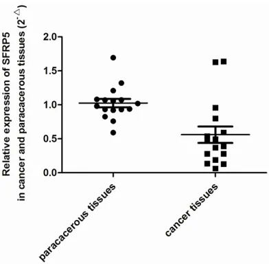
Low expression of SFRP5 in PADC by qRT-PCR and Western blot
[image:3.612.90.525.72.285.2]We used qRT-PCR and Western blot to measure the expression of SFRP5 from mRNA and pro-tein level. PDAC tissues showed lower mRNA expression levels of SFRP5 compared with paracancerous tissues (P<0.001, Figure 2). The protein expression of SFRP5 were detected Figure 1. Immunohistochemistry analysis Sfrp5 expression in PDAC tissues and paracancerous tissues. A, D. High expression of Sfrp5 in adjacent noncancerous tissues; B, E. Low expression of Sfrp5 in PC tissues; C, F. High expres-sion of Sfrp5 in adjacent noncancerous tissues and Low expresexpres-sion of Sfrp5 in PC tissues. A, B with ×200 magnifica-tion, D, E with ×400 magnificamagnifica-tion, C, F with ×100 magnification.
[image:3.612.91.287.354.546.2]tric cancer [11], colorectal cancer [12] and breast cancer [13]. The SFRP5 function as tumor suppressor gene has an important impli-cation in carcinogenesis, where they are down-regulated in many tumors [14]. In 168 primary breast carcinomas, JurgenVeeck et al, found that down regulation of SFRP5 was associated with reduced overall survival (OS) (P=0.045) and was an independent risk factor affecting OS in a multivariate Cox proportional hazard model. They indicated that SFRP5 might be a novel biomarker potentially useful in clinical breast cancer treatment [15]. Her-Young Su [16] suggested that epigenetic silencing of SFRP5 leaded to oncogenic activation and con-tributed to ovarian cancer progression and che-from six pairs of PDAC tissues, and found that
SFRP5 protein level was lower in PDAC tissues than paracancerous tissues (Figure 3).
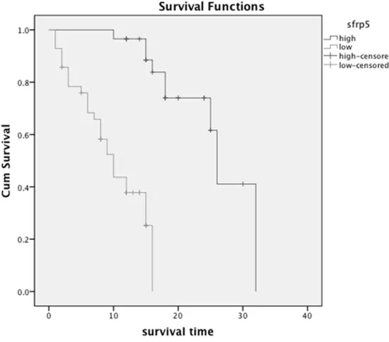
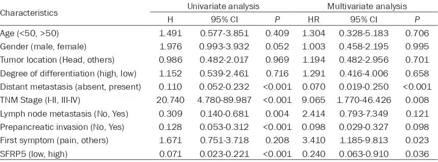
Univariate and multivariate analysis
Kaplan-Meier analysis and log-rank test were used to explore whether SFRP5 was a prognos-tic factor (Figure 4). We found the correlation of the SFRP5 expression with overall survival in 71 PADC patients. The results of univariate and multivariate analysis for overall survival are summarized in Table 2. Univariate analysis showed that distant metastasis, TNM Stage, Lymph node metastasis, Prepancreatic inva-sion as well as SFRP5 could influence overall survival. The multivariate analysis showed that
distant metastasis, TNM Sta- ge and SFRP5 were indepen-dent prognostic factors for overall survival. In a word, low expression of SFRP5 was an independent prognostic fac-tor for PADC patients.
Association of SFRP5 expres-sion with survival.
Discussion
PDAC is the most frequent type of pancreatic cancer accounting for almost 90% of all pancreatic tumors [6]. The most notable feature of PDAC is poor prognosis, as well as aggressive tumor growth [7]. Presently pancre-atectomy remained the only potential for cure. Those who received pancreatectomy had a median survival of 12.6 months, who did not receive the surgery had the median survival of 3.5 months [8]. Therefore, the effective thera-peutic and early detection strategies are greatly needed for PDAC.
In recent years, downregula-tion of SFRP5 had been reported in many tumors such as hepatocellular carcinoma [9], prostate cancer [10], gas-Figure 3. WB analysis of Sfrp5 expression in PDAC tissues and
[image:4.612.93.377.74.179.2]paracancer-ous tissues.
[image:4.612.94.371.231.474.2]moresistance. It was reported that SFRP5 was methylated in 60% of prostate cancer cell lines [17]. All the research indicates that SFRP5 will be a potential biomarker for PDAC.
However, little is known about the role of SFRP5 in PDAC. Low expression of SFRP5 was found in the majority of the 60 PDAC samples by a research group. It was observed that the expression loss of SFRP5 in PDAC samples were significantly higher than those in the para-cancerous tissue samples [18]. In our study, the mRNA and protein level of SFRP5 had been explored to be lower in PDAC tissues than paired paracancerous tissues. We also found that the expression of SFRP5 was associated with distant metastasis, TNM stage and pre- pancreatic invasion.
Early PDAC diagnostic criteria are as follow: tumor diameter less than 2 cm, confined to the pancreatic parenchyma, no peripancreatic invasion and metastasis [19]. Early diagnosis of this study was only 5.6% (4/71), but the average survival time for PDAC patients has reached 26 months of those who received radi-cal surgery. In the early diagnosis of PDAC, the current clinical application is CA19-9 and imag-ing examination [20]. Over the past 20 years, studies found that many PDAC biomarkers, including CA242 [21], TATI [22], POA [23] and MMP7 [24]. Although the sensitivity and speci-ficity of these markers have reached more than 70% [25], but the effect is not as good as CA19-9, therefore, the study on these markers need to be continued further. The researches on PDAC had accumulated a database form, which combined with serum proteomics and
molecu-lar bioinformatics were expected to find a high sensitivity and specificity of biomarker. Further functions of SFRP5 would be pivotal in finding possible novel therapeutics for cancer through targeting SFRP5. In our study, SFRP5 is the independent prognostic factor in clinical patient samples. In the future, the role of SFRP5 is needed to prove its prognostic value.
Acknowledgements
This research was supported by the Central University Fundamental Research Funds for Lanzhou University (No. lzujbky-2012-163/167) and Gansu provincial Natural Science Foun- dation (No. 1208RJZA219).
Disclosure of conflict of interest
None.
Address correspondence to: Wence Zhou, Second Department of The General Surgery, The First Hospital of Lanzhou University, Lanzhou University, Lanzhou 730000, China. E-mail: zhouwc129@163. com (WCZ); tianm13@lzu.edu.cn (MT)
References
[1] Siegel RL, Kimberly D, Ahmedin J. Global can-cer Statistics 2015. CA Cancan-cer J Clin 2015; 65: 5-29.
[2] Maitra A, Hruban RH. Pancreatic cancer. Annu Rev Pathol 2008; 3: 157-188.
[3] Rao TP, Kühl M. An updated overview on Wnt signaling pathways: a prelude for more. Circ Res 2010; 106: 1798-806.
[image:5.612.93.522.86.245.2][4] Niehrs C. The complex world of WNT receptor signaling. Nat Rev Mol Cell Biol 2012; 13: 767-779.
Table 2. Univariate and multivariate analysis of prognostic factors for overall survival in PADC patients
Characteristics Univariate analysis Multivariate analysis
H 95% CI P HR 95% CI P
Age (<50, >50) 1.491 0.577-3.851 0.409 1.304 0.328-5.183 0.706
[5] Surana R, Sikka S, Cai W, Shin EM, Warrier SR, Tan HJ, Arfuso F, Fox SA, Dharmarajan AM, Kumar AP. Secreted frizzled related proteins: Implications in cancers. Biochim Biophys Acta 2014; 1845: 53-65.
[6] Fesinmeyer MD, Austin MA, Li CI, De Roos AJ, Bowen DJ. Differences in survival by histologic type of pancreatic cancer. Cancer Epidemiol Biomarkers Prev 2005; 14: 1766-1773. [7] Hidalgo M. Pancreatic cancer. N Engl J Med
2010; 362: 1605-1617.
[8] Bilimoria KY, Bentrem DJ, Ko CY, Ritchey J, Stewart AK, Winchester DP, Talamonti MS. Validation of the 6th edition AJCC Pancreatic Cancer Staging System: report from the National Cancer Database. Cancer 2007; 110: 738-44.
[9] Takagi H, Sasaki S, Suzuki H, Toyota M, Maruyama R, Nojima M, Yamamoto H, Omata M, Tokino T, Imai K, Shinomura Y. Frequent epi-genetic inactivation of SFRP genes in hepato-cellular carcinoma. J Gastroenterol 2008; 43: 378-389.
[10] Jemal A, Siegel R, Ward E, Hao Y, Xu J, Thun MJ. Cancer statistics, 2009. CA Cancer J Clin 2009; 59: 225-249.
[11] Cheng YY, Yu J, Wong YP, Man EP, To KF, Jin VX, Li J, Tao Q, Sung JJ, Chan FK, Leung WK. Frequent epigenetic inactivation of secreted frizzled-related protein 2 (SFRP2) by promoter methylation in human gastric cancer. Br J Cancer 2007; 97: 895-901.
[12] Qi J, Zhu YQ, Luo J, Tao WH. Hypermethylation and expression regulation of secreted frizzled-related protein genes in colorectal tumor. World J Gastroenterol 2006; 12: 7113-7117. [13] Veeck J, Niederacher D, An H. Aberrant
meth-ylation of the Wnt antagonist SFRP1 in breast cancer is associated with unfavourable prog-nosis. Oncogene 2006; 25: 3479-3488. [14] Yang Z, Wang Y, Fang J, Chen F, Liu J, Wu J,
Wang Y. Expression and aberrant promoter methylation of Wnt inhibitory factor-1 in hu-man astrocytomas. J Exp Clin Cancer Res 2010; 29: 23-26.
[15] Veeck J, Geisler C, Noetzel E, Alkaya S, Hartmann A, Knüchel R, Dahl E. Epigenetic in-activation of the secreted frizzled-related protein-5 (SFRP5) gene in human breast can-cer is associated with unfavorable prognosis. Carcinogenesis 2008; 29: 991-998.
[16] Su HY, Lai HC, Lin YW, Liu CY, Chen CK, Chou YC, Lin SP, Lin WC, Lee HY, Yu MH. Epigenetic silencing of SFRP5 is related to malignant phe-notype and chemoresistance of ovarian can-cer through Wnt signaling pathway. Int J Cancan-cer 2010; 127: 555-567.
[17] Zi XL, Guo Y, Simoneau AR, Hope C, Xie J, Holcombe RF, Hoang BH. Expression of Frzb/ secreted Frizzled-Related Protein 3, a secreted Wnt antagonist, in human androgen-indepen-dent prostate cancer PC-3 cells suppresses tumor growth and cellular invasiveness. Cancer Res 2005; 65: 9762-9770.
[18] Jones S, Zhang X, Parsons DW, Lin JC, Leary RJ, Angenendt P, Mankoo P, Carter H, Kamiyama H, Jimeno A, Hong SM, Fu B, Lin MT, Calhoun ES, Kamiyama M, Walter K, Nikolskaya T, Nikolsky Y, Hartigan J, Smith DR, Hidalgo M, Leach SD, Klein AP, Jaffee EM, Goggins M, Maitra A, Iacobuzio-Donahue C, Eshleman JR, Kern SE, Hruban RH, Karchin R, Papadopoulos N, Parmigiani G, Vogelstein B, Velculescu VE, Kinzler KW. Core signaling pathways in human pancreatic cancers revealed by global genomic analyses. Science 2008; 321: 1801-1806 . [19] Bilimoria KY, Bentrem DJ, Ko CY, Ritchey J,
Stewart AK, Winchester DP, Talamonti MS. Validation of the 6th edition AJCC Pancreatic Cancer Staging System: report from the National Cancer Database. Cancer 2007; 110: 738-744.
[20] Ardengh JC, de Paulo GA, Ferrari AP. Pancreatic carcinomas smaller than 3.0 cm: endosonog-raphy (EUS) in diagnosis, staging and predic-tion of resectability. HPB (Oxford) 2003; 5: 226-230
[21] Lamerz R. Role of tumour markers: cytogenet-ics. Ann Oncol 1999; 10: 145-149.
[22] Pasanen PA, Eskelinen M, Partanen K, Pikkarainen P, Penttilä I, Alhava E. Tumor-associated trypsin inhibitor in the diagnosis of pancreatic carcinoma. J Cancer Res Clin Oncol 1994; 120: 494-497.
[23] Shahangian S, Fritsche JH Jr, Hughes JI, Gelder FB. Pancreatic oncofetal antigen and carbohy-drate antigen 19-9 in sera of patients with can-cer of the pancreas. Clin Chem 1989; 35: 405-408.
[24] Kuhlmann KF, van Till JW, Boermeester MA, de Reuver PR, Tzvetanova ID, Offerhaus GJ, Ten Kate FJ, Busch OR, van Gulik TM, Gouma DJ, Crawford HC. Evaluation of matrix metallo-proteinase 7 in plasma and pancreatic juice as a biomarker for pancreatic cancer. Cancer Epidemiol Biomarkers Prev 2007; 16: 886-891.