Original Article
A prognostic scoring model based on the HPV status of
oropharyngeal carcinoma patients treated with
postoperative radiotherapy in China
Wenyan Wu1,2,3*, Zhen Wang2,3,4*, Zengtong Zhou1,2,3, Jiang Li2,3,5
Departments of 1Oral Mucosal Diseases, 4Oral & Maxillofacial-Head & Neck Oncology, 5Oral Pathology, Shanghai Ninth People’s Hospital, College of Stomatology, Shanghai Jiao Tong University School of Medicine, Shanghai 200011, P. R. China; 2National Clinical Research Center for Oral Diseases, Shanghai 200011, P. R. China; 3Shanghai Key Laboratory of Stomatology & Shanghai Research Institute of Stomatology, Shanghai 200011, P. R. China. *Equal contributors.
Received January 6, 2019; Accepted March 22, 2019; Epub May 1, 2019; Published May 15, 2019
Abstract: Objectives: Prognostic models that can predict prognosis and guide postoperative radiotherapy (PRT) and that are based on the human papillomavirus (HPV) status of patients with oropharyngeal carcinoma (OPSCC) in China are rare. Methods: Survival was analyzed by performing a Kaplan-Meier analysis and log-rank test. A Cox re-gression analysis was performed for the multivariate analyses. A prognostic scoring model was constructed
accord-ing to the regression coefficient obtained from the Cox regression model. Results: A prognostic model that included
gender, clinical stage, histologic stage, metastasis, and HPV status was created and used to divide patients into
high-risk (PI ≥ -0.008) and low-risk (PI < -0.008) groups. The results showed that the patients who received PRT had
a longer overall survival time than those who did not receive PRT (47.31 months vs. 28.31 months). Furthermore, the patients who received PRT in the high-risk group had a longer survival time when the survival was greater than 20 months (P = 0.024), and PRT may indicate a worse prognosis in the low-risk group (P = 0.071). Conclusion: This model will contribute to the formulation of individualized treatment programs for OPSCC patients. PRT should be administered to high-risk patients.
Keywords: Oropharyngeal squamous cell carcinoma, human papillomavirus, prognostic model, postoperative ra-diotherapy
Introduction
Human papillomavirus (HPV) infection is a ma- jor risk factor responsible for a large subset of oropharyngeal squamous cell carcinomas (OP- SCCs). HPV infection has been observed in 20% to 90% of OPSCC patients in the United States and Europe. HPV-associated OPSCC was identi-fied as a solid tumor distinct from HPV-negative OPSCC due to its unique epidemiological char-acteristics, biologic behaviors, and clinical fea-tures [1-4]. However, the prevalence of HPV in OPSCC ranges only from 0%-34% in Asia [5-9]. The prognosis of OPSCC depends on the tumor stage and HPV status [10], and HPV status has been shown to be an independent prognostic factor [11, 12]. In particular, in all stages, non-smoking patients with HPV-associated OPSCC
with HPV-associated OPSCC have a moderate risk of tumor progression, invasion, and metas-tasis and smoking patients with HPV-negative OPSCC have the highest risk [13]. Gender, age, TNM stage and smoking status also likely play important roles in the development of OPSCC [14, 15]. Several studies have established prog-nostic models of OPSCC based on clinical and laboratory indices [16, 17]. Unsurprisingly, com-bining the most important prognostic factors, including the HPV status and TNM stage, in a multivariate model results in a more compre-hensive and highly predictive model than a uni-variate survival analysis.
is not practical [16, 17]. The current prognostic models for OPSCC, particularly those for HPV-associated OPSCC, are all based on data from Europe and the United States. Epidemiological investigations have shown that low HPV detec-tion rates and tobacco use are common among Chinese OPSCC HPV-positive patients [9, 18, 19]. However, a prognostic model based on cl- inicopathologic characteristics and HPV stat- us in China has not been published to date. Furthermore, the therapeutic strategies used differ between western countries and China. In China, early-stage OPSCC is typically managed with surgery with or without adjuvant radiother-apy (RT), and advanced-stage OPSCC is treated with RT or chemoradiotherapy. In western coun-tries, early-stage and HPV-related OPSCC have been treated with curative RT or chemoradio-therapy alone for many years [16, 20]. HPV-associated OPSCC patients are more sensitive to RT and chemotherapy and typically exhibit a lower recurrence rate, longer survival time, and better prognosis than those with HPV-negative OPSCC [11, 21-23]. However, the risks of RT complications that can have a significant im- pact on the patient’s quality of life, such as xerostomia, osteoradionecrosis of the mandi-ble, trismus, ischemic stroke and dysphagia, may be underestimated [13, 24, 25].
Therefore, a multi-factorial, comprehensive pr- ognostic model for OPSCC may have a signifi-cant impact on predicting prognosis and indi-vidualizing postoperative RT protocols in Chi- nese patients with OPSCC.
Methods
Clinical specimens and patient information
In total, 188 primary OPSCC patients diag-nosed between January 2008 and April 2014 at the Department of Oral Pathology of the Shanghai Ninth People’s Hospital, Shanghai Jiao Tong University School of Medicine were enrolled, and all postoperative tumor speci-mens were collected. The primary location of the tumor was determined according to the clinical diagnosis, imaging results, and patho-logic examination.
Prognostic factors and HPV testing
The demographic data, including sex, age, diag-nosis, and tumor site, were obtained from the
medical records and operative and pathology reports. The tumor stage was assigned based on either the clinical or pathologic staging according to the 2005 WHO TNM (Tumor, No- dal, Metastasis) Classification. All patients we- re followed prospectively by a head and neck surgeon from the date of the original diagnosis. The clinical status of the patients including information about the surgery, chemotherapy and RT, were recorded at each follow-up visit, which occurred at approximately 3-month inter-vals from the time of diagnosis.
The following prognostic factors were recorded at baseline: age, gender, histologic grade, T stage, N stage, smoking status, alcohol use, site of the primary tumor and HPV16/18 status, which was defined as both HPV DNA PCR and p16INK4A positivity (positive cells in ≥ 70% of the tumor).
Radiotherapy
All patients received primary tumor resection. The patients with advanced OPSCC underwent cervical lymph node dissection and postopera-tive RT (PRT) at a total dose of 50-60 Gy (2 Gy per fraction) depending on whether more than 2 regional lymph nodes, extracapsular spread, or positive margins on microscopic mucosal resection were observed.
Statistical analysis
appropriate PI cutoff point, which was used to divide the patients into the high-risk and low-risk groups. To minimize bias and approximate a randomized trial, propensity score-based me-
thods were used to balance the characteris- tics (chemotherapy treatment) between the RT-treated and non-treated patients and evalu-ate the effect of RT treatment on OS [26]. Finally, we included 177 of the initial 188 patients in this analysis. All statistical tests were two-tailed, and P < 0.05 was considered statistically significant.
Results
Patient characteristics
As shown in Table 1, of the 188 OPSCC patients, 60.11% (113/188) of the patients were young-er than 60 years, and 39.89% (75/188) of the patients were 60 years or older. In total, 89.36% (168/188) of the patients were men, and 10.64% (20/188) of the patients were women. The tumors were rarely located on the tonsils (5.32%, 10/188) and were mainly located at the base of the tongue (39.36%, 74/188), the pharyngeal wall (28.72%, 54/188), and the soft palate (26.60%, 50/188). Information regard-ing the smokregard-ing and drinkregard-ing habits were miss-ing in 10 of the patients; therefore, based on data from 178 OPSCC patients, smokers ac- counted for the majority of patients (61.70%, 116/188), and drinkers and non-drinkers each accounted for 47% (89/188). In total, 51.06% (96/188) of the OPSCC cases were in the early clinical stages (I-II), and 48.94% (92/188) of the patients were in the late clinical stages (III-IV). According to the pathologic examination, 10.11% (19/188) of the cases were classified as histologic grade I, 69.68% (131/188) of the cases were classified as histologic grade II, and 20.21% (38/188) of the cases were classified as histologic grade III. Most patients (88.83%, 167/188) were categorized as TNM stages T1-T2, 45.21% (85/188) of the cases had cervi-cal lymph node involvement and most patients (97.87%, 184/188) did not have me- tastasis. In addition, only 20.74% (39/188) of the patients received chemotherapy; however, 53.19% (100/188) of the patients received PRT.
HPV status
[image:3.612.91.284.94.647.2]According to the HPV16/18 E6/E7 PCR analy-sis, only 11.70% (22/188) of the patients were infected with HPV16, whereas no patients with HPV18 infection were identified. In addition, only 21.28% (40/188) of the patients were
Table 1. Distribution of clinicopathologic fea-tures in 188 primary OPSCC cases
Characteristic Overall Percentage (%) Age
< 60 113 60.11
≥ 60 75 39.89
Gender
Male 168 89.36
Female 20 10.64
Tumor Site
Tonsil 10 5.32
Soft palate 50 26.60
Base of tongue 74 39.36 Pharyngeal wall 54 28.72 Smoking
Non-smoker 62 32.98
Smoker 116 61.70
Unknown 10 5.32
Alcohol Consumption
Non-drinker 89 47.34
Drinker 89 47.34
Unknown 10 5.32
Clinical Stage
I-II 96 51.06
III-IV 92 48.94
Histologic Grade
I 19 10.11
II 131 69.68
III 38 20.21
T Stage
I-II 167 88.83
III-IV 21 11.17
Nodal Stage
Negative 103 54.79
Positive 85 45.21
Metastasis
Negative 184 97.87
Positive 4 2.13
Chemotherapy
No 149 79.26
Received 39 20.74
Radiation
No 88 46.81
OPSCC patients: female sex, non-smoking status, clinical st- age I-II, histologic grade I, N0 stage and HPV16 positivi- ty (P = 0.003, 0.048, 0.003, 0.009, 0.004, 0.009, respec-tively). According to the multi-variate analysis, the clinical stage (P = 0.022), histologic grade II, histologic grade III (P = 0.041, P = 0.004), metastasis (P = 0.011), and HPV16 positiv-ity (P = 0.011) were indepen-dent prognostic factors (Table 4). The regression coefficients of each factor were as follows (Table 3): PI = -1.270 (if female) + 0.586 (if clinical stage III-IV) + 1.090 (if histologic grade II) + 1.644 (if histologic grade III) + 1.434 (metastasis) - 1.843 (if HPV16 positive).
Model validation
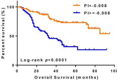
The above model was used to calculate the prognostic index for each patient. In total, 21 val-ues were obtained for the PI (range from -3.74 to 2.84). The AUC of the ROC curve was 0.74 (95% Confidence interval (CI): 0.67-0.81) (Figure 1), and the Youden index was the highest at PI = -0.008 (Figure 2). In ad- dition, we classified the pati- ents into 2 risk groups accord-ing to the above cut-off value. Patients with a PI < -0.008 were assigned to the low-risk group, and patients with a PI ≥ -0.008 were assigned to the high-risk group. In total, 107 and 81 patients were assigned to the low- and high-risk groups, respectively. The patients in the p16INK4A positive, and the p16INK4A
positivi-ty was significantly associated with HPV positiv-ity (P < 0.001) [9] (Table 2).
Survival analysis and establishment of the OPSCC prognostic model
As shown in Table 3, the following factors were associated with a better OS among the 188
high-risk group had a significantly higher risk of death, with a hazard ratio (HR) of 4.60 (95% CI: 2.21-9.58) (Figure 2).
Significance of the PI model for PRT
The necessity of PRT for OPSCC is controver-sial, and significant acute and late toxicities of RT can occur; therefore, evaluation of PRT as
Table 2. Relationship between the HPV and p16INK4A status in 188 primary OPSCC cases
p16INK4A
Overall Statistically significant
Negative Positive
HPV
Negative 148 18 166 < 0.001
Positive 0 22 22
HPV, human papillomavirus; OPSCC, oropharyngeal squamous cell carcinoma.
Table 3. Kaplan-Meier and Cox regression analyses of 188 pri-mary OPSCC cases
Variable
Kaplan-Meier Cox Regression
p-value p-value Hazard Ratio 95% Confidential Interval
Age 0.833
Gender 0.003 0.081 0.281 0.067-1.171
Tumor Site 0.378
Smoking 0.048
Alcohol 0.499
Clinical Stage 0.003 0.022 1.797 1.087-2.970 Histologic Grade I 0.009
Histologic Grade II 0.041 2.973 1.046-8.453 Histologic Grade III 0.004 5.178 1.671-16.043
T Stage 0.183
Nodal Stage 0.004
Metastasis 0.072 0.019 4.196 1.271-13.850
HPV16 0.009 0.011 0.158 0.038-0.658
P16INK4A 0.43
Table 4. Multivariate analysis of independent prognostic factors in 188 primary OPSCC cases
Variable OS
HR (95% CI) p-value
Gender 0.281 (0.067-1.171) 0.081
HPV16 0.158 (0.038-0.658) 0.011
Clinical Stage III~IV 1.797 (1.087-2.970) 0.022 Histologic Grade II 2.973 (1.046-8.453) 0.041 Histologic Grade III 5.178 (1.671-16.043) 0.004
Figure 2. Survival curves of patients based on the PI. The Youden index divided into high-risk (PI ≥ -0.008)
and low-risk (PI < -0.008) group. Patients had a high -er risk in high-risk group with a hazard ratio of 4.60 (95% CI: 2.21-9.58).
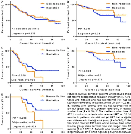
an optional treatment strategy based on the prognostic scoring model was particularly im- portant. To exclude the effect of chemotherapy on prognosis, we included the propensity sc- ores of 177 patients in this analysis. As shown in Figure 3A, among the 177 selected patients, no significant difference was observed in the OS between patients who received PRT (N = 100) and those who did not receive PRT (N = 77) (P = 0.938), suggesting that PRT does not prolong the OS. Similarly, as shown in Figure 3B, among the low-risk patients (PI < -0.008, N = 101), no difference was observed in the OS between the patients who received PRT (N = 54) and those who did not receive PRT (N = 47) (P = 0.38). However, a significant difference was observed in the high-risk patient group (PI ≥ -0.008, N = 76) (Figure 3C, P = 0.096); the median OS time was 28.31 months (95% CI: 10.69-35.31) in the patients who did not receive PRT (N = 30) and 47.31 months (95% CI: 18.34-47.62) in the patients who received PRT (N = 30). Furthermore, we compared the OS time of patients who received PRT and those who did not receive PRT with survival times longer than 20 months. Interestingly, in the low-risk patient subgroup (PI < -0.008), PRT was associated with a worse survival time (Figure 3D, P = 0.071). In the high-risk patient subgroup (PI ≥ -0.008), when the OS time was longer than 20 months, the patients who received PRT had a longer survival time than the patients who did not receive PRT (Figure 3E, P = 0.024). These results suggest that PRT is not beneficial for all patients. Therefore, our prognostic index should be used to guide treat-ment protocols regarding whether PRT is
nec-essary. PRT should not be recommended for low-risk patients, while high-risk patients should receive postoperative adjuvant RT.
Discussion
Because HPV infection is the underlying cause of a significant proportion of OPSCCs, these infections have played an important role in the recent epidemiological changes observed among OPSCC cases. HPV-associated OPSCC is more sensitive to RT, and HPV-associated OPSCC patients who receive PRT have better survival rates than patients with HPV-negative OPSCC [23, 27, 28]. However, some studies suggest that PRT might not be suitable as an alternative therapy for all high-risk OPSCC patients, such as for patients with certain clini-cal or pathologiclini-cal characteristics and because of the higher incidence rates of acute and late toxicity due to RT; such patients should consid-er othconsid-er thconsid-erapeutic options [29, 30]. These findings drew our attention and prompted the current study. In China, there is a low detection rate of HPV infection and high smoking and drinking rates in OPSCC patients, as shown in our previous study [9]. Furthermore, clinical stage, histologic grade, metastasis and HP- V16 positive status were independent progn- ostic factors for Chinese OPSCC patients. Th- erefore, the distinguishing feature of Chinese OPSCC patients is that HPV-positive status was not the main prognostic factor; clinicopatho-logic features should receive more attention in Chinese OPSCC patients. A prognostic scoring model based on gender, clinical stage, histo-Figure 1. ROC curve of the PI in primary OPSCC
[image:5.612.323.520.71.207.2]logical grade, metastasis and HPV status was established and was used to classify patients into high- and low-risk groups. The results showed that female patients of clinical stage I-II, histologic grade I, N0 metastasis and HPV16 positive showed better OS and progno-sis. A significantly higher mortality risk was observed in the high-risk group than in the low-risk group of OPSCC patients, with a HR of 4.60. According to our findings, this propensity score model is a reasonable model for individualizing postoperative OPSCC radiation protocols. Interestingly, the patients who received PRT (47.31 months) had a better OS than the patients who did not receive PRT (28.31 mo-
[image:6.612.87.522.70.514.2]nths) in the high-risk group, particularly when the survival time was longer than 20 months (P = 0.024). However, PRT may be a risk factor for decreased survival time in the low-risk group (P = 0.071). Therefore, according to this prognos-tic model, we recommend postoperative adju-vant RT for high-risk OPSCC patients only. Postoperative radiation is a standard treatm- ent option for patients with OPSCC in China. Radiation therapy can kill or inhibit tumor cells, reduce or delay the chance of local recurrence or metastasis, and improve prognosis. However, in the current study, we found that PRT was not generally effective and may even worsen the Figure 3. Survival curves of patients who received or did not receive postoperative radiation therapy (PRT). A. Pa-tients who received and had not received PRT had no
significant difference in overall survival time (P = 0.938);
B. Patients who received and had not received PRT in
low-risk group had no significant difference in overall
survival time (P = 0.38); C. The median survival time of 47.31 months in patients who received PRT vs. 28.31
prognosis of low-risk patients. RT can cause permanent and severe morbidity, such as dys-phagia and secretory dysfunction of the sali-vary glands [31, 32]. Therefore, identifying pa- tients who are truly suitable for RT is critical. Recently, RT has been shown to be more effec-tive for HPV-related OPSCC than HPV-negaeffec-tive OPSCC [22]. Using the propensity scores to compare the groups in our study population, the high-risk group, in which the majority of the cases were HPV negative, achieved a better survival time with PRT, while PRT had little or even negative effects in the low-risk group. This prognostic model has the advantage of com-prehensively evaluating individual clinicopatho-logic characteristics and HPV status to deter-mine whether the patients are candidates for PRT.
To the best of our knowledge, this is the first postoperative prognostic scoring model for patient prognosis and choice of individual treat-ment schedule in China. However, this study has limitations, such as the lack of OPSCC patients who received RT alone. More data should be collected and validated in the future to improve the utility of this OPSCC prognostic scoring model.
In conclusion, we established an OPSCC post-operative prognostic scoring model with high accuracy that includes the following 5 risk fac-tors: gender, clinical stage, histologic grade, metastasis and HPV status. This scoring model can supplement the TNM staging and can be applied for prognostic assessments to provide guidance regarding the formulation of individu-alized therapeutic strategies for PRT in Chinese OPSCC patients.
Acknowledgements
The study was supported by the National Na- tural Science Foundation of China (grant no. 81372910, 81202132).
Disclosure of conflict of interest
None.
Address correspondence to: Zengtong Zhou, De- partment of Oral Mucosal Diseases, Shanghai Ninth People’s Hospital, College of Stomatology, Shanghai Jiao Tong University School of Medicine, Shanghai 200011, P. R. China; National Clinical Research
Center for Oral Diseases, Shanghai 200011, P. R. China; Shanghai Key Laboratory of Stomatology & Shanghai Research Institute of Stomatology, Sh- anghai 200011, P. R. China. E-mail: zhouzengtong@ hotmail.com; Jiang Li, Departments of Oral Pa- thology, Shanghai Ninth People’s Hospital, College of Stomatology, Shanghai Jiao Tong University School of Medicine, Shanghai 200011, P. R. China; National Clinical Research Center for Oral Diseases, Shanghai 200011, P. R. China; Shanghai Key La- boratory of Stomatology & Shanghai Research Institute of Stomatology, Shanghai 200011, P. R. China. E-mail: lijiang182000@126.com
References
[1] Marur S, D’Souza G, Westra WH and Forastiere AA. HPV-associated head and neck cancer: a virus-related cancer epidemic. Lancet Oncol 2010; 11: 781-9.
[2] Ang KK, Harris J, Wheeler R, Weber R, Ro- senthal DI, Nguyen-Tân PF, Westra WH, Chung CH, Jordan RC, Lu C, Kim H, Axelrod R, Sil- verman CC, Redmond KP, Gillison ML. Human papillomavirus and survival of patients with oropharyngeal cancer. N Engl J Med 2010; 363: 24-35.
[3] Adelstein DJ and Rodriguez CP. Human papil-lomavirus: changing paradigms in oropharyn-geal cancer. Curr Oncol Rep 2010; 12: 115-120.
[4] D’Souza G, Kreimer AR, Viscidi R, Pawlita M, Fakhry C, Koch WM, Westra WH and Gillison ML. Case-control study of human papillomavi-rus and oropharyngeal cancer. N Engl J Med 2007; 356: 1944-1956.
[5] Li W, Tran N, Lee SC, O’Brien CJ, Tse GM, Scolyer RA, Hong A, Milross C, Yu KH and Rose BR. New evidence for geographic variation in the role of human papillomavirus in tonsillar carcinogenesis. Pathology 2007; 39: 217-222. [6] Maruyama H, Yasui T, Ishikawa-Fujiwara T, Mo-
rii E, Yamamoto Y, Yoshii T, Takenaka Y, Na- kahara S, Todo T, Hongyo T and Inohara H. Human papillomavirus and p53 mutations in head and neck squamous cell carcinoma among Japanese population. Cancer Sci 2014; 105: 409-417.
[7] Huang H, Zhang B, Chen W, Zou SM, Zhang YX and Qiao YL. [Detection of human papillomavi-rus in oropharyngeal squamous cell carcino-ma]. Zhongguo Yi Xue Ke Xue Yuan Xue Bao 2012; 34: 545-549.
[9] Wang Z, Xia RH, Ye DX and Li J. Human pap- illomavirus 16 infection and TP53 mutati- on: two distinct pathogeneses for orophar- yngeal squamous cell carcinoma in an Easte- rn Chinese population. PLoS One 2016; 11: e0164491.
[10] Mehanna H, Olaleye O and Licitra L. Oropha- ryngeal cancer - is it time to change manage-ment according to human papilloma virus sta-tus? Curr Opin Otolaryngol Head Neck Surg 2012; 20: 120-124.
[11] Ang KK, Harris J, Wheeler R, Weber R, Rosen- thal DI, Nguyen-Tan PF, Westra WH, Chung CH, Jordan RC, Lu C, Kim H, Axelrod R, Silverman CC, Redmond KP and Gillison ML. Human pap-illomavirus and survival of patients with oro-pharyngeal cancer. N Engl J Med 2010; 363: 24-35.
[12] Fakhry C, Zhang Q, Nguyen-Tan PF, Rosenthal D, El-Naggar A, Garden AS, Soulieres D, Trotti A, Avizonis V, Ridge JA, Harris J, Le QT and Gillison M. Human papillomavirus and overall survival after progression of oropharyngeal squamous cell carcinoma. J Clin Oncol 2014; 32: 3365-3373.
[13] Monnier Y and Simon C. Surgery versus radio-therapy for early oropharyngeal tumors: a nev-er-ending debate. Curr Treat Options Oncol 2015; 16: 42.
[14] Camilon PR, Stokes WA, Nguyen SA and Len-
tsch EJ. The prognostic significance of age in
oropharyngeal squamous cell carcinoma. Oral Oncol 2014; 50: 431-436.
[15] Wang MB, Liu IY, Gornbein JA and Nguyen CT. HPV-positive oropharyngeal carcinoma: a sys-tematic review of treatment and prognosis. Otolaryngol Head Neck Surg 2015; 153: 758-769.
[16] Velazquez ER, Hoebers F, Aerts HJ, Rietbergen MM, Brakenhoff RH, Leemans RC, Speel EJ, Straetmans J, Kremer B and Lambin P. Ext- ernally validated HPV-based prognostic nomo-gram for oropharyngeal carcinoma patients yields more accurate predictions than TNM staging. Radiother Oncol 2014; 113: 324-330. [17] Rietbergen MM, Witte BI, Velazquez ER, Snij-
ders PJ, Bloemena E, Speel EJ, Brakenhoff RH, Kremer B, Lambin P and Leemans CR. Diff- erent prognostic models for different patient populations: validation of a new prognostic model for patients with oropharyngeal cancer in Western Europe. Br J Cancer 2015; 112: 1733-1736.
[18] Huang H, Zhang B, Chen W, Zhou SM, Zhang YX, Gao L, Xu ZG, Qiao YL and Tang PZ. Human papillomavirus infection and prognostic pre-dictors in patients with oropharyngeal squa-mous cell carcinoma. Asian Pac J Cancer Prev 2012; 13: 891-896.
[19] Lam EW, Chan JY, Chan AB, Ng CS, Lo ST, Lam VS, Chan MM, Ngai CM, Vlantis AC and Ma RK. Prevalence, clinicopathological characteristi- cs, and outcome of human papillomavirus-as-sociated oropharyngeal cancer in southern Chinese patients. Cancer Epidemiol Biomar- kers Prev 2016; 25: 165-73.
[20] O’Sullivan B, Huang SH, Siu LL, Waldron J, Zhao H, Perez-Ordonez B, Weinreb I, Kim J, Ringash J and Bayley A, Dawson LA, Hope A, Cho J, Irish J, Gilbert R, Gullane P, Hui A, Liu FF,
Chen E, Xu W. Deintensification candidate sub -groups in human papillomavirus-related oro-pharyngeal cancer according to minimal risk of distant metastasis. J Clin Oncol 2013; 31: 543-50.
[21] Rischin D, Young RJ, Fisher R, Fox SB, Le QT, Peters LJ, Solomon B, Choi J, O’Sullivan B,
Kenny LM and McArthur GA. Prognostic signifi -cance of p16INK4A and human papillomavirus in patients with oropharyngeal cancer treated on TROG 02.02 phase III trial. J Clin Oncol 2010; 28: 4142-4148.
[22] Shoushtari A, Meeneghan M, Sheng K, Mos- kaluk CA, Thomas CY, Reibel JF, Levine PA, Jameson MJ, Keene K and Read PW. Intensity-modulated radiotherapy outcomes for oropha-ryngeal squamous cell carcinoma patients
stratified by p16 status. Cancer 2010; 116:
2645-2654.
[23] Heiduschka G, Grah A, Oberndorfer F, Kadletz L, Altorjai G, Kornek G, Wrba F, Thurnher D and Selzer E. Improved survival in HPV/p16-posi-tive oropharyngeal cancer patients treated with postoperative radiotherapy. Strahlenther Onkol 2015; 191: 209-216.
[24] Nichols AC, Yoo J, Hammond JA, Fung K, Win- quist E, Read N, Venkatesan V, MacNeil SD, Ernst DS, Kuruvilla S, Chen J, Corsten M, Odell M, Eapen L, Theurer J, Doyle PC, Wehrli B, Kw- an K and Palma DA. Early-stage squamous cell carcinoma of the oropharynx: radiotherapy vs. trans-oral robotic surgery (ORATOR)--study pro-tocol for a randomized phase II trial. BMC Cancer 2013; 13: 133.
[25] Machtay M, Moughan J, Trotti A, Garden AS, Weber RS, Cooper JS, Forastiere A and Ang KK. Factors associated with severe late toxicity af-ter concurrent chemoradiation for locally ad-vanced head and neck cancer: an RTOG analy-sis. J Clin Oncol 2008; 26: 3582-3589. [26] Kurth T, Walker AM, Glynn RJ, Chan KA, Ga-
ziano JM, Berger K and Robins JM. Results of multivariable logistic regression, propensity matching, propensity adjustment, and propen-sity-based weighting under conditions of non-uniform effect. Am J Epidemiol 2006; 163: 262-270.
re-pair pathways in HPV-positive head and ne- ck squamous cell carcinoma contributes to cellular radiosensitivity. Oncotarget 2017; 8: 29963-29975.
[28] Nichols AC, Faquin WC, Westra WH, Mroz EA, Begum S, Clark JR and Rocco JW. HPV-16 in-fection predicts treatment outcome in oropha-ryngeal squamous cell carcinoma. Otolaryngol Head Neck Surg 2009; 140: 228-234. [29] Garden AS, Harris J, Trotti A, Jones CU, Carr-
ascosa L, Cheng JD, Spencer SS, Forastiere A, Weber RS and Ang KK. Long-term results of concomitant boost radiation plus concurrent cisplatin for advanced head and neck carcino-mas: a phase II trial of the radiation therapy oncology group (RTOG 99-14). Int J Radiat Oncol Biol Phys 2008; 71: 1351-1355.
[30] Yokota T, Onitsuka T, Kusafuka K, Ogawa H, Onozawa Y, Nakagawa M, Iida Y, Kamijo T, Nishimura T, Nakajima T, Boku N and Yasui H. Is postoperative adjuvant chemoradiotherapy necessary for high-risk oropharyngeal squa-mous cell carcinoma? Int J Clin Oncol 2014; 19: 38-44.
[31] Denis F, Garaud P, Bardet E, Alfonsi M, Sire C, Germain T, Bergerot P, Rhein B, Tortochaux J, Oudinot P and Calais G. Late toxicity results of the GORTEC 94-01 randomized trial compar-ing radiotherapy with concomitant radioche-motherapy for advanced-stage oropharynx car-cinoma: comparison of LENT/SOMA, RTOG/ EORTC, and NCI-CTC scoring systems. Int J Radiat Oncol Biol Phys 2003; 55: 93-98. [32] Caudell JJ, Schaner PE, Meredith RF, Locher
JL, Nabell LM, Carroll WR, Magnuson JS, Spe- ncer SA and Bonner JA. Factors associated