Original Article
The expression of metastasis-associated in colon
cancer-1, Snail, and KAI1 in esophageal
carcinoma and their clinical significance
Wenqing Song1,2*, Xiaolin Wang1,2*, Ruixue Yang1,2*, Shiwu Wu1,2, Danna Wang1,2
1Department of Pathology, The First Affiliated Hospital of Bengbu Medical University, Anhui, China; 2Department of Pathology, Bengbu Medical University, Anhui, China. *Equal contributors.
Received December 13, 2018; Accepted January 16, 2019; Epub March 1, 2019; Published March 15, 2019
Abstract: Objective: Metastasis-associated in colon cancer-1 (MACC1) is a key transcriptional regulator of mesen-chymal-epithelial transition (MET) gene and so involved in the hepatocyte growth factor/MET signaling pathway. Snail has been reported to be associated with tumor epithelial-mesenchymal transition (EMT) and involved in the process of invasion and metastasis. KAI1 is a suppressor gene of tumor metastasis. The aim of this study is to ex-plore the associations of MACC1, Snail, and KAI1 expression in esophageal squamous cell carcinoma (ESCC) and clinicopathologic characteristics of ESCC patients and their associations with each other. Methods: Immunohisto-chemistry was conducted to detect the expression of MACC1, Snail, and KAI1 in 214 whole-ESCC-tissue samples and corresponding normal esophageal mucosa tissues. All clinicopathologic, demographic, and follow-up data were collected. Results: MACC1 and Snail were significantly up-regulated in ESCC samples when compared with control samples; KAI1 was significantly down-regulated in ESCC group when compared with control group. Furthermore, positive expression of MACC1 and Snail was positively associated with tumor stages, lymph-node-metastasis (LNM) stages, and tumor-node-metastasis (TNM) stages. Positive expression of KAI1 was negatively associated with tumor grade, tumor stage, and LNM stages as well as TNM stage. The MACC1- or Snail-positive expression group had more unfavorable overall survival (OS) time than did the MACC1- or Snail-negative group; the positive expression of KAI1 group had significantly longer OS time than did the KiSS-1 negative group. Multivariate analysis of OS showed that overexpression of MACC1 and Snail, and down expression of KAI1 and tumor stages as well as TNM stages were independent prognostic factors for patients with ESCC. Conclusions: Levels of expression of MACC1, Snail, and KAI1 are associated with the duration of OS in patients with ESCC. MACC1, Snail, and KAI1 should be considered as use-ful biomarkers and therapeutic targets in ESCC.
Keywords: ESCC, MACC1, Snail, KAI1, EMT
Introduction
Esophageal carcinoma is one of the most com-mon cancers in China, with an estimated 478 thousand new cases and 375 thousand deaths in 2015 [1], which also makes it one of the most common cancer-related deaths. Relapse and metastasis are the main reasons of cancer treatment failure. This may be related to the activation of gene of tumor metastasis or inac-tivation of suppressor of tumor metastasis. Metastasis-associated in colon cancer 1 (MA- CC1) which is originally found in colon cancer cell lines is considered as an oncogene [2]. MACC1 is a key transcriptional regulator of
and metastasis [7, 8]. The molecular hallmark of EMT is down- or lost-regulation of the cell to cell adhesion molecule E-cadherin (CDH1) and up-regulation of the mesenchymal molecular,
[image:2.612.92.328.82.652.2]The records of 214 patients (median age: 60.3 years; ranges 43-78 years) with ESCC in our hospital from January 2011 to December 2012 were collected. This study is retrospective. Pa-
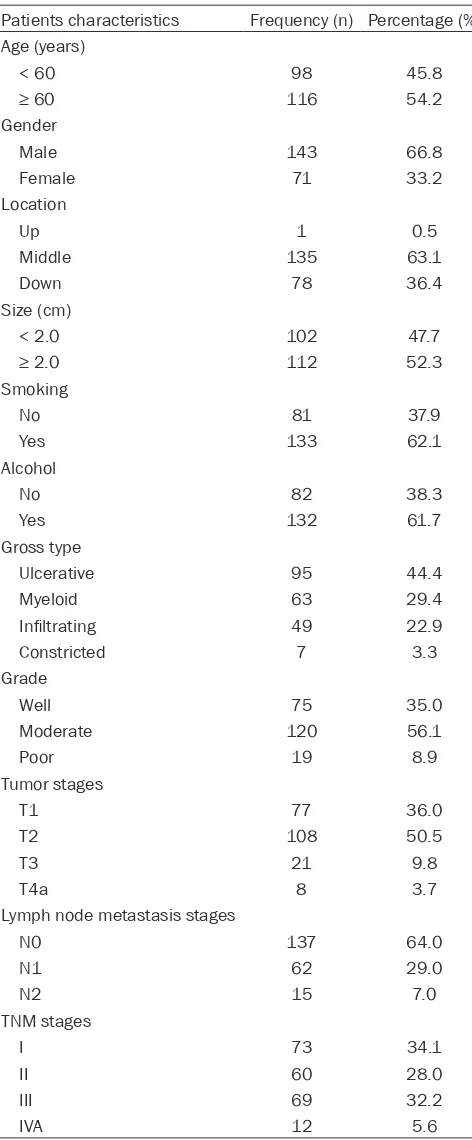
Table 1. Patients characteristics
Patients characteristics Frequency (n) Percentage (%) Age (years)
< 60 98 45.8
≥ 60 116 54.2
Gender
Male 143 66.8
Female 71 33.2
Location
Up 1 0.5
Middle 135 63.1
Down 78 36.4
Size (cm)
< 2.0 102 47.7
≥ 2.0 112 52.3
Smoking
No 81 37.9
Yes 133 62.1
Alcohol
No 82 38.3
Yes 132 61.7
Gross type
Ulcerative 95 44.4
Myeloid 63 29.4
Infiltrating 49 22.9
Constricted 7 3.3
Grade
Well 75 35.0
Moderate 120 56.1
Poor 19 8.9
Tumor stages
T1 77 36.0
T2 108 50.5
T3 21 9.8
T4a 8 3.7
Lymph node metastasis stages
N0 137 64.0
N1 62 29.0
N2 15 7.0
TNM stages
I 73 34.1
II 60 28.0
III 69 32.2
IVA 12 5.6
such as N-cadherin (CDH2), vimentin, and Snail [9, 10]. Snail is a key tran-scriptional regulator which promotes EMT. Overexpression of Snail directly activates EMT and promotes tumor metastasis through suppressing the transcription of E-cadherin. Overexpre- ssion of Snail also promotes tumor cell proliferation [11]. Recent studies have demonstrated that Snail plays impor-tant roles in serials of fundamental biologic behaviors, such as EMT, cells growth, and metastasis [11-13].
KAI1, also named CD82, was originally considered as a suppressor of tumor metastasis in prostate cancer cell lines in 1999. KAI1 gene belongs to tetraspanin superfamily which consists of four transmembrane domains [14]. Normal expression of KAI1 can sup-press cell growth, motility, migration, and strengthen cell to cell adhesion by strengthening stabilization of E-cad- herin-β-catenin complex [15-17]. It has been also confirmed that overexpres-sion of KAI1 could suppress secondary metastases without interfering primary tumor growth [18]. KAI1 also plays im- portant roles in the process of tumor initiation, invasion, and metastasis in various cancers.
The aim of this study is to evaluate the expression of MACC1, Snail, and KAI1 in the esophageal squamous cell carcinoma (ESCC) tissues of patients and their relationships between clini- copathologic characteristics and prog-nosis of patients with ESCC. Immuno- histochemical staining was used to as- sess the expression of MACC1, Snail, and KAI1 in ESCC specimens and the corresponding adjacent normal esoph-ageal mucosa specimens of patients with ESCC.
Methods
tients who had any history of chemo-therapy or radio-therapy were excluded. All ESCC patients provided written consent for their samples to be used. The study was approved by the Beng- bu Medical University ethics committee before it started and performed in accordance with the Declaration of Helsinki guidelines. Patients’ clinicopathologic, demographic, and follow-up data (at 4-month intervals by telephone, mobile phone or social applications) were collected. Overall survival (OS) time was calculated from surgery date to death day or December 2017 (mean OS time: 45.3 months; range: 10-71 months). TNM stages, tumor stages, and LNM stages were assessed in accordance with the 8th edition of the guidelines issued by the American Joint Committee on Cancer (AJCC). Tumor grades were assessed in accordance with the standards issued by the World Health Organization (WHO). Specific clinicopathologic characteristics are shown in Table 1.
Immunohistochemistry
All ESCC tissues and the correspondence nor-mal esophageal mucosa tissues were fixed in 10% buffered formalin, embedded in paraffin, and then cut into 4 μm thick slices. Subsequ- ently, all slices were deparaffinized with xylene and dehydrated with graded ethanol. Immuno-
histochemical staining was carried out in ac- cordance with ElivisionTM Plus detection kit in- structions (Lab Vision, USA). Endogenous per-oxidase activity was quenched by methanol containing 3% H2O2 solution. Then, citrate buf-fer solution was used to repair antigen and goat serum was used for blocking. MACC1 (rabbit polyclonal antibody, Santa Cruz Biote- chnology, USA), Snail (mouse monoclonal anti-body, Abcam, USA), and KAI1 (mouse monoclo-nal antibody, Santa Cruz Biotechnology, USA) primary antibodies were added and then incu-bated overnight at 4°C. Lastly, reagent A (en- hancer) and reagent B were added. All slices were to develop in diaminobenzidine substrate and counterstained with hematoxylin.
Evaluation of immunostaining
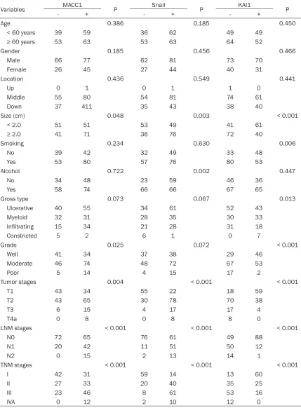
[image:3.612.90.528.72.288.2]Table 2. The associations between expression of MACC1, Snail, and KAI1 and clinicopathological characteristics of esophageal squamous cell carcinoma (ESCC)
Variables MACC1 P Snail P KAI1 P
- + - + - +
Age 0.386 0.185 0.450
< 60 years 39 59 36 62 49 49
≥ 60 years 53 63 53 63 64 52
Gender 0.185 0.456 0.466
Male 66 77 62 81 73 70
Female 26 45 27 44 40 31
Location 0.436 0.549 0.441
Up 0 1 0 1 1 0
Middle 55 80 54 81 74 61
Down 37 411 35 43 38 40
Size (cm) 0.048 0.003 < 0.001
< 2.0 51 51 53 49 41 61
≥ 2.0 41 71 36 76 72 40
Smoking 0.234 0.630 0.006
No 39 42 32 49 33 48
Yes 53 80 57 76 80 53
Alcohol 0.722 0.002 0.447
No 34 48 23 59 46 36
Yes 58 74 66 66 67 65
Gross type 0.073 0.067 0.013
Ulcerative 40 55 34 61 52 43
Myeloid 32 31 28 35 30 33
Infiltrating 15 34 21 28 31 18
Constricted 5 2 6 1 0 7
Grade 0.025 0.072 < 0.001
Well 41 34 37 38 29 46
Moderate 46 74 48 72 67 53
Poor 5 14 4 15 17 2
Tumor stages 0.004 < 0.001 < 0.001
T1 43 34 55 22 18 59
T2 43 65 30 78 70 38
T3 6 15 4 17 17 4
T4a 0 8 0 8 8 0
LNM stages < 0.001 < 0.001 < 0.001
N0 72 65 76 61 49 88
N1 20 42 11 51 50 12
N2 0 15 2 13 14 1
TNM stages < 0.001 < 0.001 < 0.001
I 42 31 59 14 13 60
II 27 33 20 40 35 25
III 23 46 8 61 53 16
IVA 0 12 2 10 12 0
staining is 2; moderate staining is 3; strong
Table 3. Correlation among expression of MACC1, Snail, and KAI1 in ESCC
Variable MACC1 r P Snail r P
- + - +
MACC1 0.244 < 0.001*
- 51 41
+ 38 84
KAI1 -0.503 < 0.001@ -0.608 < 0.001@
- 22 91 15 98
+ 70 31 74 27
*: positive association; @: negative association.
Statistical analysis
All data were analyzed using SPSS 19.0 soft-ware (Chicago, IL, US) for Windows. Countable data was conducted using the Chi-square test or Fisher’s exact test for comparisons between two groups. Univariate OS analysis was con-ducted using the Kaplan-Meier method with log-rank test. Multivariate OS analysis was con-ducted using Cox regression model test. P < 0.05 was considered a significant difference.
Results
Associations between MACC1, Snail, and KAI1 in the specimens of ESCC for patients and
clinicopathologic characteristics
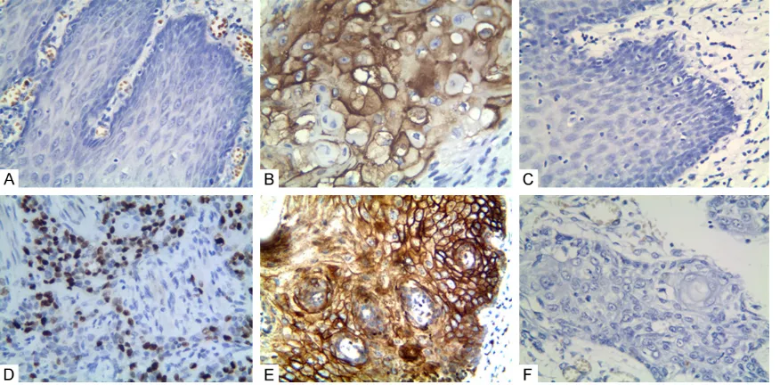
As shown in Figure 1A and 1B, positive expres-sion of MACC1 protein was mainly located at the cytoplasm. The positive rate of MACC1 ex- pression in the ESCC group (57.0%, 122/214) was significantly higher than that in the con- trol group (7.9%, 17/214; P < 0.001). Moreover, the positive rate of MACC1 in ESCC was posi-tively associated with tumor size, grade of dif-ferentiation, tumor stage, lymph node metasta-sis (LNM) stage, and tumor-node-metastametasta-sis (TNM) stage (Table 2). There was no associa-tion between positive rate of MACC1 expres-sion and ESCC patient’s age, gender, tumor location, type, smoking, or alcohol.
As shown in Figure 1C and 1D, positive expres-sion of Snail protein was mainly located at the nucleus. The positive rate of Snail expression in the cancer group (58.4%, 125/214) was sig-nificantly higher than that in the control group (7.0%, 15/214; P < 0.001). Furthermore, the positive rate of Snail in cancer was positively associated with tumor size, alcohol, tumor
stag-es, LNM stagstag-es, and TNM stages. There was no association between positive rate of Snail expression and patient’s age, gender, tumor loca-tion, type, grades of differentialoca-tion, and smoking (Table 2).
As shown in Figure 1E and 1F, posi-tive expression of KAI1 was mainly located at the membrane and cyto-plasm. The positive rate of KAI1 expression in cancer group (47.2%, 101/214) was significantly lower th- an that in the control group (93.5%, 200/214; P < 0.001). The positive rate of KAI1 in cancer was inversely associated with tumor type, size, smoking, grades of differentiation, LNM stages, tumor stages, and TNM stages. And there was no association between KAI1 expression and patient’s age, gender, alcohol, and tumor location (Table 2).
Associations among MACC1, Snail, and KAI1 in ESCC
There was a negative association between KAI1 expression and MACC1 expression (r = -0.503, P < 0.001) or Snail expression (r = -0.608, P < 0.001). There was a positive asso-ciation between MACC1 expression and Snail expression (r = 0.244, P < 0.001) (Table 3).
Univariate and multivariate analyze
Discussion
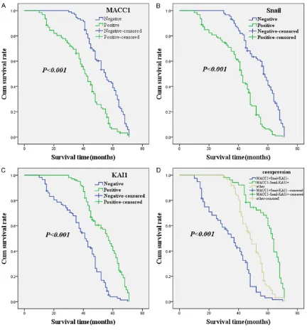
Esophageal cancer is the second most com-mon malignant tumor of the digestive system in China [1]. Esophageal squamous cell carcino-Figure 2. Kaplan-Meier analysis curve of the survival rate of patients with ESCC. The y-axis means the percentage of patients; the x-axis means their survival in months. A: OS analysis of all patients in relation to MACC1 (log-rank = 36.601, P < 0.001); B: OS analysis of all patients in relation to Snail expression (log-rank = 56.305, P < 0.001); C: OS analysis of all patients in relation to KAI1 expression (log-rank = 54.476, P < 0.001); A-C analyses, the green line represents patients with positive MACC1, or Snail, or KAI1; the blue line representing the negative MACC1, or Snail, or KAI1 group. D: OS survival of all patients in relation to the combination of KAI1, MACC1, and Snail expression (log-rank = 85.730, P < 0.001). The green line represents negative KAI1 and positive MACC1 and Snail. The blue line represents positive KAI1 and negative MACC1 and Snail. The brown line represents other positive or negative proteins.
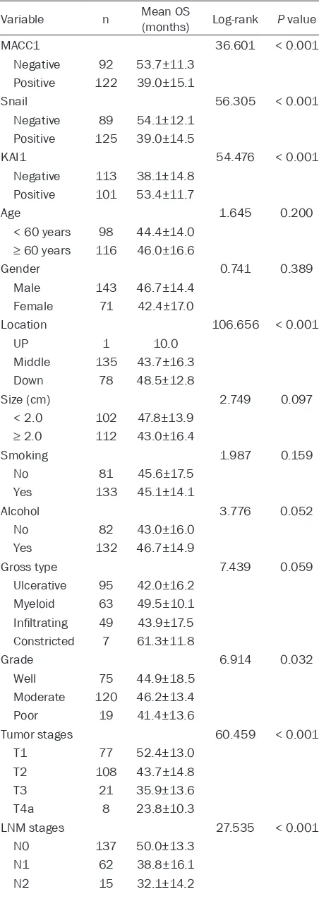
[image:6.612.91.523.71.538.2]Table 4. Results of univariate analyses of overall sur-vival (OS) time
Variable n Mean OS (months) Log-rank P value
MACC1 36.601 < 0.001
Negative 92 53.7±11.3 Positive 122 39.0±15.1
Snail 56.305 < 0.001
Negative 89 54.1±12.1 Positive 125 39.0±14.5
KAI1 54.476 < 0.001
Negative 113 38.1±14.8 Positive 101 53.4±11.7
Age 1.645 0.200
< 60 years 98 44.4±14.0 ≥ 60 years 116 46.0±16.6
Gender 0.741 0.389
Male 143 46.7±14.4
Female 71 42.4±17.0
Location 106.656 < 0.001
UP 1 10.0
Middle 135 43.7±16.3
Down 78 48.5±12.8
Size (cm) 2.749 0.097
< 2.0 102 47.8±13.9
≥ 2.0 112 43.0±16.4
Smoking 1.987 0.159
No 81 45.6±17.5
Yes 133 45.1±14.1
Alcohol 3.776 0.052
No 82 43.0±16.0
Yes 132 46.7±14.9
Gross type 7.439 0.059
Ulcerative 95 42.0±16.2 Myeloid 63 49.5±10.1 Infiltrating 49 43.9±17.5 Constricted 7 61.3±11.8
Grade 6.914 0.032
Well 75 44.9±18.5
Moderate 120 46.2±13.4
Poor 19 41.4±13.6
Tumor stages 60.459 < 0.001
T1 77 52.4±13.0
T2 108 43.7±14.8
T3 21 35.9±13.6
T4a 8 23.8±10.3
LNM stages 27.535 < 0.001
N0 137 50.0±13.3
N1 62 38.8±16.1
N2 15 32.1±14.2
ma (ESCC) which accounts for approxi-mately 90% is the most common type of esophageal cancer. ESCC is a high hete- rogeneity disease which influences its comprehensive evaluation of biomarkers. It has demonstrated that MACC1 should promote cells growth, motility, and migra-tion and be involved in the process of in- vasiveness and metastasis of cancers [2, 3]. In this study, the results showed that positive rate of MACC1 expression in ES- CC was positively associated with tumor size, grade of differentiation, tumor stage, LNM stage, and TNM stage. Moreover, patients with MACC1 positive expression had an unfavorable OS time when com-pared with patients with MACC1 nega- tive. These indicated that MACC1 should play an important role in invasion and metastasis of ESCC and be considered a useful biomarker for prediction of progno-sis [2-6, 19-21].
Epithelial-mesenchymal transition (EMT) is a process by which cells gain the ability to invade through the basement membrane appears as part of normal embryonic de- velopment and tumor development [22, 23]. EMT has been characterized by losing epithelial features and gaining mesenchy-mal features. It can allow tumor cells leave the primary tumor to promote invasion and metastasis. Snail is a critical transcription-al regulator of EMT. In this study, we found that overexpression of Snail was positively associated with tumor size, grade of differ-entiation, tumor stage, LNM stage, as well as TNM stage. Furthermore, we also found that overexpression of Snail for patients had an unfavorable OS time when com-pared with negative expression of Snail for patients. The above results suggested that Snail should be considered a useful and effective biomarker for invasion and metastasis as well as in prediction of prog-nosis [12, 24, 25].
differ-entiation, tumor stages, LNM stages, and TNM stages. Kaplan-Meier analysis indicated that positive expression of KAI1 for patients had a favorable OS time when compared with nega-tive expression of KAI1 with patients. These results demonstrated that aberrant expression (down or lost) of KAI1 should promote tumor cells invasion and metastasis, therefore, should be considered a potential predictor for invasive-ness and metastasis of ESCC, as well as prog-nosis [16, 18, 26-28].
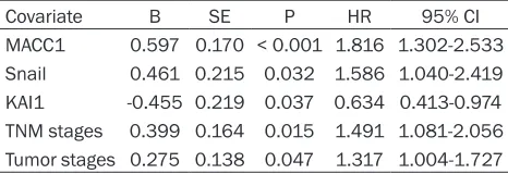
In this study, Kaplan-Meier analysis showed that positive expression of MACC1, Snail, and KAI1 significantly associated with OS time in patients with ESCC. Multivariate analysis indi-cated that tumor stages, TNM stages, positive expression of MACC1, Snail, and KAI1 should be involved in the process of invasiveness and metastasis of ESCC, as well as should be con-sidered a useful predictor of prognosis. Abnormal KAI1 expression should be involved in the initiation and recurrence of ESCC through its involvement in suppressor genes. KAI1 can suppress β-catenin tyrosine phosphorylation and stabilize E-cadherin-β-catenin complexes to inhibit tumor metastasis [17]. Furthermore, KAI1 can also inhibit the EMT process of a tumor [27]. Overexpression of MACC1 should be involved in the tumorigenesis by activation of HGF/MET signaling pathway [2, 3]. MACC1 also promotes EMT of tumor and suppresses apoptosis by activation of HGF/MET pathway [27, 29]. Overexpression of Snail directly acti-vates EMT and promotes tumor metastasis by suppressing the transcription of E-cadherin
This study was supported by the Nature Sci- ence Foundation of Anhui Province (No. 1708- 085MH230).
Disclosure of conflict of interest
None.
Address correspondence to: Shiwu Wu, Department of Pathology, Bengbu Medical University, No. 287, Changhuai Road, Anhui Province, China. Tel: +86-13705523357; E-mail: 573448542@qq.com
References
[1] Chen W, Zheng R, Baade PD, Zhang S, Zeng H, Bray F, Jemal A, Yu XQ, He J. Cancer statistics in China, 2015. CA Cancer J Clin 2016; 66: 115-32.
[2] Stein U, Walther W, Arlt F, Schwabe H, Smith J, Fichtner I, Birchmeier W, Schlag PM. MACC1, a newly identified key regulator of HGF-MET sig -naling, predicts colon cancer metastasis. Nat Med 2009; 15: 59-67.
[3] Stein U, Smith J, Walther W, Arlt F. MACC1 con -trols met: what a difference an Sp1 site makes. Cell Cycle 2009; 8: 2467-9.
[4] Ren B, Zakharov V, Yang Q, McMahon L, Yu J, Cao W. MACC1 is related to colorectal cancer initiation and early-stage invasive growth. Am J Clin Pathol 2013; 140: 707-7.
[5] Zhou L, Yu L, Zhu B, Wu S, Song W, Gong X, Wang D. Metastasis-associated in colon can-cer-1 and aldehyde dehydrogenase 1 are met-astatic and prognostic biomarker for non-small cell lung cancer. BMC Cancer 2016; 16: 876. [6] Chundong G, Uramoto H, Onitsuka T,
Shimo-kawa H, Iwanami T, Nakagawa M, Oyama T, Tanaka F. Molecular diagnosis of MACC1 sta -tus in lung adenocarcinoma by immunohisto-chemical analysis. Anticancer Res 2011; 31:
TNM stages 59.662 < 0.001
I 73 54.4±12.3
II 60 46.6±12.1
III 69 37.1±15.3
[image:8.612.90.323.185.265.2]IVA 12 31.0±13.7
Table 5. Results of multivariate analyses of overall survival (OS) time
Covariate B SE P HR 95% CI
MACC1 0.597 0.170 < 0.001 1.816 1.302-2.533 Snail 0.461 0.215 0.032 1.586 1.040-2.419 KAI1 -0.455 0.219 0.037 0.634 0.413-0.974 TNM stages 0.399 0.164 0.015 1.491 1.081-2.056 Tumor stages 0.275 0.138 0.047 1.317 1.004-1.727
tumor cells leave the primary tumor to pro-mote invasion and metastasis. Aberrant expression of KAI1 should lost or decrease its ability to suppress tumor cells invasive-ness and metastasis [27-29].
Conclusions
This study indicated that expression of MACC1, Snail, and KAI1 were associated with duration of OS time among patients with ESCC. So, MACC1, Snail, and KAI1 should considered as valuable biomarkers in ESCC and may be helpful for the predic-tion of metastasis and prognosis for ESCC.
[7] Mitra R, Chen X, Greenawalt EJ, Maulik U, Jiang W, Zhao Z, Eischen CM. Decoding critical long non-coding RNA in ovarian cancer epithelial-to-mesenchymal transition. Nat Commun 2017; 8: 1604.
[8] Yang D, Sun Y, Hu L, Zheng H, Ji P, Pecot CV, Zhao Y, Reynolds S, Cheng H, Rupaimoole R, Cogdell D, Nykter M, Broaddus R, Rodriguez-Aguayo C, Lopez-Berestein G, Liu J, Shmulev-ich I, Sood AK, Chen K, Zhang W. Integrated analyses identify a master microRNA regulato-ry network for the mesenchymal subtype in serous ovarian cancer. Cancer Cell 2013; 23: 186-99.
[9] Lamouille S, Xu J, Derynck R. Molecular mech-anisms of epithelial-mesenchymal transition. Nat Rev Mol Cell Biol 2014; 15: 178-96. [10] Kurahara H, Takao S, Maemura K, Mataki Y,
Kuwahata T, Maeda K, Ding Q, Sakoda M, Iino S, Ishigami S, Ueno S, Shinchi H, Natsugoe S. Epithelial-mesenchymal transition and mesen-chymal-epithelial transition via regulation of ZEB-1 and ZEB-2 expression in pancreatic can-cer. J Surg Oncol 2012; 105: 655-61.
[11] Zou QF, Cao LY, Yu T, Gong L, Wang LN, Zhou YL, Xiao B, Zou QM. MicroRNA-22 inhibits tu -mor growth and metastasis in gastric cancer by directly targeting MMP14 and Snail. Cell Death Dis 2015; 6: e2000.
[12] Taki M, Abiko K, Baba T, Hamanishi J, Yamagu-chi K, Murakami R, Yamanoi K, Horikawa N, Hosoe Y, Nakamura E, Sugiyama A, Mandai M, Konishi I, Matsumura N. Snail promotes ovari-an covari-ancer progression by recruiting myeloid-derived suppressor cells via CXCR2 ligand up-regulation. Nat Commun 2018; 9: 1685. [13] Hojo N, Huisken AL, Wang H, Chirshev E, Kim
NS, Nguyen SM, Campos H, Glackin CA, Ioffe YJ, Unternaehrer JJ. Snail knockdown reverses stemness and inhibits tumour growth in ovari-an covari-ancer. Sci Rep 2018; 8: 8704.
[14] Levy S, Shoham T. The tetraspanin web modu-lates immune-signalling complexes. Nat Rev Immunol 2005; 5: 136-48.
[15] Miranti CK. Controlling cell surface dynamics and signaling: how CD82/KAI1 suppresses metastasis. Cell Signal 2009; 21: 196-211. [16] Malik FA, Sanders AJ, Jiang WG. KAI-1/CD82,
the molecule and clinical implication in cancer and cancer metastasis. Histol Histopathol 2009; 24: 519-530.
[17] Abe M, Sugiura T, Takahashi M, Ishii K, Shi-moda M, Shirasuna K. A novel function of CD82/KAI-1 on E-cadherin-mediated homo-philic cellular adhesion of cancer cells. Cancer Lett 2008; 266: 163-70.
[18] Lee J, Byun HJ, Lee MS, Jin YJ, Jeoung D, Kim YM, Lee H. The metastasis suppressor CD82/ KAI1 inhibits fibronectin adhesion-induced ep -ithelial-to-mesenchymal transition in prostate cancer cells by repressing the associated
inte-[19] Qian LQ, Li XQ, Ye PH, Su HY, Wang G, Liu Y, Shen GH, Gao QG. Downregulation of MACC1 inhibits the viability, invasion and migration and induces apoptosis in esophageal carcino-ma cells through the phosphatase and ten- sin homolog/phosphoinositide 3-kinase/pro-tein kinase B signaling pathway. Oncol Lett 2017; 14: 4897-905.
[20] Yang YP, Ou JH, Chang XJ, Lu YY, Bai WL, Dong Z, Wang H, An LJ, Xu ZX, Wang CP, Zeng Z, Hu KQ. High intratumoral metastasis-associated in colon cancer-1 expression predicts poor out-comes of cryoablation therapy for advanced hepatocellular carcinoma. J Transl Med 2013; 11: 41.
[21] Zhu B, Wang Y, Wang X, Wu S, Zhou L, Gong X, Song W, Wang D. Evaluation of the correlation of MACC1, CD44, Twist1, and KiSS-1 in the metastasis and prognosis for colon carcinoma. Diagn Pathol 2018; 13: 45.
[22] Nieto MA, Huang RY, Jackson RA, Thiery JP. EMT: 2016. Cell 2016; 166: 21-45.
[23] Heerboth S, Housman G, Leary M, Longacre M, Byler S, Lapinska K, Willbanks A, Sarkar S. EMT and tumor metastasis. Clin Transl Med 2015; 4: 6.
[24] Chang HY, Tseng YK, Chen YC, Shu CW, Lin MI, Lin MI, Liou HH, Fu TY, Ger LP, Yeh MH, Liu PF. High snail expression predicts a poor progno-sis in breast invasive ductual carcinoma pa-tients with HER2/EGFR-positive subtypes. Surg Oncol 2018; 27: 314-20.
[25] Li M, Zhang X, Hu K, Shi M, Dong G, Li D, Zhang P. Prognostic role of snail in lung cancer: proto-col for a systematic review. Medicine (Balti-more) 2018; 97: e11539.
[26] Zhu J, Miao C, Liu S, Tian Y, Zhang C, Liang C, Xu A, Cao Q, Wang Z. Prognostic role of CD82/ KAI1 in multiple human malignant neoplasms: a meta-analysis of 31 studies. Onco Targets Ther 2017; 10: 5805-16.
[27] Lu G, Zhou L, Zhang X, Zhu B, Wu S, Song W, Gong X, Wang D, Tao Y. The expression of me-tastasis-associated in colon cancer-1 and KAI1 in gastric adenocarcinoma and their clinical significance. World J Surg Oncol 2016; 14: 276.
[28] Zhu B, Zhou L, Yu L, Wu S, Song W, Gong X, Wang D. Evaluation of the correlation of vascu-logenic mimicry, ALDH1, KAI1 and microvessel density in the prediction of metastasis and prognosis in colorectal carcinoma. BMC Surg 2017; 17: 47.