Original Article
Inter- and intra-observer reliability in
histologic evaluation of necrosis rate induced
by neo-adjuvant chemotherapy for osteosarcoma
Jin-Woo Kang1, Seung Han Shin1, Joon Hyuk Choi2, Kyung Chul Moon3, Jae Soo Koh4, Chan kwon Jung5,6,
Yong-Koo Park7, Kyi Beom Lee8, Yang-Guk Chung1
1Department of Orthopedic Surgery, Seoul St. Mary’s Hospital, College of Medicine, The Catholic University of Korea, Seoul, Republic of Korea; 2Department of Pathology, Yeungnam University College of Medicine, Daegu, Republic of Korea; 3Department of Pathology, Seoul National University College of Medicine, Seoul, Republic of Korea; 4Department of Pathology, Korea Cancer Center Hospital, Seoul, Republic of Korea; 5Department of Hospi-tal Pathology, 6Cancer Research Institute, College of Medicine, The Catholic University of Korea, Seoul, Republic of Korea; 7Department of Pathology, Kyung Hee University College of Medicine, Seoul, Republic of Korea; 8 Depart-ment of Pathology, Ajou University School of Medicine, Suwon, Republic of Korea
Received October 20, 2016; Accepted October 27, 2016; Epub January 1, 2017; Published January 15, 2017
Abstract: Purpose: Tumor necrosis rate following neo-adjuvant chemotherapy is one of the most important prog-nostic factors for patients with osteosarcoma, and it also provides the basis for selection of postoperative adjuvant chemotherapy. However, reported necrosis rates for the same tumors can vary among pathologists, complicating decision making for further treatment. Methods: Ten H&E stained pathology slides from 10 osteosarcoma patients treated with neo-adjuvant chemotherapy were randomly selected. Six expert pathologists were assigned to analyze the slides for tumor necrosis rate at four time points, with an interval of 3 weeks. Intraclass and interclass
correla-tion coefficients (IntraCC & InterCC) and 95% confidence intervals (CI) were calculated. Results: The overall InterCC among the 6 observers was 0.652 (95% CI 0.294-0.820) for tumor necrosis rate (range: 0-100%), suggesting good
reliability. IntraCCs were 0.799, 0.788, 0.867, 0.935, 0.962, and 0.947 respectively. The interCC among higher
careers and lower careers were 0.603 (95% CI.: 0.387-0.843) and 0.696 (95% C.I.: 0.487-0.919), respectively, which was not significantly different. Major differences in tumor necrosis estimation were due to interpretation of areas with isolated atypical cells in fibrotic stroma. Conclusion: Low inter-observer and relatively high intra-observer
reliability were observed in the histologic evaluation of necrosis rate after neo-adjuvant chemotherapy. The interCCs
between the higher career and lower career groups did not vary. These findings suggest that a valid measurement
protocol for tumor necrosis rate evaluation after chemotherapy is required to improve the clinical relevance of the
quantification of response to neo-adjuvant therapy.
Keywords: Osteosarcoma, tumor necrosis, chemotherapy response, observer variation
Introduction
Chemotherapy for osteosarcoma has dramati-cally improved the survival rate of osteosarco-ma. The 5-year survival ratefor patients with localized disease has improved from 20 to 70% in the last three decades [1]. One of the most important prognostic factors for osteosarcoma is the percentage of tumor necrosis induced by neo-adjuvant chemotherapy [2]. This factor also provides the basis for selecting postopera-tive adjuvant chemotherapy and estimating the clinical relevance of other evaluation methods
for chemotherapy response, such as diffusion MRI or 18F-FDG PET CT. Many studies suggested
Reliability of necrosis rate for osteosarcoma
patients achieved 90% or greater tumor necro -sis, which was much lower than in prior studies showing 50% or more good responders. How-ever, their 5-year overall survival rate was 65.9%, which is similar to those of previous studies [4, 12, 13]. Several studies reported the discrepancy between tumor necrosis and survival rate and have concluded that his- tologic response to chemotherapy has lost its prognostic importance, despite the contribu-tion of chemotherapy to osteosarcoma treat-ment [5, 7-11, 14-16]. These studies recom-mended further prospective randomized and controlled studies with large samples to in- vestigate the relationship between percentage of tumor necrosis and survival of osteosar- coma patients.
Large discrepancies have been reported in necrosis rates for the same tumors among pathologists. We presumed that the loss of the clinical relevance of necrosis rate might have originated from disagreement in the histologic interpretation of tumor necrosis rate. To the best of our knowledge, there has been no study evaluating this issue.
The purpose of this study was to evaluate the inter- and intra-observer reliability in histopath-ological estimation of the necrosis rate after neo-adjuvant chemotherapy for osteosarcoma and to revisit the clinical relevance of the necrosis rate induced by neo-adjuvant therapy.
Methods
Patients and pathology
The Institutional Review Board of our hospital approved this study. Ten consecutive patients who underwent two cycles of neo-adjuvant che-motherapy and sequential surgery for conven-tional osteosarcoma at Seoul St. Mary’s Hos- pital between 2012 and 2014 were enrolled. Histologic subtypes were as follows: osteoblas-tic (n=6), fibroblasosteoblas-tic (n=2), and mixed osteo -blastic and chondro-blastic (n=2). A single repre-sentative hematoxylin and eosin-stained path- ology slide of the osteosarcoma was selected from each case. One pathologist (CKJ) was responsible for slide selection. The glass slides were assigned random case numbers and sent to six pathologists.
Observers
[image:2.612.90.523.84.187.2]Six expert pathologists were assigned to ana-lyze the slides for tumor necrosis rate (Table 1). They majored in musculoskeletal oncology, and four of six observers diagnosed 10 or more
Table 1. Careers of the six observers
Observer Years* Career in musculoskeletal pathology** No. of osteosarcoma cases evaluated/year† Study abroad Y/N (period)‡
#1 1991 > 15 years 4-6 cases Yes (6 months)
#2 2002 5-10 years ≥11 cases No
#3 1995 > 15 years ≥11 cases No
#4 2002 10-15 years ≥11 cases No
#5 1982 > 15 years ≥11 cases Yes (2 years)
#6 1986 > 15 years ≥11 cases Yes (2 years)
*Year professional certification in pathology was acquired. **Career in musculoskeletal pathology. †Number of osteosarcoma
cases diagnosed each year. ‡Experience studying abroad for a sub-specialty in musculoskeletal pathology (#1 and #5, Mayo Clinic; #6, Albert Einstein College of Medicine and Hospital for Special Surgery).
[image:2.612.94.283.241.467.2]cases of osteosarcoma every year with careers of over 10 years in musculoskeletal pathology. The mean pathology career length was 22.8 years (range, 14 to 34 years). Three observers experienced abroad study of musculoskeletal oncology as a sub-specialty.
Evaluation flow design
Observers evaluated the tumor necrosis rate of each slide at 4 time points with a delay of at least 3 weeks to minimize recall bias. Every time the slides were evaluated, randomized numbers were assigned to each slide. After the second evaluation, the six observers met to share opinions and establish a consensus for estimating tumor necrosis rate. Until matched evaluation sheets of each pathologist were obtained for statistical analysis, pathologists and other authors were blinded to randomized numbers of slides and patient information (Figure 1).
Statistical analysis
Statistical analyses were conducted with SPSS 18.0 software for Windows (IBM, Armonk, NY, USA). The means, standard deviations, intra-class and interintra-class correlation coefficients
(IntraCC and interCC), and 95% confidence intervals (CIs) were calculated [17]. To evaluate the effect of experience with histologic diagno-sis of osteosarcoma on estimation of necrodiagno-sis rate, the observers were divided into two groups (higher career and lower careergroups). The interCC and intraCC of those two groups were compared. ICC values were interpreted as follows: ICC between 0.81 and 1.0 was consid-ered ‘excellent’, between 0.61 and 0.8 was considered ‘good’, between 0.41 and 0.6 was considered ‘fair to moderate’, and below 0.4 was considered ‘poor’ [18]. Significance was set at a p value of<0.05.
Results
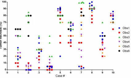
The ICC across 6 observers was 0.652 (95% CI, 0.455-0.865) through all 4 time evaluations, suggesting good reliability; ICC was 0.659 (95% CI, 0.447-0.871) at the first and second evalua -tions and 0.631 (95% CI, 0.414-0.858) at the third and fourth evaluations after a consensus meeting, which was not a significant improve -ment. Furthermore, percentages of necrosis ranged from 0 to 85% for slide #2 and from 2 to 100% for slide #7 (Figure 2). This is sufficiently different to confuse clinicians and create diffi -culty in determining further chemotherapy pro-Figure 2. Dot plot showing a dataset from four evaluations of six pathologists. The most discrepant data were from
[image:3.612.97.522.74.327.2]Reliability of necrosis rate for osteosarcoma
like change indicating partial chemotherapy effectiveness and partial tumor necrosis.
Discussion
The histologic percentage of tumor necrosis after neo-adjuvant chemotherapy in osteosar-coma patients is the most important factor for predicting prognosis and determining further chemotherapy regimens [2, 3, 13]. It also used as a basis to estimate the clinical relevance of other evaluation methods for chemotherapy response, such as diffusion MRI or PET CT. Many studies have defined a good response as 90% or more tumor necrosis [1-3]. A good responder is usually given the same chemo-therapy regimen postoperatively, while other salvage chemotherapy regimens should be considered for poor responders [1, 5, 6, 8-10]. Because chemotherapy drugs used in osteo-sarcoma are limited compared to other carcino-mas, the histologic response to standard che-motherapy has considerable importance in osteosarcoma treatment. Clinicians feel quite differently about even a 10 to 20% difference in necrosis rates, which may significantly change treatment strategy [1].
Histological evaluation of tumor necrosis is considered the gold standard method for deter-mining response to preoperative chemotherapy agents. In addition to the histological necrosis rate, many studies have introduced tools such as 18F-FDG PET/CT, MRI diffusion-weighted
imaging (DWI), and bone scan as novel meth-ods to evaluate chemotherapy response in patients with osteosarcoma [19-23]. All studies compared their results with histopathological response rate because it has a confirmative meaning for clinicians, radiologists, and radia-tion oncologists. However, if histopathological response rates lose their consistency, they will not have a standard value and will lose their usefulness.
The tumor necrosis rate is evaluated on the basis of the amount of remaining viable tumor in the resected specimen. Rosen and Huvos et al. described a method for evaluating tumor necrosis and divided tumor response into four grades [24, 25]. Salzer-Kuntschik et al. divided response into six grades. Those methods are based on random sampling of slides, which is thought to be representative of the entire tumor [26]. Each slide was analyzed and a mean per-tocols in a clinical situation. The InterCC
limit-ing those two slides among the 6 observers was 0.268 (95% CI, 0.054-0.997). From the first to the sixth observer, intrarater data were 0.799 (95% CI, 0.583-0.937), 0.788 (95% CI, 0.563-0.933), 0.867 (95% CI, 0.703-0.960), 0.935 (95% CI, 0.844-0.981), 0.962 (95% CI, 0.906-0.989), and 0.947 (95% CI, 0.845-0.985), respectively. At the first and second evaluations, intrarater data were 0.595 (95% CI, 0.051-0.877), 0.867 (95% CI, 0.570-0.965), 0.842 (95% CI, 0.485-0.958), 0.952 (95% CI, 0.831-0.988), 0.984 (95% CI, 0.938-0.996), and 0.969 (0.887-0.992), respectively. At the third and fourth evaluations after a consensus meeting, intrarater data were 0.998 (95% CI, 0.991-0.999), 0.852 (95% CI, 0.429-0.963), 0.834 (95% CI, 0.486-0.956), 0.988 (95% CI, 0.954-0.997), 0.972 (95% CI, 0.889-0.993), and 0.911 (95% CI, 0.545-0.979), respectively. There were no differences in intraCC before and after the consensus discussion.
The InterCC of the higher career group was 0.603 (95% CI, 0.387-0.843), and that of the lower career group was 0.696 (95% CI, 0.487-0.919). The intraCC was 0.799 (95% CI, 0.583-0.937), 0.867 (95% CI, 0.703-0.960), and 0.962 (95% CI, 0.906-0.989) in the higher career group and 0.788 (95% CI, 0.563-0.933), 0.935 (95% CI, 0.844-0.981), and 0.947 (95% CI, 0.845-0.985) in the lower career group. There were no statistically significant differenc -es between higher and lower career groups in both interCC and intraCC (P>0.05).
spectrum-centage of necrosis was mathematically calcu-lated by mapping on X-ray or gross specimen pictures. Raymond et al. suggested that the process by which specimens are analyzed is crude and subjective [3]. It is possible that slide
selection and necrosis judgment are subjective processes.
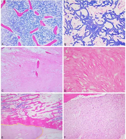
[image:5.612.87.525.71.554.2]Our study shows relatively high intra-observer reliability for histological evaluation of necrosis Figure 3. Typical histologic findings of necrotic and viable areas in osteosarcoma after neo-adjuvant chemotherapy. (A-D) Complete tumor necrosis showing acellular osteoid, necrotic or fibrotic materials. (A) Osteoblastic osteosar -coma after chemotherapy shows residual tumor osteoid and normal eosinophilic trabecular bone (×40). (B) At
high power magnification (×400), there is complete cell dropout with acellular osteoid and necrotic stroma.
Reliability of necrosis rate for osteosarcoma
rate after neo-adjuvant chemotherapy. How- ever, inter-observer reliability was lower. In so- me cases, the difference was high enough for clinicians to arrive at different treatment strate-gies. Despite long discussions, pathologist de- cisions did not coincide with each other on some points. Tumor necrosis is generally
[image:6.612.90.528.69.552.2]defin-ed as an absence of neoplastic cells. It is usu-ally represented by pathological findings such as cell dropout with residual matrix, or so called “ghost cells” [24-26]. However, pathologists are confused by some situations in which a few cells containing cytoplasmic vacuolization with bizarre change were surrounded by an area Figure 4. Isolated atypical cells with nuclear and cytoplasmic aberrations in necrotic or fibrotic stroma after osteo
-sarcoma chemotherapy. A-F. Images in right column show high-power magnification (×400) of the boxed areas in
the left column. Tumor cells are significantly dropped out. There are no mitotically active cells; however, there are a
largely replaced by fibrosis and granulation tis -sue. If these areas are considered viable, che-motherapy response may be underestimated. Michael et al. recommend considering only dense areas of mitotically-active atypical cells as viable [27]. In their study, disease-free sur-vival significantly correlated with tumor necro -sis rate when counting only dense mitotically-active atypical cells as viable (“stringent me- thod”) rather than widely distributed, rare, mito- tically inactive atypical cells. The stringent me- thod may be one of the possible solutions for improving the reliability of estimating tumor necrosis rate.
Li et al. urged caution in interpreting tumor necrosis after neo-adjuvant chemotherapy as an all-or-nothing phenomenon. They also sug-gested that a 10% viability cut-off does not indi -cate an absolute border beyond which chemo-therapy is no longer effective. They concluded that tumor necrosis showed a gradual effect on survival rate [4].
Some pathologists have suggested that it is better to select a method for interpreting tumor necrosis as an all-or-nothing phenome-non that does not acknowledge partial effec-tiveness just to improve inter-observer reliabili-ty. Many studies reported that tumor necrosis for chemotherapy response had a gradual effect on survival rate, so some have recom-mended interpretation on a spectrum that acknowledges partial effectiveness. However, this method of interpretation is more time consuming and there is no definite measure -ment protocol to reflect the degree of partial effectiveness. Many clinicians expect histo- logic evaluation of tumor necrosis to be more informative as a definitive report, and to make comparable with quantitative evaluation meth-ods such as 18F-FDG PET/CT.
All six observers in this study agreed that a consistent consensus is needed to improve inter-observer and intra-observer reliability. They also suggested that the creation of a valid measurement protocol for evaluating tumor necrosis is urgently needed. Some observers preferred to interpret tumor necrosis as a spec- trum; however, these pathologists thought that this method made it difficult to objectively cal -culate the ratio of tumor necrosis. Other observ-ers considered it better to interpret tumor necrosis as an all-or-none phenomenon beca-
use there are no unequivocal criteria for deter-mining the viability of bizarre cells. They thought these cells might represent degenerative ch- anges secondary to chemotherapy, but could also represent regenerative changes.
There is a recommendation that the best way to ensure reliable and consistent data on chemo-therapy response is to continuously refer path-ological specimens to the same pathologist. Some have argued that this is simple because most sarcoma patients are concentrated in tertiary care hospitals where clinicians and pathologists have worked well together over a long period.
There were several limitations to this study. First, it included a small number of slides and observers. Given the rarity of expert musculo-skeletal pathologists and the geographical dis-tance between them, it is difficult to obtain a large number of observers. However, to the best of our knowledge, this is the first attempt to evaluate the validity of histological evalua-tion of tumor necrosis. The second limitaevalua-tion of this study was that observers had only one con-sensus meeting to share their knowledge and experiences. Further meetings would be helpful for reaching a consensus to estimate necrosis rate. Another limitation was that the res- ponse rates were not correlated to patient sur-vival rates.
In summary, we observed lower inter-observer reliability regarding histological evaluation of necrosis rate after neo-adjuvant chemothera-py. At a consensus meeting, major differences among pathologists did not easily converge. These findings suggest that a valid measure -ment protocol for evaluating tumor necrosis rate is urgently needed in musculoskeletal tumor pathology. Pathology training programs should consider this issue to restore the clini- cal relevance of necrosis rate evaluation in osteosarcoma treatment.
Acknowledgements
Reliability of necrosis rate for osteosarcoma
Disclosure of conflict of interest
None.
Address correspondence to: Dr. Yang-Guk Chung, Department of Orthopedic Surgery, Seoul St. Mary’s Hospital, College of Medicine, The Catholic Univer- sity of Korea, 222 Banpo-daero, Seocho-gu, Seoul 06591, Republic of Korea. Tel: +82-2-2258-6116; Fax: +82-2-535-9834; E-mail: ygchung@catholic. ac.kr; Dr. Chan Kwon Jung, Department of Hos- pital Pathology, Seoul St. Mary’s Hospital, College of Medicine, The Catholic University of Korea, 222 Banpo-daero, Seocho-gu, Seoul 06591, Republic of Korea. Tel: 1622; Fax: +82-2-2258-1627; E-mail: [email protected]
References
[1] Carrle D and Bielack SS. Current strategies of chemotherapy in osteosarcoma. Int Orthop 2006; 30: 445-451.
[2] Davis AM, Bell RS and Goodwin PJ. Prognostic factors in osteosarcoma: a critical review. J Clin Oncol 1994; 12: 423-431.
[3] Raymond AK, Chawla SP, Carrasco CH, Ayala AG, Fanning CV, Grice B, Armen T, Plager C, Pa-padopoulos NE, Edeiken J, et al. Osteosarco-ma chemotherapy effect: a prognostic factor. Semin Diagn Pathol 1987; 4: 212-236. [4] Li X, Ashana AO, Moretti VM and Lackman RD.
The relation of tumour necrosis and survival in patients with osteosarcoma. Int Orthop 2011; 35: 1847-1853.
[5] Bacci G, Ferrari S, Bertoni F, Ruggieri P, Picci P, Longhi A, Casadei R, Fabbri N, Forni C, Versari M and Campanacci M. Long-term outcome for patients with nonmetastatic osteosarcoma of the extremity treated at the istituto ortopedico Rizzoli according to the istituto ortopedico Riz-zoli Osteosarcoma-2 protocol: an updated re-port. J Clin Oncol 2000; 18: 4016-4027. [6] Bacci G, Briccoli A, Ferrari S, Longhi A, Mercuri
M, Capanna R, Donati D, Lari S, Forni C and DePaolis M. Neoadjuvant chemotherapy for osteosarcoma of the extremity: long-term re-sults of the Rizzoli’s 4th protocol. Eur J Cancer 2001; 37: 2030-2039.
[7] Ferrari S, Bertoni F, Mercuri M, Picci P, Giaco-mini S, Longhi A and Bacci G. Predictive factors of disease-free survival for non-metastatic os-teosarcoma of the extremity: an analysis of 300 patients treated at the Rizzoli institute. Ann Oncol 2001; 12: 1145-1150.
[8] Smeland S, Muller C, Alvegard TA, Wiklund T, Wiebe T, Bjork O, Stenwig AE, Willen H, Holm-strom T, Folleras G, Brosjo O, Kivioja A, Jons-son K, Monge O and Saeter G. Scandinavian sarcoma group osteosarcoma study SSG VIII:
prognostic factors for outcome and the role of replacement salvage chemotherapy for poor histological responders. Eur J Cancer 2003; 39: 488-494.
[9] Le Deley MC, Guinebretière JM, Gentet JC, Pac-quement H, Pichon F, Marec-Bérard P, Entz-Werlé N, Schmitt C, Brugières L, Vanel D, Du-poüy N, Tabone MD, Kalifa C; Société Française d’Oncologie Pédiatrique (SFOP). SFOP OS94: a randomised trial comparing preoperative high-dose methotrexate plus doxorubicin to high-dose methotrexate plus etoposide and ifosfamide in osteosarcoma patients. Eur J Cancer 2007; 43: 752-761.
[10] Lewis IJ, Nooij MA, Whelan J, Sydes MR, Grimer R, Hogendoorn PC, Memon MA, Weeden S, Us-cinska BM, van Glabbeke M, Kirkpatrick A, Hauben EI, Craft AW, Taminiau AH; MRC BO06 and EORTC 80931 collaborators; European Osteosarcoma Intergroup. Improvement in his-tologic response but not survival in
osteosar-coma patients treated with intensified chemo -therapy: a randomized phase III trial of the European osteosarcoma intergroup. J Natl Cancer Inst 2007; 99: 112-128.
[11] Faisham WI, Mat Saad AZ, Alsaigh LN, Nor Azman MZ, Kamarul Imran M, Biswal BM, Bha-varaju VM, Salzihan MS, Hasnan J, Ezane AM,
Ariffin N, Norsarwany M, Ziyadi MG, Wan
Azman WS, Halim AS and Zulmi W. Prognostic factors and survival rate of osteosarcoma: a single-institution study. Asia Pac J Clin Oncol 2015; [Epub ahead of print].
[12] Bielack SS, Kempf-Bielack B, Delling G, Exner GU, Flege S, Helmke K, Kotz R, Salzer-Kuntsch-ik M, Werner M, Winkelmann W, Zoubek A, Jur-gens H and Winkler K. Prognostic factors in high-grade osteosarcoma of the extremities or trunk: an analysis of 1,702 patients treated on neoadjuvant cooperative osteosarcoma study group protocols. J Clin Oncol 2002; 20: 776-790.
[13] Bacci G, Longhi A, Versari M, Mercuri M, Bric-coli A and Picci P. Prognostic factors for osteo-sarcoma of the extremity treated with neoadju-vant chemotherapy: 15-year experience in 789 patients treated at a single institution. Cancer 2006; 106: 1154-1161.
[14] Hayashi K. [Histopathological study of the ef-fect of preoperative chemotherapy on osteo-sarcoma]. Nihon Seikeigeka Gakkai Zasshi 1994; 68: 151-161.
[15] Song WS, Jeon DG, Cho WH, Kong CB, Cho SH, Lee SY and Lee SY. Spontaneous necrosis and additional tumor necrosis induced by preoper-ative chemotherapy for osteosarcoma: a case-control study. J Orthop Sci 2015; 20: 174-179. [16] Xing D, Qasem SA, Owusu K, Zhang K, Siegal
institutions. Hum Pathol 2014; 45: 1688-1696.
[17] Chung YR, Jang MH, Park SY, Gong G, Jung WH; Korean Breast Pathology Ki-67 Study Group. Interobserver variability of Ki-67 measurement in breast cancer. J Pathol Transl Med 2016; 50: 129-137.
[18] Walter SD, Eliasziw M and Donner A. Sample size and optimal designs for reliability studies. Stat Med 1998; 17: 101-110.
[19] Byun BH, Kong CB, Lim I, Choi CW, Song WS, Cho WH, Jeon DG, Koh JS, Lee SY and Lim SM. Combination of 18F-FDG PET/CT and diffu-sion-weighted MR imaging as a predictor of histologic response to neoadjuvant chemo-therapy: preliminary results in osteosarcoma. J Nucl Med 2013; 54: 1053-1059.
[20] Hamada K, Tomita Y, Inoue A, Fujimoto T, Hashimoto N, Myoui A, Yoshikawa H and Hatazawa J. Evaluation of chemotherapy re-sponse in osteosarcoma with FDG-PET. Ann Nucl Med 2009; 23: 89-95.
[21] Hongtao L, Hui Z, Bingshun W, Xiaojin W, Zhiyu W, Shuier Z, Aina H, Yuanjue S, Daliu M, Zan S and Yang Y. 18F-FDG positron emission tomog-raphy for the assessment of histological re-sponse to neoadjuvant chemotherapy in os-teosarcomas: a meta-analysis. Surg Oncol 2012; 21: e165-170.
[22] Kubo T, Shimose S, Fujimori J, Furuta T and Ochi M. Quantitative (201)thallium scintigra-phy for prediction of histological response to neoadjuvant chemotherapy in osteosarcoma; systematic review and meta-analysis. Surg On-col 2015; 24: 194-199.
[23] Uhl M, Saueressig U, Koehler G, Kontny U, Nie-meyer C, Reichardt W, Ilyasof K, Bley T and Langer M. Evaluation of tumour necrosis dur-ing chemotherapy with diffusion-weighted MR imaging: preliminary results in osteosarcomas. Pediatr Radiol 2006; 36: 1306-1311.
[24] Huvos AG, Rosen G and Marcove RC. Primary osteogenic sarcoma: pathologic aspects in 20 patients after treatment with chemotherapy en bloc resection, and prosthetic bone replace-ment. Arch Pathol Lab Med 1977; 101: 14-18. [25] Rosen G, Caparros B, Huvos AG, Kosloff C, Ni-renberg A, Cacavio A, Marcove RC, Lane JM, Mehta B and Urban C. Preoperative chemo-therapy for osteogenic sarcoma: selection of postoperative adjuvant chemotherapy based on the response of the primary tumor to preoperative chemotherapy. Cancer 1982; 49: 1221-1230.
[26] Salzer-Kuntschik M, Brand G and Delling G. [Determination of the degree of morphological regression following chemotherapy in malig-nant bone tumors]. Pathologe 1983; 4: 135-141.
[27] Cascio MJ, O’Donnell RJ, Goldsby RE and Hor-vai AE. Are areas of isolated atypical cells or