www.impactjournals.com/oncotarget/ Oncotarget, 2017, Vol. 8, (No. 28), pp: 46635-46651
Antimicrobial peptides with selective antitumor mechanisms:
prospect for anticancer applications
Berthony Deslouches
1,2and Y. Peter Di
11Department of Environmental and Occupational Health, Graduate School of Public Health, University of Pittsburgh,
Pittsburgh, PA, USA
2Department of Microbiology and Molecular Genetics, School of Medicine, University of Pittsburgh, Pittsburgh, PA, USA Correspondence to: Berthony Deslouches, email: tdesl19@pitt.edu
Y. Peter Di, email: peterdi@pitt.edu
Keywords: antimicrobial peptides, anticancer peptides, host defense peptides, antitumor peptides, cationic peptides
Received: December 16, 2016 Accepted: March 20, 2017 Published: March 31, 2017
Copyright: Deslouches et al. This is an open-access article distributed under the terms of the Creative Commons Attribution License 3.0 (CC BY 3.0), which permits unrestricted use, distribution, and reproduction in any medium, provided the original author and source are credited.
ABSTRACT
In the last several decades, there have been significant advances in anticancer therapy. However, the development of resistance to cancer drugs and the lack of specificity related to actively dividing cells leading to toxic side effects have undermined these achievements. As a result, there is considerable interest in alternative drugs with novel antitumor mechanisms. In addition to the recent approach using immunotherapy, an effective but much cheaper therapeutic option of pharmaceutical drugs would still provide the best choice for cancer patients as the first line treatment. Ribosomally synthesized cationic antimicrobial peptides (AMPs) or host defense peptides (HDP) display broad-spectrum activity against bacteria based on electrostatic interactions with negatively charged lipids on the bacterial surface. Because of increased proportions of phosphatidylserine (negatively charged) on the surface of cancer cells compared to normal cells, cationic amphipathic peptides could be an effective source of anticancer agents that are both selective and refractory to current resistance mechanisms. We reviewed herein the prospect for AMP application to cancer treatment, with a focus on modes of action of cationic AMPs.
INTRODUCTION
Despite unprecedented successes in the field of
medicine in the last fifty years, cancer remains a serious
threat to human survival [1–4]. Chemotherapeutics,
in combination with or in addition to surgery and
radiotherapy, play an important role in increasing life
expectancy of cancer patients [5–9]. Tumors are clones of
rapidly dividing cells unregulated by normal mechanisms
of growth suppression. Chemotherapy aims at interfering
with this uncontrolled process of cell division [10, 11].
However, many cancer drugs typically lack specificity to
transformed cells [12–14]. Consequently, they also kill
healthy cells undergoing rapid proliferation resulting in
toxic side effects. Another limitation of chemotherapy is
the development of resistance by tumor cells [15–17].
Thus, a more effective alternative could be other
classes of drugs with the property to specifically target
cancer cells without toxicity to normal cells. One more
requirement is a lower tendency for development of
resistance against such drugs compared to conventional
chemotherapeutics. AMPs are an untapped resource
with low propensity to elicit development of resistance
by its target and to display toxicity to healthy cells
undergoing rapid proliferation [18–20]. AMPs are
an essential component of the host innate immunity
[21–23]. Although they display considerable diversity
in both primary and secondary structures, [24–27] the
cationic amphipathic motif is a typical structural feature
of AMPs and an important determinant of antimicrobial
functions [28–31]. While AMP investigations have
been largely focused on antimicrobial properties, there
is increasing evidence that AMPs display antitumor
functions, perhaps in the context of a multifunctional
host defense system of multicellular organisms.
Alternatively, the antitumor property could be a
“side-activity” not necessarily implicated in the natural
selection of the common cationic amphipathic structure
of AMPs. We reviewed herein the potential of AMPs for
application to antitumor therapy as anticancer peptides
Review
(ACPs) with novel mechanisms. Broad structural and
functional properties of AMPs not pertaining to selective
action against cancer cells and antitumor efficacy are
reviewed elsewhere [32–39].
Basis for antitumor property of AMPs: selective
recognition of cancer cells via electrostatic
interactions
Most AMPs are short (typically 10 to 50 residues
long) cationic peptides with an amphipathic structure
[40–48]. They are structurally diverse in both amino acid
compositions and secondary structures (α-helix, β-sheets,
extended helix, and loop) (Figure 1). There are multiple
reviews of AMP structures, [49–52] which we do not
discuss here. AMPs generally recognize their target via
electrostatic interactions with negatively charged lipids on
cell membranes [53–56]. Because these interactions are
not mediated by specific receptors, conversion of L to D
enantiomers does not necessarily disrupt the binding capacity
of AMPs as shown by Papo and others [57–59]. AMPs
display strong interactions with bacterial membranes due
to high density of electronegative charges on the bacterial
surface, such as lipopolysaccharide (LPS) on the outer
membrane of gram-negative bacteria [60–63] or lipoteichoic
acid (LTA) on gram-positive bacterial membranes [64–68].
Of note, there is no consensus sequence for binding activities,
as cationic AMPs (typical charge of +2 or more) of all types
of secondary structures (α-helix, β-sheets, loop alone or
in combination; Figure 1) and diverse primary sequences
with different positive charges are able to recognize their
microbial targets [69–73]. It is, therefore, evident that the
cationic amphipathic motif is the main requirement for
activity whereas the primary sequence determines specificity
or spectrum of activity. While AMPs may display toxicity to
mammalian cells, toxic concentrations are commonly one
log of magnitude higher compared to minimum inhibitory
concentrations against bacteria [74, 75]. Hence, it is logical
to predict that AMPs with promising pre-clinical data will
display a minimum therapeutic index (maximum tolerated
dose/minimum therapeutic dose) required for efficacy in
specific applications.
Some AMPs specifically target tumor or cancer
cells because transformed cells generally incorporate
phosphatidylserine (PS, 3–9% of the total amount of
phospholipids) in the outer leaflet of the plasma membrane
(Figure 2) [76–78]. PS is usually found on the inner leaflet
of the cytoplasmic membrane of normal mammalian cells.
However, it can be transferred to the outer leaflet of the
plasma membrane of cells undergoing apoptosis, which
disrupts the asymmetry observed for normal mammalian
cell membranes [79, 80]. Hence, this change in asymmetry
is typically shared by both apoptotic cells and several types
of cancer cells and facilitates recognition and clearance
of these cells by monocytes [81–83]. Other factors that
may contribute to elevated negative charges on cancer
cells include heparin sulfates, [84–86] and O-glycosylated
mucins on the surface of tumor cells [87–90]. However,
the density of electronegative charge (due to a single
phosphate group on PS) on cancer cells is relatively lower
compared to negative charges (due to multiple phosphate
groups on LPS, LTA, in addition a phosphate group on
PS) on bacterial cell membranes. As a result, AMP affinity
for cancer cells is inherently weaker than the affinity for
bacteria. Of note, electronegative charges do not always
enhance activity. As shown by Fadnes et al. (2009),
Cell surface heparin sulfate inhibits the bovine AMP
lactoferricin by sequestering the peptide away from the
lipid membrane [91]. Cancer cell membranes display other
properties that may facilitate killing by AMPs compared
to normal cell membranes. Some transformed cells may
incorporate lower levels of cholesterol in their membranes,
enhancing fluidity. For instance, cell membranes of human
leukemia and lung cancers display increased fluidity
due to a lower level of cholesterol in their membranes
compared to membranes of normal leukocytes and
pulmonary cells [92–94]. This increase in membrane
fluidity may potentiate lytic effects of AMPs as in the case
of cecropins and other peptides [95–97]. Conversely, some
cancer cells incorporate elevated cholesterol levels as part
of lipid rafts (e.g., prostate cancer) in their membranes
compared to normal cell membranes [98, 99]. Therefore,
the role of cholesterol content in cationic ACP activity
against cancer cells remains unclear. For instance, some
enveloped viruses are susceptible to AMPs (e.g., LL37
activity against herpes simplex and influenza viruses)
although they incorporate high cholesterol content (lipid
rafts) in their membranes [100, 101]. Another interesting
property of cancer cells, which may enhance AMP
binding, is the increase in surface area with increasing
number of microvilli [102, 103]. Upon binding to cancer
cells, AMPs may either disrupt the membrane or penetrate
the cell and attack the mitochondria leading to apoptosis.
The defensins (29–45 amino acids long), an important
class of Cys-rich antimicrobial peptides (β-sheets,
Figure 1), were among the first AMPs to be discovered
and to demonstrate antitumor activity. Although these
AMPs have been isolated from different species including
plants, [104, 105] the α-(Cys1-Cys6, Cys2-Cys4 and
Cys3-Cys5 bridges, with Cys residues numbered based
on location from the N-terminus) and β-defensins
(Cys1-Cys5, Cys2-Cys4 and Cys3-Cys6 bridges) synthesized in
humans are the most studied defensins to date [104–109].
Antitumor activities of α-defensins, notably the human
neutrophil peptides (HNP) 1–3, have been demonstrated
via both membranolytic and apoptotic mechanisms as
well as inhibition of neovascularization required for
tumor growth [110, 111]. However, the HNPs also kill
normal cells such as fibroblasts, epithelial cells, and
leucocyte. Similarly, the plant defensins also display a
lack of selectivity towards tumor cells [112–115]. As
a result, the defensins are generally not efficient ACP
therapeutics, or they require structural optimization to
achieve antitumor selectivity. We examined hereafter the
antitumor properties of AMPs derived from animals based
on two sets of mechanisms, selective plasma membrane
disruption or non-membranolytic cytotoxicity.
Evidence for selective membrane disruption
One of the earliest indications that AMPs may be a
source of anti-tumor therapy is a study by Cruciani and
colleagues [116]. Discovered by Michael Zasloff, the
magainins are a family of AMPs with broad-spectrum
antibacterial activity found in the African frog Xenopus
larvae [117–119]. Cruciani et al. showed that magainin
2 and its analogues demonstrated activity selectively
against both hematopoietic and solid tumor cells. The
cytotoxic effects of the magainins were rapid (within 1
hour) at a concentration as low at 12 µg/mL and were
not observed against normal lymphocytes even at up to
200 µg/mL. The peptides induced ion channels leading to
leakage of Na
+, K
+, and Cl
-ions. The cytotoxic effects of
the peptides were abrogated when the electrical gradient
was eliminated prior to peptide exposure, indicating the
membrane potential is essential to the peptide activity.
Exposure of the mitochondria to the peptide resulted in
inhibition of respiration and leakage of glucose through
the peptide-induced channels [120]. Subsequently, an
interesting link was established between two independent
discoveries [the Cecropins (insect) and magainins (frog)]
by Boman and Zasloff, respectively [119, 121, 122]. In
this study a hybrid between Cecropin A and magainin 2
(CA-MA-2, KWKLFKKI-P-KFLHSAKKF-NH2) was
constructed with three more derivatives based on proline
substitutions in the hinge region [123]. These investigators
discovered that the activities against several tumor cell
lines were enhanced compared to toxicity to erythrocytes
and primary fibroblasts. The cationic peptide CA-MA-2
displayed no detectable hemolysis and cytotoxicity against
the primary cell NIH-3T3 fibroblast at concentrations up
to 100 µM. In contrast, the 50% inhibition concentration
(IC50) against several tumor cell lines was as low as 20
µM. it is important to note that antimicrobial activity was
always higher than the anti-tumor effects, suggesting
that these AMPs could be further optimized specifically
for enhanced antitumor properties. Disruption of large
phosphatidylcholine (PC)/phosphatidylserine
(PS)-based unilamellar (mixed PC-PS) vesicles by CA-MA-2
indicated a membrane perturbation mechanism, although
it would have been informative for the investigators to
include the parent peptides Cecropin A and Magainin
2 in this study. Another example of cell membrane
disruption was more directly demonstrated by the AMP
chrysophsin-1 (FFGWLIKGAIHAGKAIHGLI) [124].
Using fluorescent, scanning and transmission electron
microscopy, combined with LDH release, the investigators
showed convincingly that the cationic amphipathic peptide
disrupted the plasma membrane of several cancer cell lines
at much lower concentrations compared to the CA-MA-2
peptide. Cancer cell death by apoptosis was ruled out as
caspase expression and activities were not affected by
chrysophsin-1.
While these previous studies show great promise,
they fell short of demonstrating anti-tumor efficacy
in vivo. In addition, AMP activity against cancer cells is
much lower than their antimicrobial activity. Hence, two
main problems remained to be addressed: (1) the limited
anti-tumor activity and (2) specificity toward tumor
cells. Both limitations can be overcome if the specificity
of AMPs towards tumor cells is enhanced. The strength
of AMP interaction with cancer cells may affect both
activity and specificity. Hence, in 2011 Liu and colleagues
published an elegant study addressing this limitation. They
reasoned that linking an AMP to a cancer-homing peptide
would enhance specificity and activity against cancer
cells. Isolated from frog skin, Bombesin appeared to be
a good candidate as the 14-residue peptide recognizes a
variety of human cancer cells. The question was which
AMP would be the best choice for this experiment?
Magainin II (MG2), also derived from frog skin, was
one of the most studied AMPs at the time. In fact, the
magainins are likely to become the first classical family
of AMPs to be used clinically as the derivative pexiganan
(cream, 0.08%) is currently in phase III clinical trials
in patients with mild infections of diabetic foot ulcers
[125–127]. Hence, MG2 was linked to Bombesin
(MG2B,
GIGKFLHSAKKFGKAFVGEIMNSGG-QRLGNQWAVGHLM). The MG2B peptide displayed
higher cytolytic effects compared to MG2. In fact, the
in vivo efficacy of MG2B was demonstrated in mice
bearing MCF-7 tumor grafts. With a daily intratumoral
injection of MG2B (20 mg/kg) for 5 days, there was a
significant reduction of the tumor size in mice [128].
There are several other examples of membranolytic
effects of ACPs, including the use of the ACP gomesin
(ZCRRLCYKQRCVTYCRGR) in a cream formulation
for successful topical treatment in mice [129, 130].
Importantly, one of the most significant anti-cancer
effects of a membranolytic ACP was demonstrated by
Papo and colleagues (Figures 2 and 3) [131–133]. The
investigators constructed a D-enantiomer of an engineered
ACP K6L9. With a daily dose of the peptide (9 mg/kg)
injected systemically every other day for a total of nine
doses, immunodeficient mice implanted with both breast
and prostate metastatic cancers were protected against
malignant disease. The tumors became necrotic, and
the density of the tumor-induced neovascularization was
significantly reduced. The selective binding of the ACP
to the negatively charged PS and cytoplasmic membrane
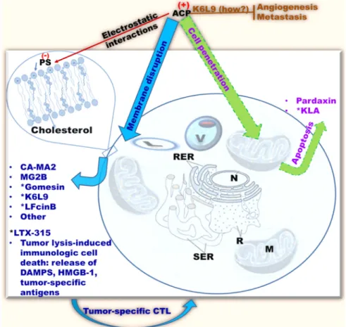
Figure 2: Common antitumor mechanisms of cationic AMPs classified as ACPs.
ACPs (anticancer peptides), or cationic AMPs with anticancer properties, selectively recognize cancer cells by electrostatic interactions with negatively charged phospholipids on the surface of eukaryotic cells [e.g., PS (phosphatidylserine)]. Some ACPs demonstrate in vivo efficacy (e.g., *MGB2, Gomesin, K6L9, LTX-315); ACPs tend to kill cancer cells by membrane perturbation (blue/cyan), although some (e.g., KLA, Pardaxin) may penetrate the target cell and disrupt the mitochondrial membrane resulting in apoptosis (green/purple). (How?), mechanism unclear; brown perpendicular bar, inhibition; CTL, cytotoxic T-Lymphocytes; M, mitochondria, SER, smooth endoplasmic reticulum; RER, rough endoplasmic reticulum; R, ribosomes; N, nucleus; V, vacuole; L, lysosome; only cholesterol and other lipids are shown; membrane proteins are omitted for clarity.depolarization were also demonstrated. The ACP
D-K6L9 (LKLLKKLLKKLLKLL) is the most successful
demonstration of the systemic anticancer efficacy of
an ACP to date [58]. Notably, D-K6L9 is made of only
two amino acids. As natural AMPs work in the context
of a multifunctional immune system, they are more
efficient at protecting the host against disease than they
are at curing established illnesses (more preventive than
therapeutic). Hence, it is logical to engineer AMPs with
more optimized structures for therapeutic applications as
opposed to using AMPs as they are produced in nature.
We predict that the success of AMP engineering will be
facilitated by the establishment of a definitive framework
for distinguishing the unique role of the cationic from that
of the hydrophobic domain in selectivity toward the target
versus host cells. Once a guideline for selective killing
of different target cells is established, it will then be
possible to design AMPs with enhanced therapeutic index
with less trial and error. In the case of the peptide K6L9,
the investigators use the principle of amphipathicity to
design a 15-residue AMP (charge = +6) with an idealized
amphipathic helix (Figure 3). In addition, the use of
D-amino acids enhances stability, addressing a concern
for lability of AMPs [58, 132–134]. This is an important
example of how AMP engineering can lead to enhanced
results. Yet, the most advanced (in terms of clinical trials)
cationic ACP is not D-K6L9. Over the last two decades,
there has been an increasing interest in bovine lactoferricin
(LFcinB). LFcinB is derived from the natural milk protein
bovine lactoferrin (LFB) by pepsin digestion [135–139]. In
addition to its antimicrobial properties, [140–143] LFcinB
demonstrates enhanced activities against cancer cells
compared to normal mammalian cells [139, 144, 145].
Importantly, high-resolution imaging revealed a
membranolytic mechanism [146]. These studies reached
a turning point when Eliassen and colleagues (2006)
began to examine the in vivo efficacy of LFcinB in mice,
demonstrating the growth inhibition of neuroblastoma
xenografts [147]. While the peptide could be localized in
the mitochondria with caspase activation, the lytic effects
on the cytoplasmic membrane represented the primary
cytotoxic mechanism, as pan-inhibition of the caspase
enzymatic cascade could not reverse the cytotoxic effects
of the peptide. These studies led to further structural
optimization using 3,3-diphenylalanine (Dip) substitutions
for Trp at specific positions of the 9-mer template. These
investigations resulted in enhanced selectivity (IC50
below 5–10 µM) against a variety of tumors compared
to normal cells. More importantly, these studies revealed
a cytolytic-immunogenic dual cell death mechanism
[148–156]. Lysis of the tumor induced the release of
danger-associated molecular patterns (DAMPs) including
high mobility group box protein 1 (HMGB1, reviewed
by Frank et al., 2015 [157]) in addition to antigens from
the dead tumor cells. As potent stimulators of immune
responses, HMGB1 potentiated A20 lymphoma-specific
activation of immature dendritic cells with subsequent
generation of tumor-specific cytotoxic T-lymphocytes
and tumor cell lysis. The immunogenic cell death was
further confirmed by the adoptive transfer of syngeneic
A20 lymphoma-specific CTLs and resulting protection
of immunodeficient mice from tumor implants. Further,
LTX-315-treated mice in remission from the tumor were
protected from a second challenge by the syngeneic tumor
and not by a different tumor type, indicating the specificity
of the immunogenic cell death. Hence, the potent
derivative LTX-315 was selected for advanced preclinical
studies. LTX-315 is now (2016) in phase 1 clinical trial
as described later. The study of LTX-315 is yet another
illustration of how natural AMPs can be optimized for
specific clinical applications.
Non-membranolytic mechanisms of ACP
The ACP K6L9, while exerting a direct
membranolytic effect on cancer cells, demonstrates
anti-angiogenic and anti-metastatic effects, an anti-tumor
mechanism that remains to be elucidated. Another common
anti-cancer mechanism is the potentiation of apoptosis in
cancer cells by ACP [158–160]. While ACPs selectively
interact with cancer compared to normal cell membranes,
some AMPs may both perturb the membrane and penetrate
the cells while other non-lytic ACPs may simply traverse
the membrane and access the intracellular compartment. In
both cases, the ACP disrupts the mitochondria (as expected
because of the bacterial origin of mitochondria) and
induces programmed cell death. This apoptotic mechanism
is demonstrated by swelling of the mitochondria,
disruption of the mitochondrial membrane, translocation of
PS to the surface of the cell, and stimulation of apoptotic
markers (e.g., caspase enzymatic pathway). One of the
earliest examples of an apoptotic ACP was reported by
Mai et al., 2001 [161]. The authors used a KLA repeat
AMP [(KLAKLAK)2] conjugated with a transduction
peptide (RRQRRTSKLMKRGGKLAKLAKKLAKLAK)
by a glycine pacer. The intratumoral injection of
the chimeric ACP resulted in the translocation of
the peptide into the cytoplasm of the tumor cells,
disruption of the mitochondria, and stimulation of
apoptotic enzymatic cascade by caspase 3 activation.
Other ACPs have the inherent property to penetrate the
cancer cells without the need for conjugation with a
cell penetrating peptide. An example of such a peptide
(GFFALIPKIISSPLFKTLLSAVGSALSSSGGQE)
is
pardaxin, which induces apoptosis of squamous cell
carcinoma cells by caspase 3 activation [162]. In addition,
some AMPs may be used as cell penetrating peptides to
transfer an anticancer drug into cancer cells. This is the case
of the AMP PR39 (RRRPRPPYLPRPRPPPFFPPRLPP
RIPPGFPPRFPPRFP) used as a cell penetrating peptide
to transfer Stat3 siRNA (the cargo) into breast cancer cells
[163–165]. As previously mentioned, another interesting
antitumor mechanism is the immunogenic cell death
conferring long-lasting protection against future challenges
from the same tumor type. Such a mechanism is displayed
by the ACP LTX-315, [151] which was selected for
advanced preclinical studies. LTX-315 is now in phase 1
clinical trial [151, 155, 156]. Noteworthy is an antitumor
mechanism occurring via the lysosomal-mitochondrial
death pathway by the defensin Brevenin-2R (in vitro
activity) [166]. As the anticancer mechanisms of AMPs
become increasingly clear, structural optimization for
enhanced anticancer potency will be more achievable and,
therefore, clinical development more likely to succeed.
Of note, AMPs synthesized in plants are another
important source of peptide therapeutics that could be used
as anticancer agents. However, for many of these AMPs,
the anticancer mechanisms are unclear. AMPs derived from
plants are beyond the scope of this review and are reviewed
elsewhere [32]. A list of representative AMPs with selective
antitumor mechanism is shown Table 1.
Prospect for clinical use of AMPs as anticancer
agents
The clinical development of AMPs as ACPs faces
some of the same challenges to AMP clinical development as
antimicrobial agents. AMPs are not traditional drugs. Because
they have multifunctional properties, the adaptation from their
natural environment to clinical applications without structural
optimization is rather challenging. While AMPs work well
in the context of a competent immune system, it is likely
that in nature a particular AMP structure is not completely
optimized for a single function (e.g., antibacterial, antiviral,
or anticancer). The field also faces some unfounded criticisms
that have hindered support for AMP development. Some of
these criticisms are that (1) “AMPs are labile and likely to
have poor pharmacokinetic properties”, an assumption based
on the peptidic nature of AMPs; (2) “AMPs are expensive to
make”; (3) “AMPs are not good drugs” because they do not
recognize specific receptors.
Although stability is an important concern, the first
criticism is based on the assumption that all peptides have
similar stability and clearance mechanisms. One of the
shortcomings of AMPs is the lack of correlation between
in vitro susceptibility testing and efficacy in animal
models, with some exceptions including some of the AMPs
discussed in this review and elsewhere [58, 156, 167–169].
Assays establishing correlation between in vitro stability
and bioavailability in animal models might address this
concern. Currently, AMPs with optimized structures are
amenable to parenteral administration including systemic,
respiratory, intramuscular, intraperitoneal, or subcutaneous
unless otherwise contraindicated [167, 168, 170–172].
Although the peptidic nature of AMPs precludes oral
delivery, the development of specific delivery systems
protecting AMPs from degradation by digestive enzymes
may increase intestinal absorption and the feasibility of oral
administration. Several strategies can be used to enhance
PK properties of AMPs. One approach is the utilization of
D-enantiomers to increase stability, [133] although such
strategy is only indicated if decreasing peptide clearance
enhances therapeutic efficacy and does not potentiate
toxic side effects. Other strategies include end-to-end
cyclization, C-terminus amidation, pegylation [attachment
of polyethylene glycol (PEG) to a molecule], and liposomal
delivery. Cyclisation and amidation can confer peptide
stability by decreasing susceptibility to protease digestion,
as demonstrated by the modification of gomesin and other
AMPs [173, 174]. As a principle, all AMPs used in our
laboratory are amidated [18, 20, 175, 176]. As shown by
the Gumbleton group and others, pegylation of AMPs can
result in lower host toxicity without affecting antimicrobial
activity [177]. Pegylation enhances the pharmacological
properties of a given drug in a number of ways [178–
183]. Because it increases hydrophilicity, PEG serves as
Figure 3: Helical wheel analysis of the engineered AMP K6L9 designed by Papo et al.
[57]. The peptide was modeled to forman idealized amphipathic helix with only two amino acids; a structural optimization strategy that has been shown to enhance antimicrobial functions, now applied to antitumor properties as well. Arrow indicates direction of the hydrophobic moment. Structural motifs: yellow, hydrophobic; blue, cationic.
a shield that protects against protease digestion, prolongs
circulation time, and reduces the glomerular filtration rate.
An interesting example of AMP pegylation is a recent
design by Kelly et al., 2016 as shown in Figure 4 [183].
A pro-peptide was designed using the AMP P18 covalently
attached to a linker region that is sensitive to the cysteine
Table 1: Representative antimicrobial peptides with selective antitumor mechanisms
AMP name
Amino acid sequence
Source
Tumor target Mechanism
Reference
Alpha-defensin-1 ACYCRIPACIAGERRYG
TCIYQGRLWAFCC
Human
HTC/STC
Apoptosis
Xu et al., 2008
[109]
Antiangiogenic
BMAP-28
GGLRSLGRKILRAWK
KYG
Bovine
HTC
MP/Ca influx
Risso et al., 2002
[158]
Apoptosis
Brevenin-2R
KFALGKVNAKLQSLN
AKSLKQSGCC
Frog
STC
LDP
Ghavami et al.,
2008 [165]
Buforin IIb
RAGLQFPVG[RLLR]3
Frog
HTC/STC
Apoptosis
Lee et al., 2008159
CA-MA-2
KWKLFKKI-P-KFLHS
AKKF
Hybrid
STC
MP
Shin et al., 2000
[122]
Cecropin A
KWKLFKKIEKVGQNIR
DGIIKAGPAVAVVGQA
TQIAK
Silk moth
HTC
MP
Hui et al., 2002
[96]
Cecropin B
KWKVFKKIEKMGRNI
RNGIVKAGPAIAVLGE
AKAL
Silk moth
HTC/STC
MP/Apoptosis
Li et al., 2016 [94]
chrysophsin-1
FFGWLIKGAIHAGKA
IHGLI
Red sea
bream
HTC/STC
MP
Hsu et al., 2011
[123]
D-K6L9
LKLLKKLLKKLLKLL
Engineered STC
MP
Papo et al., 2006
[57]
Gomesin
*ZCRRLCYKQRCVTY
CRGR
Spider
STC
MP
Domingues et al.,
2010 [128]
KLA
RRQRRTSKLMKRGGK
LAKL-AKKLAKLAK-
(KLAKLAK)2
Engineered STC
MP
Mai et al., 2001
[160]
lactoferricin B
FKC1RRWQWRMKKLG
APSITC1VRRAF
Bovine
HTC/STC
MP/Apoptosis
Eliassen et al, 2002
[145]
LL37
LLGDFFRKSKEKIGKEFKR
IVQRIKDFLRNLVPRTES
Human
Ovarian CA
MP
Chuang et al. 2009
[210]
LTX-315
K-K-W-W-K-K-W-Dip-K
Engineered HTC/STC
MP/ICD
Haug et al.,
2016148
Phase I/II trial
Magainin 2
GIGKFLHSAKKFGKAF
VGEIMNS
Frog
HTC/STC
MP
Cruciani et al.,
1991115]
Melittin
GIGAVLKVLTTGLPALIS
WIKRKRQQ
Insect
STC
MP
Wang et al., 2009
[56]
MG2B
GIGKFLHSAKKFGKAF
VGEIMNSGG-QRLGNQ
WAVGHLM
Hyprid
AMP
MCF-7 tumor MP
Liu et al., 2011
[128]
Pardaxin
GFFALIPKIISSPLFKTLL
SAVGSALSSSGGQE
Fish
STC
MP
Han et al., 2016
[162]
*pyroglutamic acid; HTC, hematological tumor cells; STC, solid tumor cells; MP, membrane permeabilization; LDP,
lysosomal death pathway; ICD, immunological cell death..
protease cathepsin B, followed by a PEG region to confer
thermodynamic stability to the peptide. In vitro studies show
that selectivity index can be enhanced against an ovarian
carcinoma cell line (A2780). While this strategy is still
conceptual, as it requires extensive in vivo studies for proof
of concept, it shows how a pro-peptide can be designed in
combination with pegylation to enhance the PK properties
of an ACP or AMP. Another strategy could be the liposomal
formulation of AMPs [184–187]. However, one shortcoming
is that AMPs are membrane active and could, therefore,
bind and disrupt the liposome. Packaging the molecule as
an inactive pro-peptide in the liposome could theoretically
overcome this problem. One last potential caveat is whether
we are able to produce liposomes that can discriminate
between the target and normal cells. An AMP delivered inside
a eukaryotic cell will probably interact with mitochondria as
it would with a bacterial cell, based on similarities between
a bacterial cell and mitochondrial membranes. One strategy
that is already developed is to tag the lysosome with a cancer
cell-specific ligand for targeted delivery of the peptide [188].
Finally, designing AMPs as part of nanoparticles for delivery
is another strategy that may improve PK properties of AMPs
and should be explored [189–191]. All of these strategies for
enhancing PK properties should be considered only when
indicated, as AMPs are highly diverse in structure, which
affects PK properties.
The second criticism is based on an outdated notion
of the highly impractical cost of peptide production.
While cost remains a concern, peptide- and
protein-based drugs have been used clinically for decades from
hypertensive (e.g. Lisinopril) [192–196] and
anti-diabetic (e.g., insulin) [197, 198] to immune, antiviral
(e.g., fuzeon), [199, 200] antibacterial, [201–203] and
hormonal therapy [196]. Modern technology and larger
scale synthesis have also significantly reduced the
production cost of AMPs. The third concern is based on
the fact that AMPs do not select their targets via specific
receptors [204]. This nonreceptor-mediated recognition is
one of the major reasons AMPs are less likely to invoke
selection of resistance compared to current antibiotics/
anticancer drugs and the basis for broad selectivity against
diverse types of multidrug-resistant microbial pathogens
and transformed cells. Lipid-mediated selectivity of
AMPs should be considered as a major strength, not a
weakness. These misconceptions against AMPs have
considerably hampered the progress of their development,
particularly AMP engineering for structural optimization
in the United States. The success of the ACPs K6L9,
LTX-315, and other engineered antibacterial AMPs [20, 167, 168,
175, 176] illustrates the need for AMP engineering and, thus,
the establishment of a rational framework for predicting
structural determinants of the selective killing of different
target cells. Such a guideline for structural optimization,
combined with PK-enhancing strategies, would enhance
our ability to increase the therapeutic index of AMPs with
significantly less trial and error.
Despite the aforementioned challenges, there are
a few AMPs being evaluated as anticancer (in addition
to antimicrobial) agents in advanced phases of clinical
development. The human cathelicidin LL37 is currently
(2016) in Phase 2 clinical trial for melanoma (lesions at
least 10 mm and not completely resectable) by intratumoral
injections in patients with no known immune deficiency, a
collaborative effort of M.D. Anderson Cancer Center and
National Cancer Institute [205]. The first patients were
enrolled in July 2015. LL37 is the single most studied
human AMP [206–208]. It is normally found in human skin,
reproductive, and respiratory systems and known to have
multiple functional properties including (but not limited
to) antibacterial, antiviral, antifungal, immunomodulatory,
and anticancer activities [209–211]. Another AMP,
LTX-315, is in phase 1 trial for PK and efficacy treatment of
multiple types of transdermally accessible tumors. This
is a lactoferrin-derived lytic peptide that binds and lyses
tumor cells. The resulting tumor necrosis leads to enhanced
presentation of tumor antigens and induced innate and
adaptive immunity against the tumor as described above.
This clinical trial started on October 28, 2013, and the last
update (December 2016) indicates that LTX-315 is still
in phase 1 “Open-label, arm, centre,
multi-dose, dose Escalation Study” for exploration of efficacy as
monotherapy or in combination with either ipilimumab or
pembrolizumab in patients with transdermally accessible
tumors” [212]. Noteworthy are other AMPs in clinical
trial as anti-infective agents (e.g., OP-145 for otitis media
and pexiganan for diabetic foot ulcer), but not as ACPs.
Toward the goal for clinical applications, it is possible to
improve the therapeutic index of current chemotherapeutic
agents by considering combination therapy. Combining
AMPs with a chemotherapeutic agent may help decrease
dosage, which would result in lower toxicity. While the
current clinical trial of LTX-315 including combination
with immunotherapeutics (Ipilimumab or Pembrolizumab)
is a step in the right direction, combination therapy using
AMPs and chemotherapeutic agents needs to be explored
for possible synergy [183].
Figure 4: Strategy to improve the PK properties of AMPs adapted from Kelly et al.,
2016.[183]. The AMP P18 is amidated atthe C-terminus. In addition, it is covalently bound to the protease cathepsin B-sensitive linker for the release of the cancer-active drug; this linker is also covalently attached to a polyethylene glycol (PEG) polymer, which is a hydrophilic moiety that serves as a protective shield from protease degradation and drug clearance.
Concluding remarks
Since the discovery of AMPs more than three
decades ago, no other class of compounds has matched
their versatility as multifunctional compounds. AMPs have
the potential to become the only class of drugs that can be
used against polymicrobial co-infections (e.g., bacterial and
viral [213]) and cancer. However, the multifunctionality
determined by a typical AMP structure suggests that no
single property is completely optimized in natural AMPs
in the context of maintaining multiple functions. Rational
peptide engineering is essential to AMP development for
clinical applications. An important task is to dissect the
structural determinants of each property to uncouple each
of AMP functions for “application-specific optimization”.
Only when such studies are conducted in a systematic way
will we begin to significantly explore the clinical potential
of AMPs as a diverse class of therapeutics. AMP research
has been largely occurring outside of the United States.
Despite a vast literature in AMP research, this is still an
area that is critically underfunded by the National Institute
of Health (NIH). Because of the initial failure of AMPs to
reach the Clinique, the resulting bias has largely hindered the
advancement of AMP research in the United States. Hence,
there is a pressing need for the NIH and pharmaceutical
companies to support more AMP research to collect the
evidence necessary to assess whether the promise of AMPs
will ever come to fruition. As an essential component of the
immune system, AMPs warrant such exploration.
ACKNOWLEDGMENTS AND FUNDING
This research is supported by NIH awards R01
HL091938, HL-125128, and a grant (CIA-123062) from
Flight Attendant Medical Research Institute.
CONFLICTS OF INTEREST
None.
REFERENCES
1. Hashim D, Boffetta P, La Vecchia C, Rota M, Bertuccio P, Malvezzi M, Negri E. The global decrease in cancer mortality: trends and disparities. Ann Oncol. 2016; 27:926–933. 2. Torre LA, Siegel RL, Ward EM, Jemal A. Global Cancer
Incidence and Mortality Rates and Trends–An Update. Cancer Epidemiol Biomarkers Prev. 2016; 25:16–27. 3. Saika K, Machii R. Cancer mortality attributable to tobacco
by region based on the WHO Global Report. Jpn J Clin Oncol. 2012; 42:771–72.
4. Shibuya K, Mathers CD, Boschi-Pinto C, Lopez AD, Murray CJ. Global and regional estimates of cancer mortality and incidence by site: II. Results for the global burden of disease 2000. BMC Cancer. 2002; 2:37.
5. Daskivich TJ. Life Expectancy and Treatment Choice for Men with High-risk Prostate Cancer. Eur Urol. 2015; 68:59–60.
6. Arrington AK, Goldstein L, Kruper L, Vito C, Yim J, Chen SL. Life expectancy after curative-intent treatment of breast cancer: impact on long-term follow-up care. Am Surg. 2014; 80:604–09.
7. Delpierre C, Lamy S, Kelly-Irving M, Molinié F, Velten M, Tretarre B, Woronoff AS, Buemi A, Lapôtre-Ledoux B, Bara S, Guizard AV, Colonna M, Grosclaude P. Life expectancy estimates as a key factor in over-treatment: the case of prostate cancer. Cancer Epidemiol. 2013; 37:462–68.
8. Repetto L, Comandini D, Mammoliti S. Life expectancy, comorbidity and quality of life: the treatment equation in the older cancer patients. Crit Rev Oncol Hematol. 2001; 37:147–52.
9. Haybittle JL. Life expectancy as a measurement of the benefit shown by clinical trials of treatment for early breast cancer. Clin Oncol (R Coll Radiol). 1998; 10:92–94. 10. Kolberg HC, Villena-Heinsen C, Deml MM, Kraemer S,
Diedrich K, Friedrich M. Relationship between chemotherapy with paclitaxel, cisplatin, vinorelbine and titanocene dichloride and expression of proliferation markers and tumour suppressor gene p53 in human ovarian cancer xenografts in nude mice. Eur J Gynaecol Oncol. 2005; 26:398–402.
11. Pierard GE, Focan C, Lapiere CM. Cell proliferation in a malignant angioendothelioma during sequential chemotherapy. J Cutan Pathol. 1979; 6:479–485.
12. Kato Y, Sato J, Kato R, Takata R, Obara W. [Side effect and supportive care to combination of gemcitabine and cisplatin chemotherapy for the advanced urothelial cancer]. [Article in Japanese]. Nihon rinsho. 2015; 73:609–613.
13. Larsen ME, Rowntree J, Young AM, Pearson S, Smith J, Gibson OJ, Weaver A, Tarassenko L. Chemotherapy side-effect management using mobile phones. Conf Proc IEEE Eng Med Biol Soc. 2008; 2008:5152–5155.
14. Saxena A. Cancer chemotherapy and its side effect management. Nurs J India. 2006; 97:109–10.
15. Chism DD, De Silva D, Whang YE. Mechanisms of acquired resistance to androgen receptor targeting drugs in castration-resistant prostate cancer. Expert Rev Anticancer Ther. 2014; 14:1369–78.
16. Chatterjee S, Damle SG, Sharma AK. Mechanisms of resistance against cancer therapeutic drugs. Curr Pharm Biotechnol. 2014; 15:1105–12.
17. Tamburrino A, Piro G, Carbone C, Tortora G, Melisi D. Mechanisms of resistance to chemotherapeutic and anti-angiogenic drugs as novel targets for pancreatic cancer therapy. Front Pharmacol. 2013; 4:56.
18. Deslouches B, Steckbeck JD, Craigo JK, Doi Y, Burns JL, Montelaro RC. Engineered cationic antimicrobial peptides to overcome multidrug resistance by ESKAPE pathogens. Antimicrob Agents Chemother. 2015; 59:1329–33.
19. Steckbeck JD, Deslouches B, Montelaro RC. Antimicrobial peptides: new drugs for bad bugs? Expert Opin Biol Ther. 2014; 14:11–14.
20. Deslouches B, Steckbeck JD, Craigo JK, Doi Y, Mietzner TA, Montelaro RC. Rational design of engineered cationic antimicrobial peptides consisting exclusively of arginine and tryptophan, and their activity against multidrug-resistant pathogens. Antimicrob Agents Chemother. 2013; 57:2511–21.
21. Dutta P, Das S. Mammalian Antimicrobial Peptides: Promising Therapeutic Targets Against Infection and Chronic Inflammation. Curr Top Med Chem. 2016; 16:99–129.
22. Hiemstra PS, Amatngalim GD, van der Does AM, Taube C. Antimicrobial Peptides and Innate Lung Defenses: Role in Infectious and Noninfectious Lung Diseases and Therapeutic Applications. Chest. 2016; 149:545–51. 23. Shagaghi N, Palombo EA, Clayton AH, Bhave M.
Archetypal tryptophan-rich antimicrobial peptides: properties and applications. World J Microbiol Biotechnol. 2016; 32:31.
24. Schmitt P, Rosa RD, Destoumieux-Garzon D. An intimate link between antimicrobial peptide sequence diversity and binding to essential components of bacterial membranes. Biochim Biophys Acta. 2016; 1858:958–970.
25. Aguilera-Mendoza L, Marrero-Ponce Y, Tellez-Ibarra R, Llorente-Quesada MT, Salgado J, Barigye SJ, Liu J. Overlap and diversity in antimicrobial peptide databases: compiling a non-redundant set of sequences. Bioinformatics. 2015; 31:2553–59.
26. He X, Yang S, Wei L, Liu R, Lai R, Rong M. Antimicrobial peptide diversity in the skin of the torrent frog, Amolops jingdongensis. Amino Acids. 2013; 44:481–487.
27. Padhi A, Verghese B. Molecular diversity and evolution of myticin-C antimicrobial peptide variants in the Mediterranean mussel, Mytilus galloprovincialis. Peptides. 2008; 29:1094–101.
28. Dennison SR, Wallace J, Harris F, Phoenix DA. Amphiphilic alpha-helical antimicrobial peptides and their structure/ function relationships. Protein Pept Lett. 2005; 12:31–39. 29. Bulet P, Hetru C, Dimarcq JL, Hoffmann D. Antimicrobial
peptides in insects; structure and function. Dev Comp Immunol. 1999; 23:329–44.
30. Hwang PM, Vogel HJ. Structure-function relationships of antimicrobial peptides. Biochem Cell Biol. 1998; 76:235– 246.
31. Sitaram N, Nagaraj R. Host-defense antimicrobial peptides: importance of structure for activity. Curr Pharm Des. 2002; 8:727–42.
32. Guzmán-Rodríguez JJ, Ochoa-Zarzosa A, Lopez-Gomez R, Lopez-Meza JE. Plant antimicrobial peptides as potential anticancer agents. Biomed Res Int. 2015; 2015:735087. 33. Farkas A, Maróti G, Kereszt A, Kondorosi É. Comparative
Analysis of the Bacterial Membrane Disruption Effect of Two Natural Plant Antimicrobial Peptides. Front Microbiol. 2017; 8:51.
34. Gomarasca M, F C Martins T, Greune L, Hardwidge PR, Schmidt MA, Rüter C. Bacterial-derived cell-penetrating peptides deliver gentamicin to kill intracellular pathogens. Antimicrob Agents Chemother. 2017; 61:e02545–16. 35. Coelho ML, de Souza Duarte AF, do Carmo de Freire
Bastos M. Bacterial Labionin-Containing Peptides and Sactibiotics: Unusual Types of Antimicrobial Peptides with Potential Use in Clinical Settings (A Review). Curr Top Med Chem. 2016; 17:1177–1198.
36. Jung HJ, Kim Y, Lee HB, Kwon HJ. Antiangiogenic activity of the lipophilic antimicrobial peptides from an endophytic bacterial strain isolated from red pepper leaf. Mol Cells. 2015; 38:273–78.
37. Gomes KM, Duarte RS, de Freire Bastos MD. Lantibiotics produced by Actinobacteria and their potential applications (a review). Microbiology. 2017; 163:109–21.
38. Barbosa AA, Mantovani HC, Jain S. Bacteriocins from lactic acid bacteria and their potential in the preservation of fruit products. Crit Rev Biotechnol. 2017; 3:1–13.
39. Ahmad V, Khan MS, Jamal QM, Alzohairy MA, Al Karaawi MA, Siddiqui MU. Antimicrobial potential of bacteriocins: in therapy, agriculture and food preservation. Int J Antimicrob Agents. 2017; 49:1–11.
40. Pirtskhalava M, Gabrielian A, Cruz P, Griggs HL, Squires RB, Hurt DE, Grigolava M, Chubinidze M, Gogoladze G, Vishnepolsky B, Alekseyev V, Rosenthal A, Tartakovsky M. DBAASP v.2: an enhanced database of structure and antimicrobial/cytotoxic activity of natural and synthetic peptides. Nucleic Acids Res. 2016; 44:6503.
41. Waghu FH, Barai RS, Gurung P, Idicula-Thomas S. CAMPR3: a database on sequences, structures and signatures of antimicrobial peptides. Nucleic Acids Res. 2016; 44:D1094–97.
42. Liu Y, Eichler J, Pischetsrieder M. Virtual screening of a milk peptide database for the identification of food-derived antimicrobial peptides. Mol Nutr Food Res. 2015; 59:2243–2254.
43. Di Luca M, Maccari G, Maisetta G, Batoni G. BaAMPs: the database of biofilm-active antimicrobial peptides. Biofouling. 2015; 31:193–99.
44. Niarchou A, Alexandridou A, Athanasiadis E, Spyrou G. C-PAmP: large scale analysis and database construction containing high scoring computationally predicted antimicrobial peptides for all the available plant species. PLoS One. 2013; 8:e79728.
45. Zhao X, Wu H, Lu H, Li G, Huang Q. LAMP: A Database Linking Antimicrobial Peptides. PLoS One. 2013; 8:e66557.
46. Piotto SP, Sessa L, Concilio S, Iannelli P. YADAMP: yet another database of antimicrobial peptides. Int J Antimicrob Agents. 2012; 39:346–51.
47. Li Y, Chen Z. RAPD: a database of recombinantly-produced antimicrobial peptides. FEMS Microbiol Lett. 2008; 289:126–29.
48. Fjell CD, Hancock RE, Cherkasov A. AMPer: a database and an automated discovery tool for antimicrobial peptides. Bioinformatics. 2007; 23:1148–55.
49. Hancock RE, Sahl HG. Antimicrobial and host-defense peptides as new anti-infective therapeutic strategies. Nat Biotechnol. 2006; 24:1551–57.
50. Hilpert K, Elliott MR, Volkmer-Engert R, Henklein P, Donini O, Zhou Q, Winkler DF, Hancock RE. Sequence requirements and an optimization strategy for short antimicrobial peptides. Chem Biol. 2006; 13:1101–07. 51. Hancock RE, Brown KL, Mookherjee N. Host defence
peptides from invertebrates—emerging antimicrobial strategies. Immunobiology. 2006; 211:315–22.
52. Brown KL, Hancock RE. Cationic host defense (antimicrobial) peptides. Curr Opin Immunol. 2006; 18:24–30.
53. Soblosky L, Ramamoorthy A, Chen Z. Membrane interaction of antimicrobial peptides using E. coli lipid extract as model bacterial cell membranes and SFG spectroscopy. Chem Phys Lipids. 2015; 187:20–33. 54. Kamimori H, Blazyk J, Aguilar MI. Lipid
membrane-binding properties of tryptophan analogues of linear amphipathic beta-sheet cationic antimicrobial peptides using surface plasmon resonance. Biol Pharm Bull. 2005; 28:148–50.
55. Maget-Dana R. The monolayer technique: a potent tool for studying the interfacial properties of antimicrobial and membrane-lytic peptides and their interactions with lipid membranes. Biochim Biophys Acta. 1999; 1462:109–40. 56. Wang C, Chen T, Zhang N, Yang M, Li B, Lü X, Cao
X, Ling C. Melittin, a major component of bee venom, sensitizes human hepatocellular carcinoma cells to tumor necrosis factor-related apoptosis-inducing ligand (TRAIL)-induced apoptosis by activating CaMKII-TAK1-JNK/p38 and inhibiting IkappaBalpha kinase-NFkappaB. J Biol Chem. 2009; 284:3804–13.
57. Mangoni ML, Papo N, Saugar JM, Barra D, Shai Y, Simmaco M, Rivas L. Effect of natural L- to D-amino acid conversion on the organization, membrane binding, and biological function of the antimicrobial peptides bombinins H. Biochemistry. 2006; 45:4266–76.
58. Papo N, Seger D, Makovitzki A, Kalchenko V, Eshhar Z, Degani H, Shai Y. Inhibition of tumor growth and elimination of multiple metastases in human prostate and breast xenografts by systemic inoculation of a host defense-like lytic peptide. Cancer Res. 2006; 66:5371–78.
59. Wakabayashi H, Matsumoto H, Hashimoto K, Teraguchi S, Takase M, Hayasawa H. N-Acylated and D enantiomer derivatives of a nonamer core peptide of lactoferricin B showing improved antimicrobial activity. Antimicrob Agents Chemother. 1999; 43:1267–69.
60. Shang D, Zhang Q, Dong W, Liang H, Bi X. The effects of LPS on the activity of Trp-containing antimicrobial peptides against Gram-negative bacteria and endotoxin neutralization. Acta Biomater. 2016; 33:153–65.
61. Nan YH, Bang JK, Jacob B, Park IS, Shin SY. Prokaryotic selectivity and LPS-neutralizing activity of short antimicrobial peptides designed from the human antimicrobial peptide LL-37. Peptides. 2012; 35:239–47.
62. Gustafsson A, Olin AI, Ljunggren L. LPS interactions with immobilized and soluble antimicrobial peptides. Scand J Clin Lab Invest. 2010; 70:194–200.
63. Giuliani A, Pirri G, Rinaldi AC. Antimicrobial peptides: the LPS connection. Methods Mol Biol. 2010; 618:137–154. 64. Malanovic N, Leber R, Schmuck M, Kriechbaum M,
Cordfunke RA, Drijfhout JW, de Breij A, Nibbering PH, Kolb D, Lohner K. Phospholipid-driven differences determine the action of the synthetic antimicrobial peptide OP-145 on Gram-positive bacterial and mammalian membrane model systems. Biochim Biophys Acta. 2015; 1848:2437–2447.
65. Jasir A, Kasprzykowski F, Lindström V, Schalén C, Grubb A. New antimicrobial peptide active against Gram-positive pathogens. Indian J Med Res. 2004; 119:74–76.
66. Jasir A, Kasprzykowski F, Kasprzykowska R, Lindstrom V, Schalen C, Grubb A. New antimicrobial cystatin C-based peptide active against gram-positive bacterial pathogens, including methicillin-resistant Staphylococcus aureus and multiresistant coagulase-negative staphylococci. APMIS. 2003; 111:1004–1010.
67. Ruan Y, Shen T, Wang Y, Hou M, Li J, Sun T. Antimicrobial peptide LL-37 attenuates LTA induced inflammatory effect in macrophages. Int Immunopharmacol. 2013; 15:575–80. 68. Gustafsson A, Sigel S, Ljunggren L. The antimicrobial
peptide LL37 and its truncated derivatives potentiates proinflammatory cytokine induction by lipoteichoic acid in whole blood. Scand J Clin Lab Invest. 2010; 70:512–518. 69. Ng TB, Cheung RC, Wong JH, Ye XJ. Antimicrobial
activity of defensins and defensin-like peptides with special emphasis on those from fungi and invertebrate animals. Curr Protein Pept Sci. 2013; 14:515–531.
70. Crack LR, Jones L, Malavige GN, Patel V, Ogg GS. Human antimicrobial peptides LL-37 and human beta-defensin-2 reduce viral replication in keratinocytes infected with varicella zoster virus. Clin Exp Dermatol. 2012; 37:534–543. 71. Nava GM, Escorcia M, Castaneda MP. Molecular
diversity of the antimicrobial domain of beta-defensin 3 and homologous peptides. Comp Funct Genomics. 2009:983636.
72. Nan YH, Bang JK, Shin SY. Design of novel indolicidin-derived antimicrobial peptides with enhanced cell specificity and potent anti-inflammatory activity. Peptides. 2009; 30:832–38.
73. Ladokhin AS, Selsted ME, White SH. CD spectra of indolicidin antimicrobial peptides suggest turns, not polyproline helix. Biochemistry. 1999; 38:12313–19. 74. Hollmann A, Martínez M, Noguera ME, Augusto MT,
Disalvo A, Santos NC, Semorile L, Maffía PC. Role of amphipathicity and hydrophobicity in the balance between hemolysis and peptide-membrane interactions of three related antimicrobial peptides. Colloids Surf B Biointerfaces. 2016; 141:528–36.
75. Son M, Lee Y, Hwang H, Hyun S, Yu J. Disruption of interactions between hydrophobic residues on nonpolar faces is a key determinant in decreasing hemolysis and
increasing antimicrobial activities of α-helical amphipathic peptides. ChemMedChem. 2013; 8:1638–42.
76. Utsugi T, Schroit AJ, Connor J, Bucana CD, Fidler IJ. Elevated expression of phosphatidylserine in the outer membrane leaflet of human tumor cells and recognition by activated human blood monocytes. Cancer Res. 1991; 51:3062–66.
77. Gerber DE, Hao G, Watkins L, Stafford JH, Anderson J, Holbein B, Oz OK, Mathews D, Thorpe PE, Hassan G, Kumar A, Brekken RA, Sun X. Tumor-specific targeting by Bavituximab, a phosphatidylserine-targeting monoclonal antibody with vascular targeting and immune modulating properties, in lung cancer xenografts. Am J Nucl Med Mol Imaging. 2015; 5:493–503.
78. Kenis H, Reutelingsperger C. Targeting phosphatidylserine in anti-cancer therapy. Curr Pharm Des. 2009; 15:2719–23. 79. Bevers EM, Williamson PL. Getting to the Outer Leaflet:
Physiology of Phosphatidylserine Exposure at the Plasma Membrane. Physiol Rev. 2016; 96:605–45.
80. Connor J, Pak CC, Schroit AJ. Exposure of phosphatidylserine in the outer leaflet of human red blood cells. Relationship to cell density, cell age, and clearance by mononuclear cells. J Biol Chem. 1994; 269:2399–404. 81. Koopman G, Reutelingsperger CP, Kuijten GA, Keehnen
RM, Pals ST, van Oers MH. Annexin V for flow cytometric detection of phosphatidylserine expression on B cells undergoing apoptosis. Blood. 1994; 84:1415–20.
82. Martin SJ, Reutelingsperger CP, McGahon AJ, Rader JA, van Schie RC, LaFace DM, Green DR. Early redistribution of plasma membrane phosphatidylserine is a general feature of apoptosis regardless of the initiating stimulus: inhibition by overexpression of Bcl-2 and Abl. J Exp Med. 1995; 182:1545–1556.
83. Lee SH, Meng XW, Flatten KS, Loegering DA, Kaufmann SH. Phosphatidylserine exposure during apoptosis reflects bidirectional trafficking between plasma membrane and cytoplasm. Cell Death Differ. 2013; 20:64–76.
84. Cole CL, Rushton G, Jayson GC, Avizienyte E. Ovarian cancer cell heparan sulfate 6-O-sulfotransferases regulate an angiogenic program induced by heparin-binding epidermal growth factor (EGF)-like growth factor/EGF receptor signaling. J Biol Chem. 2014; 289:10488–10501.
85. Kontro H, Joenväärä S, Haglund C, Renkonen R. Comparison of sialylated N-glycopeptide levels in serum of pancreatic cancer patients, acute pancreatitis patients, and healthy controls. Proteomics. 2014; 14:1713–23.
86. Tian Y, Esteva FJ, Song J, Zhang H. Altered expression of sialylated glycoproteins in breast cancer using hydrazide chemistry and mass spectrometry. Mol Cell Proteomics. 2012; 11:M111 011403.
87. Lee CS, Taib NA, Ashrafzadeh A, Fadzli F, Harun F, Rahmat K, Hoong SM, Abdul-Rahman PS, Hashim OH. Unmasking Heavily O-Glycosylated Serum Proteins Using Perchloric Acid: Identification of Serum Proteoglycan 4 and
Protease C1 Inhibitor as Molecular Indicators for Screening of Breast Cancer. PLoS One. 2016; 11:e0149551.
88. Mu AK, Lim BK, Hashim OH, Shuib AS. Identification of O-glycosylated proteins that are aberrantly excreted in the urine of patients with early stage ovarian cancer. Int J Mol Sci. 2013; 14:7923–7931.
89. Osinaga E. Expression of cancer-associated simple mucin-type O-glycosylated antigens in parasites. IUBMB Life. 2007; 59:269–73.
90. Yoon WH, Park HD, Lim K, Hwang BD. Effect of O-glycosylated mucin on invasion and metastasis of HM7 human colon cancer cells. Biochem Biophys Res Commun. 1996; 222:694–99.
91. Fadnes B, Rekdal O, Uhlin-Hansen L. The anticancer activity of lytic peptides is inhibited by heparan sulfate on the surface of the tumor cells. BMC Cancer. 2009; 9:183. 92. Sok M, Sentjurc M, Schara M, Stare J, Rott T. Cell
membrane fluidity and prognosis of lung cancer. Ann Thorac Surg. 2002; 73:1567–1571.
93. Sok M, Sentjurc M, Schara M. Membrane fluidity characteristics of human lung cancer. Cancer Lett. 1999; 139:215–20.
94. Berger M, Motta C, Boiret N, Aublet-Cuvelier B, Bonhomme J, Travade P. Membrane fluidity and adherence to extracellular matrix components are related to blast cell count in acute myeloid leukemia. Leukem & lymphom. 1994; 15:297–302.
95. Li X, Shen B, Chen Q, Zhang X, Ye Y, Wang F, Zhang X. Antitumor effects of cecropin B-LHRH’ on drug-resistant ovarian and endometrial cancer cells. BMC Cancer. 2016; 16:251.
96. Suttmann H, Retz M, Paulsen F, Harder J, Zwergel U, Kamradt J, Wullich B, Unteregger G, Stockle M, Lehmann J. Antimicrobial peptides of the Cecropin-family show potent antitumor activity against bladder cancer cells. BMC Urol. 2008; 8:5.
97. Hui L, Leung K, Chen HM. The combined effects of antibacterial peptide cecropin A and anti-cancer agents on leukemia cells. Anticancer Res. 2002; 22:2811–16. 98. Liu Y, Chen L, Gong Z, Shen L, Kao C, Hock JM, Sun
L, Li X. Lovastatin enhances adenovirus-mediated TRAIL induced apoptosis by depleting cholesterol of lipid rafts and affecting CAR and death receptor expression of prostate cancer cells. Oncotarget. 2015; 6:3055–70. doi: 10.18632/ oncotarget.3073.
99. Zhuang L, Lin J, Lu ML, Solomon KR, Freeman MR. Cholesterol-rich lipid rafts mediate akt-regulated survival in prostate cancer cells. Cancer Res. 2002; 62:2227–31. 100. Mulder KC, Lima LA, Miranda VJ, Dias SC, Franco OL.
Current scenario of peptide-based drugs: the key roles of cationic antitumor and antiviral peptides. Front Microbiol. 2013; 4:321.
101. Rothan HA, Mohamed Z, Suhaeb AM, Rahman NA, Yusof R. Antiviral cationic peptides as a strategy for
innovation in global health therapeutics for dengue virus: high yield production of the biologically active recombinant plectasin peptide. Omics. 2013; 17:560–567.
102. Chan SC, Hui L, Chen HM. Enhancement of the cytolytic effect of anti-bacterial cecropin by the microvilli of cancer cells. Anticancer Res. 1998; 18:4467–74.
103. Kolata GB. Microvilli: a major difference between normal and cancer cells? Science. 1975; 188:819–20.
104. Lacerda AF, Vasconcelos EA, Pelegrini PB, Grossi de Sa MF. Antifungal defensins and their role in plant defense. Front Microbiol. 2014; 5:116.
105. Vriens K, Cammue BP, Thevissen K. Antifungal plant defensins: mechanisms of action and production. Molecules. 2014; 19:12280–303.
106. Lehrer RI, Barton A, Daher KA, Harwig SS, Ganz T, Selsted ME. Interaction of human defensins with Escherichia coli. Mechanism of bactericidal activity. J Clin Invest. 1989; 84:553–61.
107. Panyutich AV, Voitenok NN, Lehrer RI, Ganz T. An enzyme immunoassay for human defensins. J Immunol Methods. 1991; 141:149–55.
108. Venkataraman N, Cole AL, Ruchala P, Waring AJ, Lehrer RI, Stuchlik O, Pohl J, Cole AM. Reawakening retrocyclins: ancestral human defensins active against HIV-1. PLoS Biol. 2009; 7:e95.
109. Muñoz A, Chu M, Marris PI, Sagaram US, Kaur J, Shah DM, Read ND. Specific domains of plant defensins differentially disrupt colony initiation, cell fusion and calcium homeostasis in Neurospora crassa. Mol Microbiol. 2014; 92:1357–74.
110. Xu N, Wang YS, Pan WB, Xiao B, Wen YJ, Chen XC, Chen LJ, Deng HX, You J, Kan B, Fu AF, Li D, Zhao X, Wei YQ. Human alpha-defensin-1 inhibits growth of human lung adenocarcinoma xenograft in nude mice. Mol Cancer Ther. 2008; 7:1588–97.
111. Ren XY, Li WY, Xu M. [Expression of human beta-defensin-2 in laryngeal squamous cell carcinoma and its correlation with CD1a(+) dendritic cells infiltrating in tumor.]. [Article in Chinese]. Zhonghua Er Bi Yan Hou Tou Jing Wai Ke Za Zhi. 2009; 44:1029–33.
112. Ganz T, Oren A, Lehrer RI. Defensins: microbicidal and cytotoxic peptides of mammalian host defense cells. Med Microbiol Immunol (Berl). 1992; 181:99–105.
113. Kagan BL, Ganz T, Lehrer RI. Defensins: a family of antimicrobial and cytotoxic peptides. Toxicology. 1994; 87:131–49.
114. Lehrer RI, Lichtenstein AK, Ganz T. Defensins: antimicrobial and cytotoxic peptides of mammalian cells. Annu Rev Immunol. 1993; 11:105–28.
115. Lichtenstein AK, Ganz T, Nguyen TM, Selsted ME, Lehrer RI. Mechanism of target cytolysis by peptide defensins. Target cell metabolic activities, possibly involving endocytosis, are crucial for expression of cytotoxicity. J Immunol. 1988; 140:2686–94.
116. Cruciani RA, Barker JL, Zasloff M, Chen HC, Colamonici O. Antibiotic magainins exert cytolytic activity against transformed cell lines through channel formation. Proc Natl Acad Sci USA. 1991; 88:3792–96.
117. Moore KS, Bevins CL, Brasseur MM, Tomassini N, Turner K, Eck H, Zasloff M. Antimicrobial peptides in the stomach of Xenopus laevis. J Biol Chem. 1991; 266:19851–57.
118. Soravia E, Martini G, Zasloff M. Antimicrobial properties of peptides from Xenopus granular gland secretions. FEBS Lett. 1988; 228:337–40.
119. Zasloff M. Magainins, a class of antimicrobial peptides from Xenopus skin: isolation, characterization of two active forms, and partial cDNA sequence of a precursor. Proc Natl Acad Sci USA. 1987; 84:5449–53.
120. Westerhoff HV, Hendler RW, Zasloff M, Juretić D. Interactions between a new class of eukaryotic antimicrobial agents and isolated rat liver mitochondria. Biochim Biophys Acta. 1989; 975:361–69.
121. Merrifield RB, Vizioli LD, Boman HG. Synthesis of the antibacterial peptide cecropin A (1–33). Biochemistry. 1982; 21:5020–31.
122. Hultmark D, Engstrom A, Bennich H, Kapur R, Boman HG. Insect immunity: isolation and structure of cecropin D and four minor antibacterial components from Cecropia pupae. Eur J Biochem. 1982; 127:207–217.
123. Shin SY, Kang JH, Jang SY, Kim Y, Kim KL, Hahm KS. Effects of the hinge region of cecropin A(1–8)-magainin 2(1–12), a synthetic antimicrobial peptide, on liposomes, bacterial and tumor cells. Biochim Biophys Acta. 2000; 1463:209–18.
124. Hsu JC, Lin LC, Tzen JT, Chen JY. Characteristics of the antitumor activities in tumor cells and modulation of the inflammatory response in RAW264.7 cells of a novel antimicrobial peptide, chrysophsin-1, from the red sea bream (Chrysophrys major). Peptides. 2011; 32:900–10. 125. Cirioni O, Simonetti O, Pierpaoli E, Barucca A, Ghiselli R,
Orlando F, Pelloni M, Minardi D, Trombettoni MM, Guerrieri M, Offidani A, Giacometti A, Provinciali M. Enhanced Efficacy of Combinations of Pexiganan with Colistin Versus Acinetobacter Baumannii in Experimental Sepsis. Shock. 2016; 46:219–25.
126. Zhang XL, Jiang AM, Ma ZY, Li XB, Xiong YY, Dou JF, Wang JF. The synthetic antimicrobial peptide pexiganan and its nanoparticles (PNPs) exhibit the anti-helicobacter pylori activity in vitro and in vivo. Molecules. 2015; 20:3972–85. 127. Dipexium Pharmaceuticals I. Dipexium Pharmaceuticals
Initiates Pivotal Phase 3 Clinical Trial Of Locilex® In Patients With Mild Infections Of Diabetic Foot Ulcers. http://contentequisolvenet/dipexiumpharmaceuticals/ news/2014–07-17_Dipexium_Pharmaceuticals_Initiates_ Pivotal_Phase_54pdf, PRNewswire. 2014
128. Liu S, Yang H, Wan L, Cai HW, Li SF, Li YP, Cheng JQ, Lu XF. Enhancement of cytotoxicity of antimicrobial peptide magainin II in tumor cells by bombesin-targeted delivery. Acta Pharmacol Sin. 2011; 32:79–88.

![Figure 3: Helical wheel analysis of the engineered AMP K6L9 designed by Papo et al. [57]](https://thumb-us.123doks.com/thumbv2/123dok_us/9809487.482158/6.918.336.595.751.1009/figure-helical-wheel-analysis-engineered-amp-designed-papo.webp)
![Figure 4: Strategy to improve the PK properties of AMPs adapted from Kelly et al., 2016.[183]](https://thumb-us.123doks.com/thumbv2/123dok_us/9809487.482158/8.918.97.827.964.1000/figure-strategy-improve-pk-properties-amps-adapted-kelly.webp)
