RESEARCH
Characterization of thermophilic fungi
producing extracellular lignocellulolytic
enzymes for lignocellulosic hydrolysis
under solid-state fermentation
Paramjeet Saroj, Manasa P and Korrapati Narasimhulu
*Abstract
Background: Thermotolerant lignocellulolytic enzymes have become a subject of interest in industrial processes due to their ability to degrade lignocellulosic polysaccharides. Development of cost-effective, large-scale screening for production of desirable enzymes by thermophilic fungi is a challenge. The present investigation focused on isolat-ing, screenisolat-ing, and identifying industrially relevant thermophilic producers of lignocellulolytic enzymes from various locations in the Warangal district, Telangana, India.
Results: Fifteen thermophilic fungi were isolated from soil on their ability to grow at 50 °C and were screened for their activity of cellulase, hemicellulase, and lignin degradation based on holo zone around colonies. The appear-ance of the black color zone of diffusion in esculin agar is a positive indication for the β-glucosidases activity test. Out of fifteen isolates, Aspergillus fumigatus JCM 10253 have shown as a potential producer of extracellular enzymes for lignocelluloses degradation showing higher activity for cellulase (EI 1.50) as well as β-glucosidase (4 mg/mL), simultaneously for xylanase (EI 1.18) by plate assay methods. A. fumigatus JCM 10253 was selected for extracellular hydrolytic enzymes production under solid-state fermentation. Maximum CMCase (26.2 IU/mL), FPase (18.2 IU/mL), β-glucosidase (0.87 IU/mL), and xylanase (2.6 IU/mL) activities were obtained after incubation time of 144 h at 50 °C. The thermostability of crude cellulase showed the optimum activity at 60 °C and for FPase, β-glucosidase, and xyla-nase at 50 °C which recommended that the enzymes have a potentially significant role in the biofuel industries. Conclusion: The high titer production of active enzymes that cleave different β-1,4-glycosidic bonds still remains a challenge and is the major bottleneck for the lignocellulosic conversion. In particular, the finding of thermostable enzymes which would allow the development of more robust processes is a major goal in this field.
Keywords: Lignocellulolytic enzymes, Cellulase, Hemicellulase, β-Glucosidase, Aspergillus fumigatus, Hydrolytic enzymes, Solid-state fermentation
© The Author(s) 2018. This article is distributed under the terms of the Creative Commons Attribution 4.0 International License (http://creat iveco mmons .org/licen ses/by/4.0/), which permits unrestricted use, distribution, and reproduction in any medium, provided you give appropriate credit to the original author(s) and the source, provide a link to the Creative Commons license, and indicate if changes were made.
Open Access
*Correspondence: [email protected]
Department of Biotechnology, National Institute of Technology, Warangal, Telangana 506004, India
Background
Increasing global energy needs and greenhouse gas emis-sions have had a significant impact on ecosystem stability and the environment. Therefore, there is a need to find alternative sources of energy to replace existing fossil fuel resources. The conversion of lignocellulosic biomass
into material for the production of biofuels is a potential alternative energy source with several advantages (Arora
et al. 2015). Lignocellulose, composed mainly of
cellu-lose, hemicelluloses, and lignin, is the main component
of the plant cell wall (Lynd et al. 2002). Lignocellulose has
of the production process (Behera et al. 2014). Addition-ally, chemical hydrolysis of lignocelluloses results in the formation of toxic components that are hazardous to the environment. Microbes are known to produce enzyme complexes that degrade plant biomass, offering an
effi-cient and cost-effective alternative (Behera et al. 2013).
Cellulose is a linear polymer chain of repeating units of cellobiose joined by β-1,4 linkages. Cellulose chains interact with each other via hydrogen bonds, resulting in the formation of a rigid, insoluble crystalline structure
(Yamada et al. 2013). Three specific enzymes act
syner-gistically to degrade cellulose into glucose, including endoglucanase (EC 3.2.1.4), exoglucanase (EC 3.2.1.91),
and β-glucosidase (EC 3.2.1.21) (Garvey et al. 2013;
Jut-uru and Wu 2014). Endoglucanase cleaves cellulose
fib-ers into small fragments, releasing free reducing and non-reducing ends. Exoglucanase acts on non-reducing ends to release small cellobiose oligosaccharides. Finally, β-glucosidase converts cellobiose into monomeric sugars
(Singhania et al. 2013). β-Glucosidases is a rate-limiting
factor in the process of converting cellulose to glucose for bioethanol production. In the absence of β-glucosidase activity, cellobiose oligosaccharides accumulate, caus-ing end-product inhibition and limitcaus-ing hydrolysis yields
(Suwannarangsee et al. 2012). Hence, commercially
avail-able cellulolytic preparations are usually added along with β-glucosidase to increase overall activity. Xylanases (EC 3.2.1.8) are a class of hydrolytic enzymes that can hydrolyze the polysaccharide 1,4-xylan found in
hemicel-luloses (Ramanjaneyulu et al. 2015).
Since commercial processes require high tempera-tures, thermostable enzymes are necessary for indus-trial use. Therefore, there is an increasing demand for thermophilic and thermostable producers of cellulolytic and hemicellulolytic enzymes, particularly those that are resistant to product inhibition and highly stable (Arora
et al. 2014; Mallerman et al. 2015). Recently, thermophilic
fungal enzymes have become the subject of increased interest due to their ability to produce highly efficient, thermostable extracellular enzymes for plant biomass degradation at elevated temperatures. Degradation of lignocellulose by fungi is achieved by producing extracel-lular hydrolytic and oxidative ligninolytic enzymes. The hydrolytic enzymes cellulase and hemicellulase degrade polysaccharides into simple sugars, whereas ligninolytic ligninase enzymes degrade lignin and open phenyl rings
(Sánchez 2009).
Economically feasible enzyme production can be applied to the development of technologies that utilize cellulosic material as a renewable resource. The main objective of the present study was to screen and charac-terize thermophilic fungal isolates based on their abil-ity to produce extracellular lignocellulolytic enzymes
and to degrade lignin and open phenyl rings, using plate assays. Based on the zone of inhibition, potential strains were selected for assessment of enzyme production using solid-state fermentation. Thermostability of respective enzymes was investigated.
Methods
Isolation and identification
Warangal district is a part of the northern Telangana of newly formed Telangana State, India. It is situated
approximately between the latitude of 17°19′ and 18°13′
North latitude of and 78°49′ and 80°43′ East latitudes.
The soils of the Warangal city consist of sandy loams with patches of shallow black cotton soils and at places even medium and deep cotton soils. The weather of Waran-gal district is generally dry. Summers are hot sometimes, temperature reaches 50 °C. The temperature drops to 13 °C in winter seasons during the months of December
and January (Narasimha Ramulu and Benarjee 2016).
Soil samples were collected in sterile polythene bags from different sites in the Warangal district and were stored at 4 °C. Emerson Yeast Soluble Starch (YSS) agar media (4 g/L yeast extract, 15 g/L soluble starch,
1.0 g/L K2HPO4, 0.5 g/L MgSO4·7H2O, and 20 g/L agar,
pH 7.0 ± 0.2) and malt extract agar (MEA) (20 g/L malt
extract, 20 g/L dextrose, 6 g/L peptone, and 15 g/L agar) were used for fungal isolation experiments. Pure cul-tures were obtained by serial dilution. Inoculated agar plates were incubated at 30 °C and monitored at regular intervals for 10 days to assess fungal growth. Isolated fungi were sub-cultured on Emerson YSS agar slants and stored at 4 °C. A loopful spore of fungal strain was inoc-ulated into Sabouraud Dextrose Broth (dextrose 20 g/L,
peptone 10 g/L, pH 5.6 ± 0.2) for 7 days in the incubator
shaker at 30 °C.
Screening and identification of thermophilic fungi
To assess thermostability, isolated fungi were grown at temperatures between 30 and 50 °C. Fungi were inocu-lated at the center of the plate and radial growth patterns were monitored for 7 days. Based on the growth patterns at different temperatures, fifteen thermophilic fungi were selected for plate assays. Isolated thermophilic fungi were identified based on 18S rRNA sequence analyses per-formed by Gujarat State Biotechnology Mission, Depart-ment of Science and Technology, Gandhinagar, Gujarat, India.
Plate assay for cellulase and xylanase activity
The thermophilic fungal isolates were screened for their ability to degrade cellulose and hemicelluloses using
Czapek-Dox media (30 g/L sucrose, 2 g/L NaNO3, 1 g/L
and 20 g/L agar) (Kluepfel 1988) containing 1% carboxy-methyl cellulose (CMC) as the substrate for cellulase and 1% beechwood as the substrate for xylanase (Hölker et al.
2004). The pH was adjusted to 5.0 and the media were
autoclaved. Sterile agar medium was poured into sterile Petri dishes and allowed to solidify. About 0.4-cm-diam-eter wells were made on solidified agar plates. A total of 16 μL of fungal broth was pipetted into each well and the plates were sealed with Parafilm and incubated at 50 °C for 7 days. Fifteen thermophilic fungi were then screened for cellulase and xylanase activity using Congo red (1%
aqueous) solution (Ahirwar et al. 2017). After
incuba-tion at 50 °C for 7 days, culture plates were flooded with Congo red and left for 30 min, then destained by washing twice with 1 M NaCl for 20 min on a gel rocker. Trans-parent hydrolytic zones where cellulose and xylan were degraded into simple sugars were observed. The enzy-matic index (EI) was calculated by measuring the clear-ance zone and using the following expression (Teather
and Wood 1982; Bradner et al. 1999; Ruegger and
Tauk-Tornisielo 2004; Pečiulytė 2007; Florencio et al. 2012):
The growth of fungal isolates on MEA was taken as control. The inhibition index was calculated using the ratio of the diameter of the colony on CMC or xylan agar to the diameter of the colony on control agar (Khokhar
et al. 2012). Each experiment was performed in triplicate.
Esculin gel diffusion assay
Esculin gel diffusion assay was used to assess
β-glucosidase activity (Saqib and Whitney 2006).
About 3% of agar in 0.2 M sodium acetate buffer of pH 5 was autoclaved and kept at 50 °C to avoid agar
solidifi-cation. Esculin (0.2%) was mixed with 6 mL of 1% FeCl3
and heated to 50 °C in a water bath. Both solutions were mixed thoroughly and poured into 15-cm Petri dishes. The esculin agar was allowed to solidify and wells 0.4 cm in diameter were made. A total of 16 μL of fungal broth was pipetted into each well and the plates were sealed with Parafilm and incubated at 37 °C for 5 h. After incu-bation, the plates were transferred to an ice bath to slow down enzyme activity. Plates were assessed for the appearance of a dark zone indicating esculin hydrolysis. Each experiment was performed in triplicate.
Plate assay for lignin degradation and laccase activity Thermophilic fungal isolates were screened for their abil-ity to degrade lignin polymers using lignin (alkaline) as a substrate following the method described by Tekere
Enzymatic index =
Diameter of hydrolysis zone(in cm) Diameter of the colony(in cm) .
et al. (2001). The reaction mixture contained 0.25% (w/v)
lignin (alkaline), 5 g/L glucose, 5 g/L ammonium
tar-trate, 0.5 g/L MgSO4·H2O, 1 g/L malt extract, 0.01 g/L
CaCl2·2H2O, 0.01 g/L FeCl3, 0.1 g/L NaCl, 1 mg/L
thia-mine, and 20 g/L agar and was autoclaved. Sterile lignin agar medium was poured into sterile Petri dishes and allowed to solidify. Wells 0.4 cm in diameter were made on the solidified agar plates. A total of 16 μL of test cul-ture was pipetted into each well and the plates were sealed with Parafilm and incubated at 50 °C for 7 days. After 7 days, mycelia were scratched from the plate
sur-face and flooded with a 1% FeCl3 and K3[Fe(CN)6]
solu-tion. The distance across each clearance zone, indicating degraded lignin, was measured.
Thermophilic fungi were examined for their ability to produce laccase, which is involved in lignin degradation, using guaiacol agar medium (0.02% w/v guaiacol, 1% w/v yeast extract, and 2% w/v agar). A total of 16 μL of test culture was pipetted into each well and plates were incu-bated at 50 °C in the dark for 7 days to ensure adequate
growth of cultures in the medium (Singh et al. 2013).
Enzyme production in shaking flasks
Aspergillus fumigatus JCM 10253 was selected from the fifteen isolates as a potential producer of hydro-lytic enzymes and used in further experiments.
Man-dels and Weber (1969) medium supplemented with 1%
CMC or beechwood as a carbon source and 2.5% wheat bran as a substrate was used for shaking flask fermen-tation experiments. The medium was prepared as
fol-lows: 2 g/L KH2PO4, 0.3 g/L CaCl2·2H2O, 0.3 g/L urea,
0.3 g/L MgSO4·7H2O, 1.4 g/L (NH4)2SO4, 0.25 g/L
pep-tone, 0.1 g/L yeast extract, 1 mL Tween-80, 0.005 g/L
FeSO4·7H2O, 0.0016 g/L MnSO4·H2O, 0.0014 g/L
ZnSO4·7H2O, and 0.002 g/L CoCl2·6H2O, pH 5. The
pre-pared medium was sterilized at 121 °C, 15 psi. A loopful
of spores of the isolated fungal strain A. fumigatus JCM
10253 was inoculated into the medium. Conical flasks were incubated at 50 °C with shaking at 160 rpm for 12 days.
Enzyme assays
The extracellular total protein content of the culture fil-trate was estimated using a Bradford assay (Bradford
1976). Carboxymethyl cellulase (CMCase) and filter
paper activity (FPase) assay were performed according
to the methods described by Ghose (1987). The assay for
with 1 mL 50 mM sodium citrate buffer (pH 4.8)
contain-ing Whatman No. 1 filter paper strip (1 cm × 6 cm) for
60 min at 50 °C. Xylanase assay was carried out
accord-ing to the method described by Bailey et al. (1992).
The activity was determined by incubating 0.2 mL of the crude enzyme with 1.8 mL of 1% beechwood dis-solved in 50 mM sodium citrate buffer (pH 5.0) at 50 °C for 5 min. All the above reaction was stopped by add-ing 3 mL of 3,5-dinitrosalicylic acid (DNS) reagent and heating for 10 min in a boiling water bath. The amount of reducing sugar released was measured using a UV–vis-ible spectrophotometer (Model: Jasco-V-630) at 540 nm.
β-Glucosidase activity was analyzed using p
-nitrophenyl-β-d-glucopyranoside as a substrate according to the
method described by Grover et al. (1977). The
reac-tion mixture consists of 1.4 mL of 0.1 M sodium acetate buffer (pH 5.0), 0.1 mL of crude enzyme with 0.5 mL of
0.02 M p-nitrophenyl-β-d-glucopyranoside (pNPG) and
incubated for 15 min at 37 °C. The enzyme reaction was stopped by the addition of 2.0 mL 0.2 M sodium carbon-ate and absorbance was measured at 400 nm. Enzyme activity was measured in international units (IU) per mL. The amount of enzyme required to liberate 1 µmol of
glu-cose, xylose, or p-nitrophenol from their respective
sub-strates per min under the assay conditions was defined as
1 IU of enzyme activity (Kovács et al. 2009).
Thermostability assay
The effect of temperature on enzymes activity was determined by incubating crude enzyme at different
temperatures ranging from 30 to 80 °C at the various time intervals. After incubation, the activity of the enzymes was determined by the standard assay as described earlier.
Results and discussion
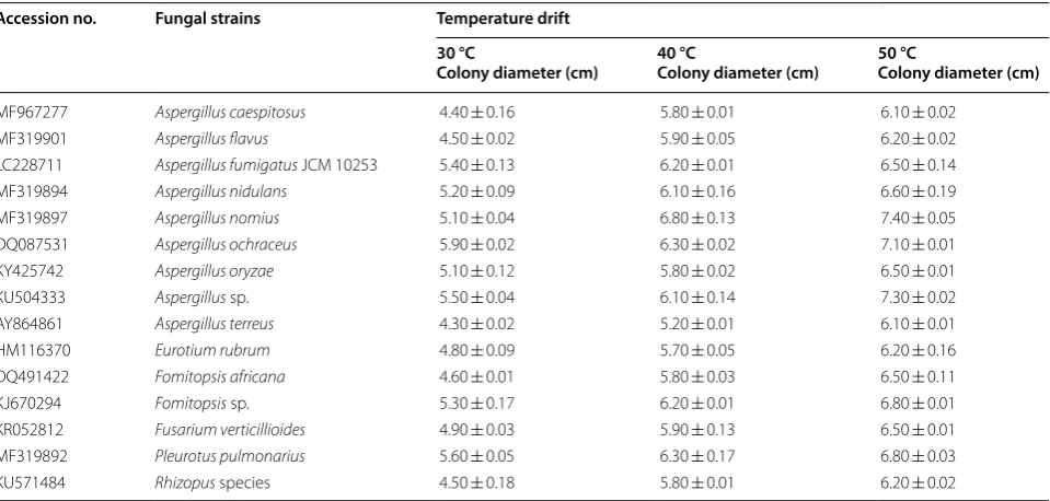
In the current study, fifteen thermophilic fungi were iso-lated from soil based on their ability to grow at high tem-peratures. Fungal identification was performed using 18S
rRNA sequencing (Table 1).
Screening of cellulase and xylanase activity
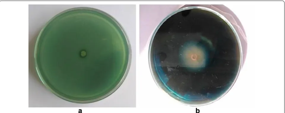
Fifteen thermophilic fungal isolates from soil samples were screened for cellulase and xylanase activity on Cza-pek-Dox medium containing CMC or beechwood as a substrate. Cellulolytic fungal strains were selected based on the diameter of hydrolysis zones surrounding the col-onies. Detection of extracellular hydrolytic enzyme activ-ity was performed using qualitative evaluation of Congo
red staining (Meddeb-Mouelhi et al. 2014). Cellulase
and xylanase activities were measured using the enzyme activity index by measuring the clearance zone
surround-ing the colonies (Figs. 1, 2). Fungal isolates with high EI
values were considered to be potential cellulase and
xyla-nase producers. Of the fifteen isolated fungi, A.
fumiga-tus JCM 10253 and Aspergillus terreus exhibited the
highest cellulase activity with EI values of 1.50 and 1.24,
respectively, followed by Aspergillus sp. (EI 1.13),
Fusar-ium verticillioides (EI 1.11), and Eurotium rubrum (EI 1.09). However, the hydrolytic activity of other isolates,
Table 1 List of the selected fungal strains isolated from soil source and its observation after temperature drift Accession no. Fungal strains Temperature drift
30 °C
Colony diameter (cm) 40 °CColony diameter (cm) 50 °CColony diameter (cm)
MF967277 Aspergillus caespitosus 4.40 ± 0.16 5.80 ± 0.01 6.10 ± 0.02
MF319901 Aspergillus flavus 4.50 ± 0.02 5.90 ± 0.05 6.20 ± 0.02
LC228711 Aspergillus fumigatus JCM 10253 5.40 ± 0.13 6.20 ± 0.01 6.50 ± 0.14
MF319894 Aspergillus nidulans 5.20 ± 0.09 6.10 ± 0.16 6.60 ± 0.19
MF319897 Aspergillus nomius 5.10 ± 0.04 6.80 ± 0.13 7.40 ± 0.05
DQ087531 Aspergillus ochraceus 5.90 ± 0.02 6.30 ± 0.02 7.10 ± 0.01
KY425742 Aspergillus oryzae 5.10 ± 0.12 5.80 ± 0.02 6.50 ± 0.01
KU504333 Aspergillus sp. 5.50 ± 0.04 6.10 ± 0.14 7.30 ± 0.02
AY864861 Aspergillus terreus 4.30 ± 0.02 5.20 ± 0.01 6.10 ± 0.01
HM116370 Eurotium rubrum 4.80 ± 0.09 5.70 ± 0.05 6.20 ± 0.16
DQ491422 Fomitopsis africana 4.60 ± 0.01 5.80 ± 0.03 6.50 ± 0.11
KJ670294 Fomitopsis sp. 5.30 ± 0.17 6.20 ± 0.01 6.80 ± 0.01
KR052812 Fusarium verticillioides 4.90 ± 0.03 5.90 ± 0.13 6.50 ± 0.01
MF319892 Pleurotus pulmonarius 5.60 ± 0.05 6.30 ± 0.17 6.80 ± 0.03
including Aspergillus caespitosus (EI 1.05), Aspergillus oryzae (EI 1.05), Rhizopus sp. (EI 1.05), Aspergillus nid-ulans (EI 1.03), Aspergillus nomius (EI 1.03), Fomitop-sis africana (EI 1.03), Fomitopsis sp. (EI 1.03), Pleurotus pulmonarius (EI 1.03), Aspergillus flavus (EI 1.02), and
Aspergillus ochraceus (EI 1.02), was relatively low. More-over, none of the fungal isolates were resistant to CMC agar and the colony growth of fungal isolates on CMC
agar was greater than that on control agar (Table 2). In
agreement with these results, Aspergillus sp. has been
reported to possess all of the necessary components of
the cellulase enzyme system (de Vries and Visser 2001;
Khokhar et al. 2012).
The most efficient xylanase (endo-1,4-β-d-xylanohydrolase) producing strains were selected based on the size of the clearance zone surrounding the fungal
colonies. A. fumigatus JCM 10253 (EI 1.18) and F.
verti-cillioides (EI 1.09) exhibited the highest xylanase
activi-ties followed by E. rubrum (EI 1.08), F. africana (EI 1.08),
Aspergillus sp. (EI 1.03), A. nomius (EI 1.03), P.
pulmonar-ius (EI 1.03), and A. terreus (EI 1.02). Other isolated
fun-gal strains, including A. oryzae (EI 1.01), A. caespitosus
Fig. 1 Detection of cellulase activity from the isolated fungal strain on agar plates. Plates showing the zone of hydrolysis by Aspergillus fumigatus
JCM 10253 with Congo red staining on the 7th day. a Plate without CMC; b plate with CMC as a substrate
Fig. 2 Detection of xylanase activity from the isolated fungal strain on agar plates. Plates showing the zone of hydrolysis by Aspergillus fumigatus
Table 2 G ro wth and enz yme ac tivities of isola
ted fungal str
ains
Did not sho
w an y ac tivit y (–) Fungal str ains β-G luc osidase ac tivit y Cellulase enz yma tic ac tivit y Xylanase enz yma tic ac tivit y
Zone of diffusion (cm)
H
ydr
oly
sis
zone diamet
er (cm) Colon y diamet er
on CMC agar (cm)
Enz yma tic inde x (EI) Colon y diamet er on c on tr ol agar (cm)
Inhibition inde
x
H
ydr
oly
sis
zone diamet
er (cm) Colon y diamet er on x ylan agar (cm) Enz yma tic inde x (EI) Colon y diamet er on c on tr ol agar (cm)
Inhibition inde
x Asper gillus caespitosus 0.50 ± 0.02 8.20 ± 0.0 7.80 ± 0.2 1.05 ± 0.0 5.80 ± 0.03 1.34 ± 0.0 5.50 ± 0.06 5.40 ± 0.05 1.01 ± 0.01 5.10 ± 0.04 1.05 ± 0.0 Asper gillus flavus 0.40 ± 0.03 7.90 ± 0.1 7.70 ± 0.0 1.02 ± 0.0 5.70 ± 0.1 1.35 ± 0.0 5.20 ± 0.0 5.10 ± 0.0 1.01 ± 0.03 4.90 ± 0.03 1.04 ± 0.0 Asper gillus
fumigatus JCM 10253
1.50 ± 0.01 8.60 ± 0.0 5.70 ± 0.2 1.50 ± 0.02 3.50 ± 0.2 1.62 ± 0.04 5.10 ± 0.0 4.30 ± 0.01 1.18 ± 0.0 3.20 ± 0.09 1.34 ± 0.02 Asper gillus nidulans 0.40 ± 0.01 8.40 ± 0.13 8.10 ± 0.2 1.03 ± 0.0 6.10 ± 0.03 1.32 ± 0.01 5.90 ± 0.01 5.80 ± 0.07 1.01 ± 0.1 5.60 ± 0.01 1.03 ± 0.0 Asper gillus nomius – 8.30 ± 0.0 8.0 ± 0.3 1.03 ± 0.02 6.30 ± 0.08 1.26 ± 0.0 6.10 ± 0.0 5.90 ± 0.02 1.03 ± 0.1 5.80 ± 0.06 1.01 ± 0.0 Asper gillus ochr ac eus 1.0 ± 0.02 7.50 ± 0.1 7.30 ± 0.2 1.02 ± 0.04 5.10 ± 0.01 1.43 ± 0.0 5.80 ± 0.0 5.70 ± 0.0 1.01 ± 0.0 4.50 ± 0.01 1.26 ± 0.0 Asper gillus or yz ae 0.60 ± 0.01 7.10 ± 0.0 6.70 ± 0.5 1.05 ± 0.01 4.80 ± 0.02 1.39 ± 0.03 5.60 ± 0.16 5.50 ± 0.0 1.01 ± 0.01 4.40 ± 0.01 1.25 ± 0.0 Asper gillus s p. – 8.30 ± 0.0 7.30 ± 0.0 1.13 ± 0.0 5.40 ± 0.09 1.35 ± 0.0 5.40 ± 0.03 5.20 ± 0.01 1.03 ± 0.0 5.10 ± 0.07 1.01 ± 0.0 Asper gillus terr eus 1.0 ± 0.03 6.10 ± 0.06 4.90 ± 0.2 1.24 ± 0.1 3.60 ± 0.09 1.36 ± 0.0 4.60 ± 0.01 4.50 ± 0.16 1.02 ± 0.06 3.40 ± 0.03 1.32 ± 0.03 Eur
otium rubrum
0.60 ± 0.02 7.80 ± 0.01 7.10 ± 0.1 1.09 ± 0.0 5.10 ± 0.01 1.39 ± 0.06 5.50 ± 0.0 5.40 ± 0.02 1.08 ± 0.04 5.10 ± 0.3 1.05 ± 0.03
Fomitopsis afric
(EI 1.01), A. ochraceus (EI 1.01), A. nidulans (EI 1.01), A. flavus (EI 1.01), Fomitopsis sp. (EI 1.01) and Rhizopus sp. (EI 1.01) exhibited relatively low xylanase activity when grown on xylan agar. Moreover, the colony growth of all isolated fungal strains on xylan agar was found to be
slightly higher than on control agar (Table 2). Hence, the
fungal isolates were found non-resistant towards xylan agar, as the colony growth on xylan agar was slightly higher than control.
The growth simulation/inhibition index was estimated by the ratio of the diameter of colonies on CMC or xylan agar to the diameter of the colonies on control agar. The index value represents the activity of each enzyme; an index value < 1 indicates substrate-inhibited fungal growth, whereas an index value > 1 indicates substrate-induced growth stimulation. Based on the inhibition
indices for cellulase, the growth of A. fumigatus JCM
10253 (1.62), F. verticillioides (1.46), A. ochraceus (1.43)
and Rhizopus sp. (1.42) was enhanced by CMC agar medium, whereas the effect of CMC agar medium was slightly reduced for the other isolates. On the other hand, based on the inhibition indices, the growth of the
xyla-nase-producing fungal strains A. fumigatus JCM 10253
(1.34), A. terreus (1.32), A. ochraceus (1.26) and A. oryzae
(1.25) was enhanced by xylan medium, whereas the effect of xylan was slightly reduced for the other isolates.
The most commonly used microbes for the production
of hydrolytic enzymes belong to the genera Aspergillus,
Trichoderma, and Penicillium (Yadav 2017). Previous
reports showed that soft rot fungi, such as Aspergillus
niger and Trichoderma reesei (Xue et al. 2017), and the
white rot fungus Phanerochaete chrysosporium
pro-duce large amounts of cellulase (Manavalan et al. 2015).
A recent study showed that some of the white rot fungi are the most promising fungi in terms of biomass deg-radation and delignification because of their potential to synthesize cellulolytic, hemicellulolytic, and ligninolytic
enzymes (Xu et al. 2018). Hydrolysis zone diameter was
correlated with colony diameter on CMC and xylan agar media. However, the hydrolytic activity of all the isolates was found to be slightly higher. The hydrolytic activity of
all isolates was found to be < 1. A. fumigatus JCM 10253
exhibited the highest hydrolytic activity, whereas the hydrolytic activity of the other isolates was slightly lower. The difference between isolates was statistically
signifi-cant (p ≤ 0.05). The method used to screen for hydrolytic
enzyme-producing fungi has previously been reported
and found to be suitable (Abdel-Sater and El-Said 2001;
Lalita and Prasher 2014; Bairagi 2016).
Screening of β-glucosidase activity by gel diffusion plate assay
Fungal isolates were deemed to be positive for β-glucosidase activity based on the appearance a black zone caused by the reaction between esculetin
(6,7-dihy-droxycoumarin) and ferric ion (Veena et al. 2011).
Escu-lin is a glycoside, which is a naturally occurring sugar derivative. Glycosidase enzymes catalyze the hydrolysis of glycosides into sugars and non-sugars that can be used as a fuel resource for energy production. The presence of β-glucosidase causes the hydrolysis of esculin, producing a glucose molecule and the aglycone component escule-tin. The non-sugar component aglycone can be used to
assess the presence of glycosidase (Ford 2014), as it is the
aglycone that reacts with ferric ions, resulting in the for-mation of a black chelate compound. Thus, the hydrolysis of the glycosidic bond by β-glycosidase via a hydrolytic reaction, resulting in the formation of an iron complex, is the key to the identification of enzyme activity. The
reac-tion of esculetin with Fe+3 or ferric salts is a highly
sensi-tive method due to the intensity of the color and the ease
of identification (Edberg et al. 1985). The use of esculin
with ferric chloride is a proven zymographic technique for the screening of β-glucosidase that is both
cost-effec-tive and gives distinct results (Kwon et al. 1994). In the
present study, 16 μL of fungal culture filtrate was inocu-lated onto esculin agar plates and incubated at 37 °C for 5 h. Different incubation periods between 2 and 5 h did not result in any significant changes in the diameter of the zone of diffusion. Therefore, a 2-h incubation period is sufficient for the gel diffusion assay. The black zone of diffusion was compared to the standard curve of almond
β-glucosidase (EC 3.2.1.21; activity ≥ 10U/mg solid;
Fig. 3).
Table 2 shows the fungal activity of the fifteen isolates
determined by gel diffusion plate assays. The zone of
dif-fusion was largest for A. fumigatus JCM 10253 (4 mg/
mL; 1.5 cm) followed by F. africana and Fomitopsis sp.
(2 mg/mL; 1.4 cm), P. pulmonarius (1.1 cm), A.
ochra-ceus (1.0 cm), E. rubrum (0.6 cm), and A. caespitosus
(0.5 cm). These results indicate that these fungi exhibit β-glucosidase activity and esculin hydrolysis. In contrast,
no zone of diffusion was visible when A. nomius,
Asper-gillus sp., or F. verticillioides were inoculated on esculin gel. The absence of a black zone clearly indicates that these fungal strains do not have the ability to hydrolyze esculin. It has been previously reported that the diam-eter of the diffusion zone in a gel diffusion assay is
Saqib and Whitney 2006). Therefore, a standard curve of β-glucosidase was prepared on esculin gel to confirm this correlation.
Plate assay for lignin degradation and laccase activity A lignin degradation plate assay was performed to assess clearance zones by staining the fungal agar plates with
a 1% w/v aqueous solution of FeCl3 and K3[Fe(CN)6], as
shown in Fig. 4. After staining, oxidized phenolic
com-ponents appear blue–green with a clear zone around
the colonies (Pointing 1999; Ali and Muid 2012). The
clearance zone of lignin degradation for each of the
fun-gal strains is shown in Table 3. A. ochraceus exhibited the
largest clearance zone around the colonies (5.5 cm)
fol-lowed by Rhizopus sp. (3.9 cm), F. africana (3.8 cm), F.
verticillioides (3.5 cm), A. nomius (3.3 cm) and A. flavus
(3.2 cm). P. pulmonarius and Fomitopsis sp. exhibited
smaller clearance zones than the other isolates.
Fungal isolates were further screened for their abil-ity to secrete extracellular laccase using guaiacol as a phenolic substrate. Extracellular laccase is constitu-tively produced in small amounts by basidiomycete
Fig. 3 Standard curve on esculin gel diffusion assay for β-glucosidase. a Different concentrations of standard β-glucosidase enzyme from sweet
almond (EC 3.2.1.21; activity: ≥ 10 U/mg solid procured from Himedia) activity on EGDA plate, b standard curve of enzyme activity
fungi, particularly wild-type strains of white rod fungi
such as Pleurotus ostreatus (Hou et al. 2004), Funalia
trogii (Birhanli and Yesilada 2006), and Trametes pube-scens (Galhaup et al. 2002). The formation of a reddish brown color on solid media when guaiacol is used as
the substrate is an indication of extracellular laccase
production by filamentous fungi (Thurston 1994;
Mad-havi and Lele 2009). In this study, the appearance of
a reddish brown color due to the redox reaction was
used to indicate laccase secretion (Fig. 5). All isolates
were shown to be positive and diameter of the zone was measured for their potential laccase activity, as
shown in Table 3. The growth diameter of P.
pulmonar-ius (4.5 cm), F. verticillioides (4.3 cm), and A. nomius
(4.2 cm) was highest on guaiacol-containing agar plates.
A. caespitosus, Aspergillus sp., and E. rubrum exhibited the lowest activity. The extracellular laccase formation observed in the present study is in agreement with pre-vious studies of various fungal strains carried out by
Revankar and Lele (2006) and Fu et al. (2013). Table 4
shows the qualitative assessment of lignocellulolytic enzymes produced by plate assay methods.
Enzyme production
Aspergillus fumigatus strain JCM 10253 was used for hydrolytic enzyme production by solid-state fermenta-tion. Solid-state fermentation system provides an intrin-sic state which is suitable for growing filamentous fungi
to produce cellulases and xylanase (Singhania et al. 2010).
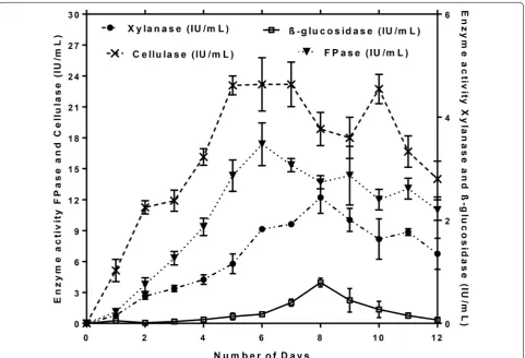
In the present study, maximum CMCase (26.2 IU/mL) and FPase (18.2 IU/mL) activity was observed at 144 h, whereas maximum β-glucosidase (0.87 IU/mL) and xyla-nase (2.6 IU/mL) activity was observed after 192 h of
incubation time at 50 °C (Fig. 6). However, in the present
study, β-glucosidase activity remained relatively low and the longer incubation period caused a decline in enzyme production. This may be due to the depletion of nutrients
Table 3 Summary of plate assay method for lignin degradation and laccase activity of fungal isolates on the 7th day
Fungal strains Lignin degradation activity Zone
of degradation (cm)
Laccase activity Zone of growth (cm)
Aspergillus caespitosus 2.50 ± 0.04 2.50 ± 0.01
Aspergillus flavus 3.20 ± 0.01 3.10 ± 0.02
Aspergillus fumigatus JCM
10253 3.10 ± 0.02 3.30 ± 0.01
Aspergillus nidulans 2.40 ± 0.01 2.80 ± 0.09
Aspergillus nomius 3.30 ± 0.07 4.20 ± 0.02
Aspergillus ochraceus 5.50 ± 0.03 3.50 ± 0.12
Aspergillus oryzae 3.10 ± 0.02 3.00 ± 0.01
Aspergillus sp. 2.50 ± 0.09 2.50 ± 0.17
Aspergillus terreus 3.30 ± 0.02 3.30 ± 0.04
Eurotium rubrum 2.50 ± 0.01 2.50 ± 0.02
Fomitopsis africana 3.80 ± 0.01 3.80 ± 0.16
Fomitopsis sp. 2.10 ± 0.01 2.70 ± 0.01
Fusarium verticillioides 3.50 ± 0.06 4.30 ± 0.03
Pleurotus pulmonarius 1.90 ± 0.03 4.50 ± 0.02
Rhizopus species 3.90 ± 0.01 3.30 ± 0.01
Table 4 Qualitative estimation of lignocellulolytic enzymes by plate assay methods
(−= no activity, += minimum, ++= medium, +++= maximum activity) Fungal strains β-Glucosidase
activity Cellulase activity Xylanase activity Lignin degradation activity
Laccase activity
Aspergillus caespitosus + ++ ++ + +
Aspergillus flavus + ++ ++ ++ ++
Aspergillus fumigatus JCM 10253 +++ +++ +++ ++ ++
Aspergillus nidulans + ++ ++ + +
Aspergillus nomius − ++ ++ ++ +++
Aspergillus ochraceus ++ ++ ++ +++ ++
Aspergillus oryzae + ++ ++ ++ ++
Aspergillus sp. − ++ ++ + +
Aspergillus terreus ++ +++ ++ ++ ++
Eurotium rubrum + ++ ++ + +
Fomitopsis africana +++ ++ ++ +++ ++
Fomitopsis sp. +++ ++ ++ + +
Fusarium verticillioides − ++ +++ ++ +++
Pleurotus pulmonarius ++ ++ ++ + +++
Rhizopus species + ++ ++ +++ ++
or the spontaneous denaturation of the enzyme
(Sand-hya et al. 2005). Solid-state fermentation is a
well-estab-lished technology for enzyme production and has various advantages, such as low costs of operation and the ability to use agro-industrial residues as the raw material
(Raim-bault 1998; Pandey et al. 2000; Adsul et al. 2014). Enzyme
production also depends on various factors, such as chemical composition, accessibility, and the physiochem-ical links between the components of the substrate (Gao
et al. 2008).
Comparative studies
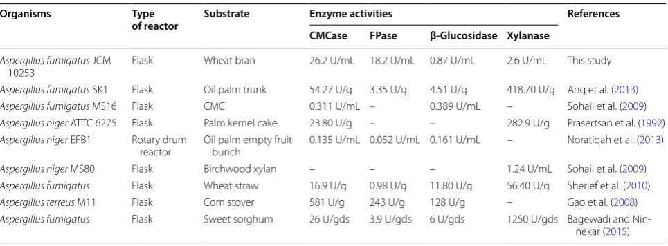
Previous studies on Aspergillus sp. in the production of
cellulase and hemicellulase as summarized in Table 5.
In the present study, A. fumigatus strain JCM 10253
produced hydrolytic enzymes with higher activity as compared to previous studies. The maximum CMCase (26.2 IU/mL), FPase (18.2 IU/mL), β-glucosidase (0.87 IU/mL), and xylanase (2.6 IU/mL) activities were
obtained. In previous reports, A. fumigatus SK1 showed
CMCase (54.3 U/g), FPase (3.35 U/g), β-glucosidase (4.5 U/g) and xylanase (418.7 U/g) activities using oil
palm trunk as a substrate (Ang et al. 2013). On the other
hand, A. fumigatus SK1 cellulase activity was 1.4-fold
higher compared to A. fumigatus MS16 and xylanase
activity was 2.7-fold high as compared to A. niger MS80
(Sohail et al. 2009; Ang et al. 2013). Grigorevski-Lima
et al. (2009) obtained 365 U L−1 activity of endoglucanase
(CMCase) from A. fumigatus at temperature 65 °C and
pH 2. It has been reported that Aspergillus sp. is
catego-rized as intense xylanase producer (Polizeli et al. 2005;
Sohail et al. 2009; Ibrahim 2008). Hence, A. fumigatus
JCM 10253 showed higher xylanase activity as compared to A. niger MS 80.
Thermostability studies
The activity of crude enzymes from A. fumigatus JCM
10253 was investigated at various temperatures rang-ing from 30 to 80 °C in an attempt to understand the effects of temperature on the enzyme activity. The tem-perature profile demonstrated the optimum temtem-perature for CMCase, FPase, β-glucosidase, and xylanase. The CMCase showed optimum activity at 60 °C and FPase, β-glucosidase, and xylanase exhibited optimum activity
at 50 °C as shown in Fig. 7a. Hence, declining in enzyme
activity with the further increase in temperature could
be due to denaturation by heat (Olajuyigbe et al. 2016).
Figure 7b depicts the crude cellulase retained 80% of its
initial activity in first 20 min of incubation at 60 °C, but this gradually decreased in the next 40 min, providing a maximum of 65–70% activity after 40 min. β-Glucosidase and FPase retained 80–90% of their activities in first 120 min of incubation at 40–50 °C. However, there was a
decline in the activity at a higher temperature (Fig. 7c, d).
Xylanase enzyme retained about 95% activity of its ini-tial activity in the first 20 min of incubation at 30–50 °C, whereas it decreases to 75% with the increase in the incubation time period. Increase in temperature showed
lower stability retaining about 30–60% activity (Fig. 7e).
Concluding remarks
Thermophilic fungi are known to produce industrially important lignocellulolytic enzymes which play a sig-nificant role in degradation processes of lignocellulosic
Table 5 Comparison of cellulases and xylanase produced by Aspergillus sp. with different substrates
Organisms Type
of reactor Substrate Enzyme activities References
CMCase FPase β-Glucosidase Xylanase
Aspergillus fumigatus JCM
10253 Flask Wheat bran 26.2 U/mL 18.2 U/mL 0.87 U/mL 2.6 U/mL This study
Aspergillus fumigatus SK1 Flask Oil palm trunk 54.27 U/g 3.35 U/g 4.51 U/g 418.70 U/g Ang et al. (2013)
Aspergillus fumigatus MS16 Flask CMC 0.311 U/mL – 0.389 U/mL – Sohail et al. (2009)
Aspergillus niger ATTC 6275 Flask Palm kernel cake 23.80 U/g – – 282.9 U/g Prasertsan et al. (1992)
Aspergillus niger EFB1 Rotary drum
reactor Oil palm empty fruit bunch 0.135 U/mL 0.052 U/mL 0.161 U/mL – Noratiqah et al. (2013)
Aspergillus niger MS80 Flask Birchwood xylan – – – 1.24 U/mL Sohail et al. (2009)
Aspergillus fumigatus Flask Wheat straw 16.9 U/g 0.98 U/g 11.80 U/g 56.40 U/g Sherief et al. (2010)
Aspergillus terreus M11 Flask Corn stover 581 U/g 243 U/g 128 U/g – Gao et al. (2008)
Aspergillus fumigatus Flask Sweet sorghum 26 U/gds 3.9 U/gds 6 U/gds 1250 U/gds Bagewadi and
to the Aspergillus genus. A. fumigatus JCM 10253 showed high cellulase activity and simultaneous escu-lin and xylose hydrolysis in plate assays. This suggests
Fig. 7 Optimum temperature (a) and thermostability of crude CMCase (b), FPase (c), β-glucosidase (d) and xylanase (e) of Aspergillus fumigatus JCM 10253
that thermophilic fungus is a producer of extracellular enzymes via lignocellulosic biomass degradation. Maxi-mum CMCase (26.2 IU/mL), FPase (18.2 IU/mL), β-glucosidase (0.87 IU/mL), and xylanase (2.6 IU/mL) activity was obtained after incubation time of 144 h at 50 °C under solid-state fermentation. The thermostability of crude cellulase and xylanase showed optimum activi-ties at 60 and 50 °C, respectively. This study represents an approach to scaling up studies in a bioreactor for com-mercial production to solve the problems associated with the bioconversion of lignocellulosic biomass into biofu-els by selecting highly efficient producers of extracellular hydrolytic and ligninase enzymes.
Abbreviations
CMCase: Carboxymethyl cellulase; CMC: Carboxymethyl cellulose; DNS: 3,5-Dinitrosalicylic acid; EGDA: Esculin gel diffusion assay; EI: Enzymatic index; FPase: Filter paper activity; IU: International units; MEA: Malt extract agar; SSF: Solid-state fermentation; UV: Ultra violet; YSS: Yeast soluble starch.
Authors’ contributions
PS performed the research experiments and wrote the manuscript. MP helped in the experiments and manuscript writing. KN guided both authors during the experiments and manuscript preparation. All authors read and approved the final manuscript.
Acknowledgements
The authors acknowledge the support provided by the Department of Bio-technology, National Institute of Technology Warangal, Telangana, India.
Competing interests
The authors declare that they have no competing interests.
Availability of data and materials
All data generated or analyzed during this study are included in this article.
Consent for publication Not applicable.
Ethics approval and consent to participate Not applicable.
Funding Not applicable.
Publisher’s Note
Springer Nature remains neutral with regard to jurisdictional claims in pub-lished maps and institutional affiliations.
Received: 10 January 2018 Accepted: 18 June 2018
References
Abdel-Sater M, El-Said A (2001) Xylan-decomposing fungi and xylanolytic activity in agricultural and industrial wastes. Int Biodeterior Biodegrada-tion 47:15–21
Adsul M, Sharma B, Singhania RR, Saini JK, Sharma A, Mathur A, Gupta R, Tuli DK (2014) Blending of cellulolytic enzyme preparations from different fungal sources for improved cellulose hydrolysis by increasing synergism. RSC Adv 4:44726–44732
Ahirwar S, Soni H, Prajapati BP, Kango N (2017) Isolation and screening of thermophilic and thermotolerant fungi for production of hemicellulases from heated environments. Mycology 8(3):125–134
Ali A, Muid S (2012) Effect of media and temperature on growth and prelimi-nary detection of ligninolytic and cellulolytic activity of Trametes spp. Borneo J Resour Sci Technol 2:33–40
Ang S, Shaza E, Adibah Y, Suraini A, Madihah M (2013) Production of cellulases and xylanase by Aspergillus fumigatus SK1 using untreated oil palm trunk through solid state fermentation. Process Biochem 48:1293–1302 Arora R, Behera S, Sharma NK, Singh R, Yadav YK, Kumar S (2014) Biochemical
conversion of rice straw (Oryza sativa L.) to bioethanol using thermotoler-ant isolate K. marxianus NIRE-K3. In: Sharma NR, Thakur RC, Sharma M, Parihar L, Kumar G (eds) Proceedings of exploring & basic sciences for next generation frontiers. Elsevier, New Delhi, pp 143–146
Arora R, Behera S, Kumar S (2015) Bioprospecting thermophilic/thermotolerant microbes for production of lignocellulosic ethanol: a future perspective. Renew Sustain Energy Rev 51:699–717
Bagewadi ZK, Ninnekar HZ (2015) Production, purifiation and characterization of endoglucanase from Aspergillus fumigatus and enzymatic hydrolysis of lignocellulosic waste. Int J Biotechnol Biomed Sci 1:25–32
Bailey MJ, Biely P, Poutanen K (1992) Interlaboratory testing of methods for assay of xylanase activity. J Biotechnol 23:257–270
Bairagi S (2016) Isolation, screening and selection of fungal strains for potential cellulase and xylanase production. Int J Pharm Sci Invent 5(3):1–6 Behera S, Arora R, Kumar S (2013) Bioprospecting the cellulases and xylanases
thermozymes for the production of biofuels. In: Paper presented at AICHE annual meeting, San Francisco, 3–8 November, 2013
Behera S, Arora R, Nandhagopal N, Kumar S (2014) Importance of chemical pretreatment for bioconversion of lignocellulosic biomass. Renew Sustain Energy Rev 36:91–106
Birhanli E, Yesilada O (2006) Increased production of laccase by pellets of Funa-lia trogii ATCC 200800 and Trametes versicolor ATCC 200801 in repeated-batch mode. Enzyme Microb Technol 39:1286–1293
Bradford MM (1976) A rapid and sensitive method for the quantitation of microgram quantities of protein utilizing the principle of protein-dye binding. Anal Biochem 72:248–254
Bradner J, Gillings M, Nevalainen K (1999) Qualitative assessment of hydrolytic activities in Antarctic microfungi grown at different temperatures on solid media. World J Microbiol Biotechnol 15:131–132
de Vries RP, Visser J (2001) Aspergillus enzymes involved in degradation of plant cell wall polysaccharides. Microbiol Mol Biol Rev 65:497–522
Edberg SC, Trepeta RW, Kontnick CM, Torres AR (1985) Measurement of active constitutive beta-d-glucosidase (esculinase) in the presence of sodium desoxycholate. J Clin Microbiol 21:363–365
Florencio C, Couri S, Farinas CS (2012) Correlation between agar plate screening and solid-state fermentation for the prediction of cel-lulase production by Trichoderma strains. Enzyme Res. https ://doi. org/10.1155/2012/79370 8
Ford M (2014) Medical microbiology. Oxford University Press, Oxford Fu K, Fu S, Zhan H, Zhou P, Liu M, Liu H (2013) A newly isolated wood-rot
fungus for laccase production in submerged cultures. BioResources 8:1385–1397
Galhaup C, Wagner H, Hinterstoisser B, Haltrich D (2002) Increased production of laccase by the wood-degrading basidiomycete Trametes pubescens. Enzyme Microb Technol 30:529–536
Gao J, Weng H, Zhu D, Yuan M, Guan F, Xi Y (2008) Production and characteri-zation of cellulolytic enzymes from the thermoacidophilic fungal Asper-gillus terreus M11 under solid-state cultivation of corn stover. Bioresour Technol 99:7623–7629
Garvey M, Klose H, Fischer R, Lambertz C, Commandeur U (2013) Cellulases for biomass degradation: comparing recombinant cellulase expression platforms. Trends Biotechnol 31:581–593
Ghose T (1987) Measurement of cellulase activities. Pure Appl Chem 59:257–268
Grigorevski-Lima A, Da Vinha F, Souza D, Bispo A, Bon E, Coelho R, Nascimento R (2009) Aspergillus fumigatus thermophilic and acidophilic endoglu-canases. Appl Biochem Biotechnol 155:18–26
Hölker U, Höfer M, Lenz J (2004) Biotechnological advantages of laboratory-scale solid-state fermentation with fungi. Appl Microbiol Biotechnol 64:175–186
Hou H, Zhou J, Wang J, Du C, Yan B (2004) Enhancement of laccase production by Pleurotus ostreatus and its use for the decolorization of anthraquinone dye. Process Biochem 39:1415–1419
Ibrahim C (2008) Development of applications of industrial enzymes from Malaysian indigenous microbial sources. Bioresour Technol 99:4572–4582 Juturu V, Wu JC (2014) Microbial cellulases: engineering, production and
applications. Renew Sustain Energy Rev 33:188–203
Khokhar I, Haider MS, Mushtaq S, Mukhtar I (2012) Isolation and screen-ing of highly cellulolytic filamentous fungi. J Appl Sci Environ Manag 16:223–226
Kluepfel D (1988) Screening of prokaryotes for cellulose-and hemicellulose-degrading enzymes. In: Wood WA, Kellogg ST (eds) Methods in enzymol-ogy, vol 160. Elsevier, Amsterdam, pp 180–186
Kovács K, Szakacs G, Zacchi G (2009) Comparative enzymatic hydrolysis of pre-treated spruce by supernatants, whole fermentation broths and washed mycelia of Trichoderma reesei and Trichoderma atroviride. Bioresour Technol 100:1350–1357
Kwon KS, Lee J, Kang HG, Hah YC (1994) Detection of β-glucosidase activity in polyacrylamide gels with esculin as substrate. Appl Environ Microbiol 60:4584–4586
Lalita, Prasher IB (2014) Qualitative screening of lignocellulolytic enzymes in wood rotting Agaricomycetes from North Western Himalayas. J Adv Bot Zool. https ://doi.org/10.15297 /JABZ.V1I3.07
Lynd LR, Weimer PJ, Van Zyl WH, Pretorius IS (2002) Microbial cellulose utilization: fundamentals and biotechnology. Microbiol Mol Biol Rev 66:506–577
Madhavi V, Lele S (2009) Laccase: properties and applications. BioResources 4:1694–1717
Mallerman J, Papinutti L, Levin L (2015) Characterization of β-glucosidase produced by the white rot fungus Flammulina velutipes. J Microbiol Biotechnol 25:57–65
Manavalan T, Manavalan A, Heese K (2015) Characterization of lignocellulolytic enzymes from white-rot fungi. Curr Microbiol 70:485–498
Mandels M, Weber J (1969) The production of cellulases. Adv Chem 95:391–414
Meddeb-Mouelhi F, Moisan JK, Beauregard M (2014) A comparison of plate assay methods for detecting extracellular cellulase and xylanase activity. Enzyme Microb Technol 66:16–19
Narasimha Ramulu K, Benarjee G (2016) Diversity and distribution of macro-phytes in Nagaram tank of Warangal district, Telangana state
Noratiqah K, Madihah M, Aisyah BS, Eva MS, Suraini A, Kamarulzaman K (2013) Statistical optimization of enzymatic degradation process for oil palm empty fruit bunch (OPEFB) in rotary drum bioreactor using crude cel-lulase produced from Aspergillus niger EFB1. Biochem Eng J 75:8–20 Olajuyigbe FM, Nlekerem CM, Ogunyewo OA (2016) Production and
charac-terization of highly thermostable β-glucosidase during the biodegrada-tion of methyl cellulose by Fusarium oxysporum. Biochem Res Int. https :// doi.org/10.1155/2016/39781 24
Pandey A, Soccol CR, Mitchell D (2000) New developments in solid state fer-mentation: I-bioprocesses and products. Process Biochem 35:1153–1169 Pečiulytė D (2007) Isolation of cellulolytic fungi from waste paper gradual
recycling materials. Ekologija 53(4):11–18
Pointing SB (1999) Qualitative methods for the determination of lignocellulo-lytic enzyme production by tropical fungi. Fungal Divers 2:17–33 Polizeli M, Rizzatti A, Monti R, Terenzi H, Jorge JA, Amorim D (2005) Xylanases
from fungi: properties and industrial applications. Appl Microbiol Biotech-nol 67:577–591
Prasertsan P, H-kittikul A, Chitmanee B (1992) Isolation and selection of cel-lulolytic fungi from palm oil mill effluent. World J Microbiol Biotechnol 8:614–617
Raimbault M (1998) General and microbiological aspects of solid substrate fermentation. Electron J Biotechnol 1:26–27
Ramanjaneyulu G, Reddy GPK, Kumar KD, Reddy BR (2015) Isolation and screening of xylanase producing fungi from forest soils. Int J Curr Micro-biol App Sci 4:586–591
Revankar MS, Lele S (2006) Enhanced production of laccase using a new isolate of white rot fungus WR-1. Process Biochem 41:581–588 Ruegger MJS, Tauk-Tornisielo SM (2004) Atividade da celulase de fungos
isolados do solo da Estação Ecológica de Juréia-Itatins, São Paulo, Brasil. Rev Bras Bot 27:205–211
Sánchez C (2009) Lignocellulosic residues: biodegradation and bioconversion by fungi. Biotechnol Adv 27:185–194
Sandhya C, Sumantha A, Szakacs G, Pandey A (2005) Comparative evaluation of neutral protease production by Aspergillus oryzae in submerged and solid-state fermentation. Process Biochem 40:2689–2694
Saqib AAN, Whitney PJ (2006) Esculin gel diffusion assay (EGDA): a simple and sensitive method for screening β-glucosidases. Enzyme Microb Technol 39:182–184
Sherief A, El-Tanash A, Atia N (2010) Cellulase production by Aspergillus fumigatus grown on mixed substrate of rice straw and wheat bran. Res J Microbiol 5:199–211
Singh S, Dutt D, Tyagi C (2013) Screening of xylanases from indigenously iso-lated white rot fungal strains for possible application in pulp biobleach-ing. Open Access Sci Rep 2:262
Singhania RR, Sukumaran RK, Patel AK, Larroche C, Pandey A (2010) Advance-ment and comparative profiles in the production technologies using solid-state and submerged fermentation for microbial cellulases. Enzyme Microb Technol 46(7):541–549
Singhania RR, Patel AK, Sukumaran RK, Larroche C, Pandey A (2013) Role and significance of beta-glucosidases in the hydrolysis of cellulose for bioethanol production. Bioresour Technol 127:500–507
Sohail M et al (2009) Distribution of hydrolytic enzymes among native fungi:
Aspergillus the pre-dominant genus of hydrolase producer. Pak J Bot 41:2567–2582
Suwannarangsee S, Bunterngsook B, Arnthong J, Paemanee A, Thamchaipenet A, Eurwilaichitr L, Laosiripojana N, Champreda V (2012) Optimisation of synergistic biomass-degrading enzyme systems for efficient rice straw hydrolysis using an experimental mixture design. Bioresour Technol 119:252–261
Teather RM, Wood PJ (1982) Use of Congo red-polysaccharide interactions in enumeration and characterization of cellulolytic bacteria from the bovine rumen. Appl Environ Microbiol 43:777–780
Tekere M, Zvauya R, Read JS (2001) Ligninolytic enzyme production in selected sub-tropical white rot fungi under different culture conditions. J Basic Microbiol 41:115–129
Thurston CF (1994) The structure and function of fungal laccases. Microbiol-ogy 140:19–26
Veena V, Poornima P, Parvatham R, Kalaiselvi K (2011) Isolation and characteri-zation of β-glucosidase producing bacteria from different sources. Afr J Biotechnol 10:14891–14906
Wood PJ, Erfle JD, Teather RM (1988) Use of complex formation between Congo Red and polysaccharides in detection and assay of polysaccharide hydrolases. In: Wood WA, Kellogg ST (eds) Methods in enzymology, vol 160. Elsevier, Amsterdam, pp 59–74
Xu X, Lin M, Zang Q, Shi S (2018) Solid state bioconversion of lignocellulosic residues by Inonotus obliquus for production of cellulolytic enzymes and saccharification. Bioresour Technol 247:88–95
Xue DS, Liang LY, Zheng G, Lin DQ, Zhang QL, Yao SJ (2017) Expression of
Piromyces rhizinflata cellulase in marine Aspergillus niger to enhance halostable cellulase activity by adjusting enzyme-composition. Biochem Eng J 117:156–161
Yadav SK (2017) Technological advances and applications of hydrolytic enzymes for valorization of lignocellulosic biomass. Bioresour Technol.
https ://doi.org/10.1016/j.biort ech.2017.05.066