R E S E A R C H A R T I C L E
Open Access
Cortical plasticity between the pain and
pain-free phases in patients with episodic
tension-type headache
Bing Chen
1, Yuan He
2, Lei Xia
1, Li-Li Guo
3and Jin-Long Zheng
1*Abstract
Background:State-related brain structural alterations in patients with episodic tension-type headache (ETTH) are unclear. We aimed to conduct a longitudinal study to explore dynamic gray matter (GM) changes between the pain and pain-free phases in ETTH.
Methods:We recruited 40 treatment-naïve ETTH patients and 40 healthy controls. All participants underwent brain structural scans on a 3.0-T MRI system. ETTH patients were scanned in and out of pain phases. Voxel-based morphometry analysis was used to determine the differences in regional gray matter density (GMD) between groups. Additional regression analysis was used to identify any associations between regional GMD and clinical symptoms.
Results:ETTH patients exhibited reduced GMD in the bilateral primary somatosensory cortex, and increased GMD
in the bilateral anterior cingulate cortex (ACC) and anterior insula for the in pain phase compared with the out of pain phase. The out of pain phase of ETTH patients exhibited no regions with higher or lower GMD compared with healthy controls. GMD in the left ACC and left anterior insula was negatively correlated with headache days. GMD in the left ACC was negatively correlated with anxiety and depressive symptoms in ETTH patients.
Conclusions:This is the first study to demonstrate dynamic and reversible GMD changes between the pain and
pain-free phases in ETTH patients. However, this balance might be disrupted by increased headache days and progressive anxiety and depressive symptoms.
Keywords:Episodic tension-type headache, Voxel-based morphometry, Gray matter density, Primary somatosensory
cortex, Anterior cingulate cortex, Anterior insula
Background
Tension-type headache (TTH) is the most prevalent and the most neglected form of primary headache worldwide [1, 2]. Epidemiological studies reported that the 1-year prevalence of infrequent episodic TTH ranges from 48 to 63.5%, whereas the prevalence of frequent and chronic TTH is 21.6 to 34%, and 0.9 to 2%, respectively [3]. The infrequent and frequent episodic types can be
combined under “episodic” TTH (ETTH) for
patho-physiological purposes [4]. Although ETTH is generally considered to be less disabling than migraine, it has a
greater socioeconomic impact [5, 6]. Patients with ETTH tend to go untreated unless headache symptoms are severe, which contributes to its progression [3, 4, 7]. The pathogenesis of ETTH remains incompletely understood. Peripheral pain mechanisms are most likely to predominate in ETTH, whereas involvement of central pain mechanisms in ETTH remains to be determined [4, 6, 8].
ETTH is characterized by a recurrence of pain and pain-free states. Recent neuroimaging studies from other cyclical recurrence of pain conditions, including episodic migraine, episodic cluster headache, and menstrual pain, demonstrated dynamic brain structural changes depending on the states of diseases. These findings suggest that this neural plasticity may be an important pathophysiological * Correspondence:zhenghayy@sina.com
1Department of Neurology, Huai’an First People’s Hospital Affiliated to
Nanjing Medical University, 223300, Beijing West Road 6#, Huai’an, Jiangsu Province, People’s Republic of China
Full list of author information is available at the end of the article
mechanism underlying these disorders [7, 9–13]. It is un-clear, however, whether the state-related brain structural alterations actually exist in patients with ETTH.
Voxel-based morphometry (VBM) is a powerful ana-lytical tool based on structural MRI data [14] that has been widely used to evaluate brain morphological alter-nations in different chronic pain syndromes [15, 16]. The only study using VBM in patients with chronic TTH demonstrated a significant gray matter (GM) decrease in parelated brain structures, with which in-creasing headache duration was positively correlated [17]. These GM changes were considered the conse-quence of central sensitization in chronic TTH [17]. To our knowledge, no VBM study has been conducted in patients with ETTH to date.
The purpose of this study was to use whole-brain VBM analysis to longitudinally explore whether dynamic GM changes existed between pain and pain-free phases and to delineate possible relationships between GM changes and clinical variables in patients with ETTH. We hypothesized that ETTH exhibited state-related GM changes and that these regions might be involved in pain processing.
Methods Subjects
An ETTH diagnosis was established according to the third edition (beta version) of the International Classifi-cation of Headache Disorders (ICHD) for ETTH [6]. Most of the patients were enrolled from our headache clinic. Some patients were enrolled from the local area by poster advertisement. Patients were 18 to 60 years old without a history of cognitive dysfunction. Only the treatment-naïve patients (at least three months) with ETTH were enrolled because previous evidence shows that treatment of chronic pain conditions [18, 19] and anti-inflammatory drugs [20] can affect brain morphom-etry. Exclusion criteria were as follows: any other type of primary or secondary headache or other pain disorders, systemic hypertension, diabetes, other systemic diseases, and previous history of head trauma, other neurologic diseases or psychiatric co-morbidities. Conventional MR images were evaluated to exclude participants with gross brain abnormalities. Finally, we enrolled 40 consecutive treatment-naïve patients with ETTH for the study. The ETTH patients were scanned twice during the pain and pain-free periods, separately. Patients were considered in the pain phase when they were experiencing acute head-ache attacks. Patients who were attack-free at least 3 days before and after the scans were considered to be in the pain-free phase [9]. The patients were not allowed to take any analgesic drugs until the end of the study unless they could not bear the headache. The visual analog scale (VAS) [21] was used to evaluate the rating of pain
intensity during attacks in patients with ETTH. All par-ticipants of the groups were evaluated with the Zung Rating Anxiety Scale (SAS) [22] and the Zung Self-Rating Depression Scale (SDS) [23]. Forty healthy age-and gender-matched controls were enrolled from the local area by poster advertisement. The controls were free of a history of any form of headache in addition to referring to the exclusion criteria of ETTH. The study protocol was approved by the local ethics committee. Prior to study in-clusion, all participants received a complete description of the study and granted written informed consent according to the Declaration of Helsinki.
Brain MRI acquisition and analysis
Image acquisition
High-resolution T1-weighted images, using a 3D-spoiled gradient echo sequence were obtained on a 3.0-Tesla MRI scanner (Siemens Verio, Erlangen, Germany) with a standard head coil for all participants. The scanning se-quences were as follows: repetition time (TR) = 8.5 ms; echo time (TE) = 3.93 ms; flip angle = 12°; slice thickness = 1 mm; field of view (FOV) = 240 × 240 mm2; matrix size = 256 × 256; in-plane resolution = 0.47 × 0.47 mm2; and number of slices = 156. A T2-weighted axial scan and a coronal fluid-attenuated inversion recovery (FLAIR) scan were also acquired to exclude brain lesions in pa-tients and controls.
Preprocessing
For longitudinal imaging data (pain ETTH vs. pain-free ETTH), we conducted the VBM analysis using the Compu-tational Anatomy Toolbox (CAT12) (http://www.neuro.uni-jena.de/cat12/CAT12-Manual.pdf). Image pre-processing used the default settings involving intra-subject realign-ment, bias correction, segmentation, and normalization. A flexible factorial model was applied for the statistical analysis in one group with 2 time points.
Statistical analyses
For demographic and clinical data, the Statistical Pack-age for the Social Sciences software version 19.0 (SPSS Inc., Chicago, IL) was used for statistical evaluation. Continuous variables were examined using 2-tailed paired or 2-samplet tests. For categorical data, χ2tests were applied.
For cross-sectional GM density (GMD) analysis, a gen-eral linear model in SPM8 was applied within and between groups (pain-free ETTH vs. healthy controls; pain ETTH vs. healthy controls) to assess the possible morphological changes with covariation for the age, the interval time between scans, total intracranial volume (TIV), and SAS and SDS scores. For longitudinal GMD analysis (pain ETTH vs. pain-free ETTH), a flexible factorial model was used. The statistical significance level was set at p< 0.05, corrected by AlphaSim (per-voxelp< 0.001 with cluster size greater than 33 contigu-ous voxels). We performed further analyses to explore the correlation between regional GMD over the entire brain and clinical features (disease duration, headache
days per month, VAS score, SAS score, and SDS score) in ETTH patients with age and TIV as covariates. Apvalue less than 0.05 after correction for multiple comparisons was considered significant.
Results Clinical data
All participants completed the study. Table 1 summa-rizes the demographic and clinical data of study partici-pants. There were no significant differences in sex, age, education level, or handedness between ETTH patients and healthy controls (all p> 0.05). The interval time between scans in ETTH patients was 6.56 (2.72) days. Compared with healthy controls, ETTH patients had significantly higher scores on the SAS and SDS for both in pain and out of pain phases (all p< 0.05). The SAS and SDS scores in ETTH patients were significantly increased during the pain phase compared with the pain-free phase (bothp< 0.05).
Whole-brain VBM data
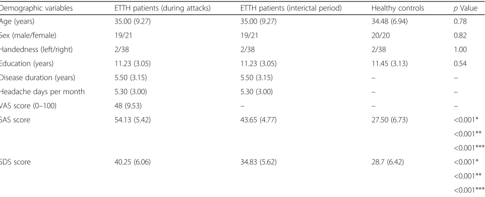
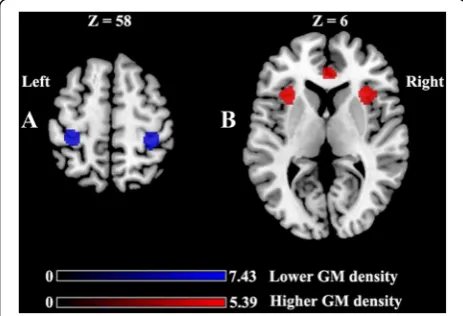
No significant differences were identified between pa-tients and controls for the total volume of GM, WM, or TIV. As demonstrated in Table 2 and Fig. 1, significant GMD reductions in the bilateral primary somatosensory cortex (S1) (A) and significant GMD increases in the bi-lateral anterior cingulate cortex (ACC) and the bibi-lateral anterior insula (B) were observed between the pain phase and pain-free phase in patients with ETTH. Com-pared to healthy controls, patients with ETTH in the out
Table 1Demographic variables and clinical characteristics of study participants
Demographic variables ETTH patients (during attacks) ETTH patients (interictal period) Healthy controls pValue
Age (years) 35.00 (9.27) 35.00 (9.27) 34.48 (6.94) 0.78
Sex (male/female) 19/21 19/21 20/20 0.82
Handedness (left/right) 2/38 2/38 2/38 1.00
Education (years) 11.23 (3.05) 11.23 (3.05) 11.45 (3.13) 0.54
Disease duration (years) 5.50 (3.15) 5.50 (3.15) – –
Headache days per month 5.30 (3.00) 5.30 (3.00) – –
VAS score (0–100) 48 (9.53) – – –
SAS score 54.13 (5.42) 43.65 (4.77) 27.50 (6.73) <0.001*
<0.001**
<0.001***
SDS score 40.25 (6.06) 34.83 (5.62) 28.7 (6.42) <0.001*
<0.001**
<0.001***
Values are expressed as mean (SD). Thepvalues were calculated using appropriate statistical tests (2-tailed pairedttest or 2-samplettest for continuous data andχ2tests for categorical data)
ETTHepisodic tension-type headache,SASself-rating anxiety scale,SDSself-rating depression scale,VASvisual analog scale *2-tailed paired t test for SAS or SDS scores between pain phase and out of phase in ETTH patients
of pain phase exhibited similar GMD changes. In con-trast, the ETTH patients out of pain phase showed no region with higher or lower GMD compared with healthy controls. Furthermore, the whole brain correlation analyses revealed that GMD in the left ACC and left an-terior insula was negatively correlated with headache days per month (r=−0.782,p= 0.002 andr=−0.646,p= 0.007, respectively). In addition, GMD in the left ACC was nega-tively correlated with the SAS score (r=−0.841,p= 0.001) and the SDS score (r=−0.579, p= 0.021) in ETTH pa-tients during the pain phase. No correlation was identified between regional GMD and disease duration or regional GMD and the VAS score in ETTH patients.
Discussion
To the best of our knowledge, this is the first longitudinal study that primarily investigated whether treatment-naïve ETTH patients have dynamic changes of brain GMD in different pain states. Our results demonstrated a lower GMD in the bilateral S1 and a higher GMD in the bilateral ACC and anterior insula in ETTH patients during the pain phase compared with the pain-free phase. In con-trast, no GMD changes were observed in ETTH patients
during the pain-free period. Our study exhibited a dynamic cortical plasticity in patients with ETTH. Fur-thermore, the correlation analyses indicated that these GMD changes could be affected by the headache days per month and anxiety and depressive symptoms.
Convergent evidence from anatomical, imaging, and le-sion data reveals that the S1, the ACC, and the anterior in-sula are key regions implicated in complex nociceptive processing [27, 28]. The S1 is responsible for detecting the presence and magnitude of a pain stimulus and is involved in pain perception [27]. The ACC participates in the emotional-motivational aspect of pain [29]. Often working together with the ACC, the anterior insula is proposed to involve in the integration of polymodal sensory informa-tion as well as the integrainforma-tion of emoinforma-tional and cognitive processes [29, 30]. A recent neuroimaging meta-analysis revealed common activations during pain for healthy subjects and patients with chronic pain in the ACC and the anterior insula regardless of modality, body part, or clinical experience [29]. This finding further supported the central role of the ACC and the anterior insula in human pain processing [29]. Painful stimulation during the pain phase in ETTH patients contribute to these brain functional changes, which could further lead to the struc-tural reorganization observed in our study.
Of note, the structural abnormalities were not ob-served in the pain-free period in ETTH patients. This structural reorganization and dynamic change may be ascribed to the transmission of sensory input and pain perception [31]. Although little is known regarding the neurobiological basis of this dynamic pattern in ETTH, fast adjusting reversible neuronal processes, such as den-drite spine and synapse turnover, are more likely respon-sible for these rapid morphometric changes [9]. This feature may reflect a defensive adaptation designed to orient cortical attention towards stimuli that threaten the body’s integrity [31, 32] or reflect a balance of de-scending pain modulatory circuits [31, 33, 34].
However, this adaptation or balance might be dis-rupted as the headache days increased and anxiety and depressive symptoms progressed. Our correlation ana-lyses demonstrated that ETTH patients with longer
Table 2Summary of gray matter density differences in ETTH patients between the pain and pain-free phases
Brain regions Brodmann areas Maximum MNI coordinates (x, y, z) Voxels T value
Pain phase < pain-free phase
Right primary somatosensory cortex 3/4 32,–36, 62 300 7.43
Left primary somatosensory cortex 3/4 −34,−34, 58 155 6.61
Pain phase > pain-free phase
Bilateral anterior cingulate cortex 32/24 10, 38, 16 493 5.35
Left anterior insula 13 34, 20, 6 255 5.39
Right anterior insula 13 −34, 21, 7 218 5.35
ETTHepisodic tension-type headache,MNIMontreal Neurological Institute
Fig. 1a: lower GM density in the bilateral primary somatosensory
headache days per month had lower GMD in the ACC and anterior insula in our study. Increased headache days may contribute to the progression from episodic to chronic TTH, which leads to the central sensitization with GM reductions in multiple cerebral regions, such as the ACC and the anterior insula [17]. This correlation also contributes to explaining why we observed in-creased GMD in the ACC and anterior insula in ETTH not like that most studies reported decreases here in chronic pain diseases, including chronic TTH [16, 35]. Increased GMD in ETTH may reflect a defensive adap-tation, whereas decreased GMD may indicate decom-pensation as disease develops to the chronic form. This dynamic change in these areas calls on us to pay more attention to TTH in the episodic form. In addition, anxiety and depressive symptoms are common in ETTH patients [36]. The comorbidity may confer a worse prog-nosis in TTH patients [36, 37]. Although their pathophysi-ology remains unknown, their relationship may be bidirectional [36]. ACC is the common neuroanatomical site implicated in mental illness, including depression, anxiety and other psychiatric disorders [38]. Our data demonstrated negative correlations between GMD in the ACC and the SAS and SDS scores. This information calls attention to a timely recognition of these symptoms and the need to offer proper treatment in ETTH patients.
Some limitations should be mentioned when interpret-ing the findinterpret-ings of our study. First, VBM has inherent limitations. For example, VBM detects only linear, spatially limited differences [39]. Second, our study did not investigate a control group longitudinally in the same time intervals using the same preprocessing and statistics, although the interval time was short, which might bias our results. Third, this study is the first to evaluate the brain structural changes in ETTH; further studies would benefit from integrating both structural and functional networks associated with the patho-physiological underpinnings of ETTH.
Conclusions
This is the first study to demonstrate dynamic and revers-ible GMD changes in the S1, ACC, and anterior insula be-tween pain and pain-free phases in ETTH patients, which suggests cerebral adaptation to pain stimuli with a balance of pain modulatory circuits. However, this balance might be disrupted by increased headache days and progressive anxiety and depressive symptoms. Future studies are war-ranted to determine whether this structural plasticity is a characteristic of ETTH.
Abbreviations
ACC:Anterior cingulate cortex; DARTEL: Diffeomorphic Anatomic Registration Through Exponentiated Lie Algebra; ETTH: Episodic tension-type headache; FOV: Field of view; GM: Gray matter; GMD: Gray matter density; ICHD: International Classification of Headache Disorders; MNI: Montreal Neurological Institute;
S1: Primary somatosensory cortex; SAS: The Zung Self-Rating Anxiety Scale; SDS: The Zung Self-Rating Depression Scale; SPM8: Statistical Parametric Mapping version 8; SPSS: Statistical Package for the Social Sciences; TE: Echo time; TIV: Total intracranial volume; TR: Repetition time; TTH: Tension-type headache; VAS: Visual analog scale; VBM: Voxel-based morphometry; WM: White matter
Acknowledgments
The authors thank all patients and volunteers for their participation in this study.
Funding
No funding supported this work.
Authors’contributions
BC performed the experiments, analyzed the data and outlined the manuscript. YH contributed to the data analysis and the manuscript. LX contributed to the study design and the manuscript. L-LG contributed to the data acquisition and analysis and the manuscript. J-LZ designed the study, supervised the experimental work and the data analysis and refined the manuscript. All authors read and approved the final manuscript.
Competing interests
The authors declare that they have no competing interests.
Ethics approval and consent to participate
The study protocol was approved by the local ethics committee of Huai'an First People’s Hospital Affiliated to Nanjing Medical University. Informed consent was obtained from all participants.
Author details
1
Department of Neurology, Huai’an First People’s Hospital Affiliated to Nanjing Medical University, 223300, Beijing West Road 6#, Huai’an, Jiangsu Province, People’s Republic of China.2Department of Gastrointestinal Surgery, Huai’an Hospital Affiliated to Xuzhou Medical College and Huai’an Second People’s Hospital, Huai’an, People’s Republic of China.3Department of Medical Imaging, Huai’an First People’s Hospital Affiliated to Nanjing Medical University, Huai’an, People’s Republic of China.
Received: 31 August 2016 Accepted: 9 November 2016
References
1. Stovner L, Hagen K, Jensen R, Katsarava Z, Lipton R, Scher A, Steiner T, Zwart JA (2007) The global burden of headache: a documentation of headache prevalence and disability worldwide. Cephalalgia 27(3):193–210 2. Bendtsen L, Jensen R (2006) Tension-type headache: the most common,
but also the most neglected, headache disorder. Curr Opin Neurol 19(3): 305–309
3. Semenov IA (2015) Tension-type headaches. Dis -Month 61(6):233–235 4. Chen Y (2009) Advances in the pathophysiology of tension-type headache:
from stress to central sensitization. Curr Pain Headache Rep 13(6):484–494 5. Dowson A (2015) The burden of headache: global and regional prevalence
of headache and its impact. Int J Clin Prac Suppl 182:3–7
6. Headache Classification Committee of the International Headache S (2013) The International Classification of Headache Disorders, 3rd edition (beta version). Cephalalgia 33(9):629–808
7. Lai TH, Protsenko E, Cheng YC, Loggia ML, Coppola G, Chen WT (2015) Neural plasticity in common forms of chronic headaches. Neural Plast 2015:205985 8. Fumal A, Schoenen J (2008) Tension-type headache: current research and
clinical management. Lancet Neurol 7(1):70–83
9. Coppola G, Di Renzo A, Tinelli E, Iacovelli E, Lepre C, Di Lorenzo C, Di Lorenzo G, Di Lenola D, Parisi V, Serrao M et al (2015) Evidence for brain morphometric changes during the migraine cycle: a magnetic resonance-based morphometry study. Cephalalgia 35(9):783–791
10. Naegel S, Holle D, Desmarattes N, Theysohn N, Diener HC, Katsarava Z, Obermann M (2014) Cortical plasticity in episodic and chronic cluster headache. Neuroimage Clin 6:415–423
12. Coppola G, Tinelli E, Lepre C, Iacovelli E, Di Lorenzo C, Di Lorenzo G, Serrao M, Pauri F, Fiermonte G, Bianco F et al (2014) Dynamic changes in thalamic microstructure of migraine without aura patients: a diffusion tensor magnetic resonance imaging study. Eur J Neurol 21(2):287–e213 13. Chou KH, Yang FC, Fuh JL, Huang CC, Lirng JF, Lin YY, Lee PL, Kao HW, Lin
CP, Wang SJ (2014) Altered white matter microstructural connectivity in cluster headaches: a longitudinal diffusion tensor imaging study. Cephalalgia 34(13):1040–1052
14. Ashburner J, Friston KJ (2000) Voxel-based morphometry–the methods. Neuroimage 11(6 Pt 1):805–821
15. Pan PL, Zhong JG, Shang HF, Zhu YL, Xiao PR, Dai ZY, Shi HC (2015) Quantitative meta-analysis of grey matter anomalies in neuropathic pain. Eur J Pain 19(9):1224–1231
16. Smallwood RF, Laird AR, Ramage AE, Parkinson AL, Lewis J, Clauw DJ, Williams DA, Schmidt-Wilcke T, Farrell MJ, Eickhoff SB et al (2013) Structural brain anomalies and chronic pain: a quantitative meta-analysis of gray matter volume. J Pain 14(7):663–675
17. Schmidt-Wilcke T, Leinisch E, Straube A, Kampfe N, Draganski B, Diener HC, Bogdahn U, May A (2005) Gray matter decrease in patients with chronic tension type headache. Neurology 65(9):1483–1486
18. Seminowicz DA, Shpaner M, Keaser ML, Krauthamer GM, Mantegna J, Dumas JA, Newhouse PA, Filippi CG, Keefe FJ, Naylor MR (2013) Cognitive-behavioral therapy increases prefrontal cortex gray matter in patients with chronic pain. J Pain 14(12):1573–1584
19. Riederer F, Gantenbein AR, Marti M, Luechinger R, Kollias S, Sandor PS (2013) Decrease of gray matter volume in the midbrain is associated with treatment response in medication-overuse headache: possible influence of orbitofrontal cortex. J Neuroscience 33(39):15343–15349
20. Walther K, Bendlin BB, Glisky EL, Trouard TP, Lisse JR, Posever JO, Ryan L (2011) Anti-inflammatory drugs reduce age-related decreases in brain volume in cognitively normal older adults. Neurobiol Aging 32(3):497–505 21. Price DD, Bush FM, Long S, Harkins SW (1994) A comparison of pain
measurement characteristics of mechanical visual analogue and simple numerical rating scales. Pain 56(2):217–226
22. Zung WW (1971) A rating instrument for anxiety disorders. Psychosomatics 12(6):371–379
23. Zung WW, Richards CB, Short MJ (1965) Self-rating depression scale in an outpatient clinic. Further validation of the SDS. Arch Gen Psychiatry 13(6):508–515 24. Ashburner J, Friston KJ (2005) Unified segmentation. NeuroImage 26(3):839–851 25. Ashburner J, Friston KJ (2009) Computing average shaped tissue probability
templates. NeuroImage 45(2):333–341
26. Ashburner J (2007) A fast diffeomorphic image registration algorithm. NeuroImage 38(1):95–113
27. Bushnell MC, Duncan GH, Hofbauer RK, Ha B, Chen JI, Carrier B (1999) Pain perception: is there a role for primary somatosensory cortex? Proc Natl Acad Sci U S A 96(14):7705–7709
28. Duerden EG, Albanese MC (2013) Localization of pain-related brain activation: a meta-analysis of neuroimaging data. Hum Brain Mapp 34(1):109–149 29. Jensen KB, Regenbogen C, Ohse MC, Frasnelli J, Freiherr J, Lundstrom JN
(2016) Brain activations during pain: a neuroimaging meta-analysis of patients with pain and healthy controls. Pain 157(6):1279–1286 30. Gu X, Gao Z, Wang X, Liu X, Knight RT, Hof PR, Fan J (2012) Anterior insular
cortex is necessary for empathetic pain perception. Brain 135(Pt 9):2726–2735 31. Teutsch S, Herken W, Bingel U, Schoell E, May A (2008) Changes in brain
gray matter due to repetitive painful stimulation. Neuroimage 42(2):845–849 32. Burns E, Chipchase LS, Schabrun SM (2016) Primary sensory and motor
cortex function in response to acute muscle pain: A systematic review and meta-analysis. Eur J Pain (London, England). 20(8):1203–213 33. Heinricher MM (2016) Pain modulation and the transition from acute to
chronic pain. Adv Exp Med Biol 904:105–115
34. Ossipov MH, Morimura K, Porreca F (2014) Descending pain modulation and chronification of pain. Curr Opin Support Palliat Care 8(2):143–151 35. Cauda F, Palermo S, Costa T, Torta R, Duca S, Vercelli U, Geminiani G, Torta
DM (2014) Gray matter alterations in chronic pain: A network-oriented meta-analytic approach. Neuroimage Clin 4:676–686
36. Crystal SC, Robbins MS (2010) Epidemiology of tension-type headache. Curr Pain Headache Rep 14(6):449–454
37. Kikuchi H, Yoshiuchi K, Ando T, Yamamoto Y (2015) Influence of psychological factors on acute exacerbation of tension-type headache: Investigation by ecological momentary assessment. J Psychosom Res 79(3):239–242
38. Goodkind M, Eickhoff SB, Oathes DJ, Jiang Y, Chang A, Jones-Hagata LB, Ortega BN, Zaiko YV, Roach EL, Korgaonkar MS et al (2015) Identification of a common neurobiological substrate for mental illness. JAMA psychiatry 72(4):305–315
39. Davatzikos C (2004) Why voxel-based morphometric analysis should be used with great caution when characterizing group differences. Neuroimage 23(1):17–20
Submit your manuscript to a
journal and benefi t from:
7 Convenient online submission
7 Rigorous peer review
7 Immediate publication on acceptance
7 Open access: articles freely available online
7 High visibility within the fi eld
7 Retaining the copyright to your article