252
_______________________________________________________________
_______________________________________________________________
DIABETIC NEUROPATHY: EPIDEMIOLOGY, PATHOPHYSIOLOGY
AND MANAGEMENT
Kundan G. Ingale*
1,Neeraj S Vyawahare
2,
N. D. Bendale
1, D. T. Gautam
1, B.Y. Rane
1,
S. W. Rangari
1, R. L. Bakal
11*Department of Pharmacology, KYDSCT’s College of Pharmacy, Sakegaon, Bhusawal-425 201, India. 2Dr. D. Y. Patil College of Pharmacy, Akurdi, Pune -411035, India.
ABSTRACT
Neuropathies of the peripheral and autonomic nervous systems affect upto half of all people with diabetes, and are major risk factors for foot ulceration and amputation. The aetiology is multi factorial includes metabolic changes in diabetes may directly affect neural tissue, but importantly, neurodegenerative changes are precipitated by compromised nerve vascular supply. Experiments in animal models of diabetic neuropathy suggest that similar metabolic sequelae affect neurons and endothelium. These include elevated polyol pathway activity, oxidative stress, the formation of advanced glycation and lipoxidation end products and various proinflammatory changes such as elevated protein kinase C, nuclear factor κB and p38 mitogen activated protein kinase signaling. These mechanisms do not work in isolation but strongly interact in a mutually facilitatory fashion. Nitrosative stress and the induction of the enzyme poly (ADP-ribose) polymerase form one important link between physiological stress or such as reactive oxygen species and the pro-inflammatory mechanisms. Recently, evidence points to endoplasmic stress and the unfolded protein response as forming another crucial link. This review focuses on the aetiopathogenesis of neurovascular changes in diabetic neuropathy, elucidated in animal studies, and on putative therapeutic targets the majority of which have yet to be tested for efficacy in clinical trials.
Key words: Diabetes, Diabetic Neuropathy (DN), Management, Pathophysiology.
INTRODUCTION
Diabetes poses a significant economic burden in India; corresponds to 5.0% prevalence to the world adult population [1, 2]. The number of people with diabetes is increasing due to population growth, aging, urbanization and increasing prevalence of obesity and physical inactivity. Diabetes mellitus is categorized as a metabolic disease characterized by hyperglycemia resulting from defects in insulin secretion, insulin action or both. As a result, the amount of glucose in the blood increases while the cells are starved of energy [3]. It is predicted that by 2030, India, China and the United States will have the largest number of people with diabetes [4]. Sustained higher levels of blood glucose cause damage to nerves and blood vessels, leading to complications such as heart disease and stroke, the leading causes of death among people with diabetes [5, 6]. By 2030 diabetes will be a seventh leading cause of deaths worldwide. Uncontrolled diabetes with chronic hyperglycemia can eventually lead to other health problems and microvascular complications
as well, such as, nephropathy, neuropathy, vision loss or retinopathy and sexual dysfunction, further decreasing quality of life, reducing life expectancy, and increasing the economic burden [2, 7, 8]. Uncontrolled high blood glucose level for long time may develop depression, foot or leg amputation and skin complications [9, 10]. Diabetic neuropathy is an important microvascular complication of diabetes mellitus. It is a major contributor to foot ulceration and lower limb amputation in persons with diabetes [11]. As the population of diabetes is increasing worldwide, the prevalence of diabetes related diabetic neuropathy is also on the rise. Duration of diabetes mellitus is an important risk factor for all diabetes related neuropathy. People with diabetes can develop nerve problems at any time, but risk rises with age and longer duration of diabetes. Diabetic neuropathies also appear to be more common in people who have problems controlling their blood glucose, also called blood sugar, as well as those with high levels of blood fat and blood pressure and
*Corresponding Author Kundan G. Ingale E mail: kundan_ingale@rediffmail.com
International Journal
of
Pharmacy Review & Research
www.ijprr.com
253
those who are overweight. In patients with type 2 diabetes mellitus (T2DM), average medical costs of patients with neuropathy are 3.3 fold higher than those without complications [12, 13].
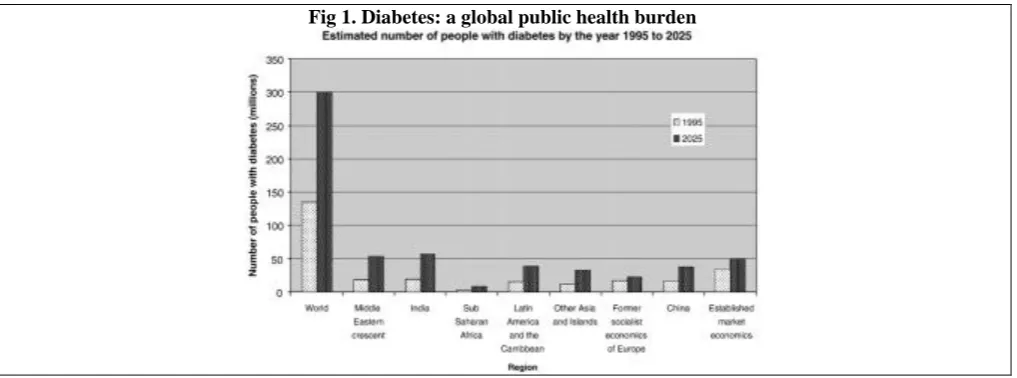
Epidemiology of Neuropathy
The increasing worldwide incidence of DM in adults constitutes a global public health burden (Fig. 1).
Diabetic neuropathy (DN) is an important complication of diabetes mellitus resulting in a great deal of morbidity. The prevalence of diabetic neuropathy is about 26.1% in Indian population [14]. Up to 7.5% of patients with non insulin dependent diabetes mellitus (NIDDM) have clinical neuropathy at the time of diagnosis and this rate increases to 50% among patients who have had diabetes for 25 years [15, 16].
Signs and symptoms
Peripheral diabetic neuropathy is characterized by symptoms such as tingling and numbness, sharp pains or insensitivity to pain, motor in coordination, loss of sense of vibration etc. Untreated, it may lead to loss of reflexes and deformities that may progress to gangrene. The condition is characterised by peripheral demyelination, decrease in the nerve conduction and degeneration of myelinated and demyelinated sensory nerve fibres [17].
Symptoms depend on the type of neuropathy and which nerves are affected. Some people with nerve damage have no symptoms at all [18, 19].
Pathophysiology of DN
The pathophysiology of diabetic neuropathy is multifactorial and no single etiology is at the forefront. The mechanisms leading to diabetic neuropathy have not been fully elucidated yet. It has been suggested that they result from hyperglycemia or the loss of insulin dependent regulation. Different pathophysiological mechanisms have been postulated. One of them implicates the polyol pathway, where increased glucose leads to an increased activity of the enzyme aldose reductase and, thus, to the production of polyols which cause a decreased activity of the sodium/potassium (Na/K) pump and a depletion of reducing equivalents such as NADPH (nicotinamide adenine dinucleotide phosphate, reduced form) with a secondary decrease in glutathione production and, as a result, the induction of intracellular oxidative stress [20]. In most publications there is a heterogenous theory that encompasses main causative factors: metabolic and microangiopathic changes. While the primary and proven cause of DN includes a prolonged hyperglycemia, among other causative factors the role of deficiency of neuronal growth factor (NGF), neurotrophins and insulin like growth factor (IGF-1), impaired nerve fiber regeneration, inflammation in response to autoimmune processes, and genetic background are also emphasized [21, 22]. Together with neurons, endothelial cells also belong to insulin-independent cells, in which transport of glucose into the cell takes place through facilitated diffusion. In Metabolic factors oxidative stress as a result of glucose oxidation and the subsequent formation of advanced glycation end
(AGE) products, disruption of polyol pathway, altered glucose metabolism and decreased antioxidant defenses are some of the implicated mechanisms in the pathogenesis of neuropathy [23]. Aldose reductase plays an important role as it diverts excess of glucose in hyperglycemic states to sorbitol by polyol pathway which is responsible for diabetic complications including neuropathy. Chronic use of aldose reductase inhibitors have shown beneficial effect on peripheral sensory neuropathy of experimentally induced diabetic rats as well as in clinical trials [24, 25]. Some of the major metabolic changes in diabetes thought to contribute to DPN and DAN. These include elevated polyol pathway activity, oxidative stress, the formation of advanced glycation end products, and various proinflammatory changes such as elevated nuclear factor κB (NFκB) and p38 mitogen activated protein kinase (MAPK) signalling. These mechanisms do not work in isolation but strongly interact in a mutually facilitatory fashion [26]. Recent studies of the intraepidermal nerve endings, as a means to analyze small fibres, showed that a major loss of these endings was associated with neuropathic pain only in patients who had little or no objective signs of neuropathy, thereby suggesting that the loss of nerve endings is not sufficient to induce pain [27, 28].
Risk factors
Modifiable risk factors are degree of metabolic control (hyperglycemia, hypoglycemia, episodes of ketoacidosis), presence of microalbuminuria and retinopathy, arterial hypertension, atherosclerosis, dyslipidemia, smoking habit, and alcohol overuse. Whereas non-modifiable factors include male gender, advanced age, duration of diabetes, height, genetic factors [29].
Polyol pathway
The polyol pathway is present in many tissues including peripheral nerve and blood vessels. Glucose is converted to sorbitol by aldose reductase, and sorbitol may be further metabolised to fructose, catalysed by sorbitol dehydrogenase. Aldose reductase is found in various tissues, such as nerve, glomerulus, retina, lens and vascular cells [30]. NADPH is an important cofactor necessary to regenerate reduced glutathione (GSH) and GSH is a scavenger of reactive oxygen species (ROS). Depletion of GSH could exacerbate intracellular oxidative stress and there by contribute to diabetic complications [31]. Therefore it seemed logical to target inhibition of the polyol pathway as a potential treatment of diabetic neuropathy. This led to the development of several aldose reductase inhibitors (ARIs). These ARIs were highly effective in preventing or correcting neuropathic changes in diabetic animal models [32]. The first step of the polyol pathway, catalysed by aldose reductase, is considerably more important for neurovascular actions than the second sorbitol dehydrogenase dependent step.
Oxidative stress and advanced glycation
254
pathophysiology, prevention and treatment of DPN. Intervention studies with antioxidant compounds have been performed in animal models of DPN and DAN as well as in diabetic neuropathic patients. Several experimental studies have shown that treatment with antioxidants in experimental diabetes can prevent and reverse nerve dysfunction and damage [35-38]. In contrast, the number of clinical studies is limited. Increased oxidative stress in the peripheral diabetic nerve may be explained by several pathways that contribute to altered nerve metabolism and microvascular dysfunction. Traditionally, the focus has been on the polyol pathway, non-enzymatic glycation and glucose auto oxidation [33, 39]. Increased polyol pathway flux leads to GSH deficits and attenuated free radical scavenging capacity as described above. Non-enzymatic glycation of antioxidant enzymes causes a further deficit in protection against oxidative damage [40, 41]. Advanced glycation end products (AGEs) are formed by non enzymatic reactions between aldehyde groups of reducing sugars with proteins, lipids or nucleic acids. Many enzymatic processes also produce ROS, and several have been identified as contributing to DPN/DAN in experimental models by the use of specific inhibitors or gene knock out [26, 31, 42, 43]. The mitochondrion is highly efficient in the utilization of oxygen and substrates mainly derived from glucose to produce cell energy in the form of ATP. Thus, electrons from oxidized substrates are transferred to oxygen by a series of reduction reactions to generate water [44]. In this process, protons are pumped from the mitochondrial matrix to cross the inner mitochondrial membrane through the respiratory complexes forming the oxidative phosphorylation chain [45, 46]. The respiratory chain consists of five multi polypeptide enzyme complexes that form the oxidative phosphorylation system: complex I (NADH-ubiquinone reductase); complex II (FADH2 succinate ubiquinone reductase); complex III (ubiquinol-cytochrome C reductase); complex IV ((ubiquinol-cytochrome C oxidase and two mobile electron transporters, ubiquinone and cytochrome C); and complex V (ATP synthase). Electrons generated from the reducing equivalents (NADH and FADH2) pass between the complexes and generate an increase in energy that allows for proton pumping to complexes I, III, and IV. Finally, the proton pump generated in the inner mitochondrial membrane is used to generate ATP [47].
Diagnosis
Various screening modalities for diabetic neuropathy include recording of symptoms or signs, nerve conduction studies, quantitative sensory testing, and autonomic testing. Quantitative assessment of vibration perception threshold (VPT) is a widely applied tool in the screening for, and staging of, diabetic sensory neuropathy, particularly in epidemiological studies. Values of VPT of more than 25 V are associated with a 6–10-fold risk of developing a foot ulcer. These findings suggest the role of severity of diabetic neuropathy in the etiology of its complications. Diabetic neuropathy assessment was done by measuring VPT using sensitometer. The VPT was
measured by a single observer by placing biothesiometer probe perpendicular to the distal plantar surface of the great toe of both legs [16]. Recently, in the diagnosis of DPN new tool include biopsy of the nerve and skin, since it has been proven that there are differences in the microscopic image of the skin of a healthy and diabetic subject. These differences include reduction of the number of cutaneous nerve fibers shown by staining for the neuronal antigen PGP 9.5, in small nerve fiber neuropathy [27].
Confocal microscopy and new immune histochemical methods were used to test non myelinated nerves of the skin and internal organs [48]. Confoscan, which is a variation of light microscopy and is characterized by increased contrast and resolution, was used to examine cornea, as a noninvasive and safe method. Significant thinning (reduction of density) of corneal nerve fibers was defined as the early marker of DPN [49]. Furthermore, an increase in tortuosity of corneal nerve fibers was observed in patients with diabetes, as compared with control group. Rosenberg's research team at the University of Helsinki has demonstrated a correlation between structural changes of corneal nerves, reduced corneal sensitivity and the intensity of DPN in patients with type1diabetes [50]. New research techniques have also shown that one of the mechanisms of acute neuropathic pain is a disorder of blood circulation in small blood vessels that supply the nerves [51]. On the basis of magnetic resonance imaging, researchers from Chicago have demonstrated the presence of structural changes in the brain of diabetic patients with chronic (present for more than a year) back pain. In 26 subjects a 5%–11% reduction in prefrontal and thalamic gray matter was found, while in the normal brain ageing processes such reduction may be upto 0.5%. Demonstrated differences are the equivalent of 10–20 years of physiological ageing processes [52].
Types
Diabetic neuropathy had been classified as peripheral, autonomic, proximal, or focal. Each affects different parts of the body in various ways.
Peripheral neuropathy (DPN), the most common type of diabetic neuropathy, with prevalence of 60-70% in diabetic population causes pain or loss of feeling in the toes, feet, legs, hands, and arms. DPN gives rise to stocking gloves pattern of sensory loss progressing proximally as diabetes duration [53].
Autonomic neuropathy (DAN) causes changes in digestion, bowel and bladder function, sexual response, and perspiration. It can also affect the nerves that serve the heart and control blood pressure, as well as nerves in the lungs and eyes [54]. Autonomic neuropathy can also cause hypoglycemia unawareness, a condition in which people no longer experience the warning symptoms of low blood glucose levels.
Proximal neuropathy causes pain in the thighs, hips, or buttocks and leads to weakness in the legs.
255 Treatment
Drug treatment
There is no definitive treatment for DN at present.
There is now a consensus among experts that the analgesic efficacy of drug treatments for neuropathic pain is independent of the aetiology of neuropathy. Analgesics like paracetamol, salicylates and non-steroidal anti-inflammatory drugs (NSAIDs). These drugs are considered ineffective or poorly effective against neuropathic pain [55, 56]. Tricyclic antidepressants [57], SSRIs, anticonvulsants, opioids and topical capsaicin have been tried in the management of painful neuropathy of which SSNRIs such as duloxetine and pregabalin have been
approved by the USFDA. These drugs have been widely
used for the treatment of neuropathic pain for symptomatic relief. The use of these drugs is limited by their cost and side effects. A systematic review concluded that SSRIs have only limited and clinically non-significant effects on neuropathic pain. The most frequent side effects of SSRIs include dizziness, somnolence, headache and nausea [58, 59].
Combination therapy
The choice of medication depends on comorbidities, contraindications and the economic status of the patient. When no desirable results are obtained with the use of one of the above mentioned first line medication, combination therapy with two drugs is preferred. If pain persists opioid should be added (tramadol 200–400 mg/day, oxycodone 20–80 mg/day; morphine 20–80 mg/day).The guidelines also outline topical medication capsaicin cream (0.075 %, 3–4 times per day). Few studies have tested the efficacy of combination drug therapies. Gabapentin associated with the SSNRI venlafaxine appeared to be more effective than gabapentin alone [60]. An additive effect was found when combining gabapentin with morphine compared with monotherapy with either gabapentin or morphine alone, [61] the efficacy of the combination against pain was greater at lower doses than those used in monotherapy. Similar results were also reported for the combination of gabapentin and a tricyclic antidepressant (nortriptyline, a metabolite of amitriptyline) in a study [62].
Non Pharmacological treatment
Non-pharmacological therapies includes acupuncture, infrared radiation, laser therapy and psychotherapy, are available, although recommendations are not possible, given that very few trials have been carried out and with conflicting results. Transcutaneous electrical nerve stimulation [63] (TENS) and external muscle stimulation [64] has been the subject of a few randomized studies. The basis for the treatment of diabetic polyneuropathy is good metabolic control of diabetes, arterial hypertension, as well as smoking cessation and avoiding alcohol abuse. In patients treated with intensive functional insulin therapy (FIT) lower incidence of neuropathy has been demonstrated and in patients with clinical demonstration of neuropathy relief of symptoms was observed. To date no satisfactory treatment targeting
the causes of neuropathy exists except for good metabolic control, which slows but does not prevent progression35.The only aldose reductase inhibitor approved
is Epalrestat which is marketed in Japan. The
unsatisfactory results associated with conventional treatments for symptoms of diabetic peripheral neuropathy (DPN) demonstrate a need for research into alternative therapies. Whole Body Vibrational (WBV) therapy is an experimental modality for pain associated with DPN [65]. Currently two published case studies support the efficacy of WBV as a treatment of DPN [66]. Both levels significant decreases in pain levels measured by visual analog pain scale (VAS). On the other hand however, caution is advised in excessive attempt to achieve normoglycemia, which may lead to reduced blood flow and nerve ischemia, thus enhancing pain and other symptoms associated with neuropathy. Therefore, an indicated desired blood glucose range is 90–180 mg/dL (5.0–10.0 mmol/L). In addition to glycemic control, α-lipoic acid (ALA) appears to be the only effective drug in the treatment for DPN. This is a mitochondrially synthesized octanoic acid derivative with a potent antioxidant effect. ALA shows a hypoglycemic effect, reduces insulin degradation and insulin resistance, increases glucose metabolism and its uptake in the liver and muscles. More pronounced therapeutic effects were observed in patients who received the drug intravenously [64]. When all of the above mentioned treatment methods have been used, spinal cord stimulation may be indicated. The effectiveness of this method was demonstrated in a study, in which 10 patients had stimulating electrodes implanted in the epidural space. Significant improvement was reported by 8 patients, which was reflected in the visual analog scale of pain, and after completion of a 12-month study, 6 patients decided on continuation of this mode of pain management [67].
Pathogenetic treatments Glycaemic control
256 Fig 1. Diabetes: a global public health burden
CONCLUSION
Neuropathy is a common complication of diabetes, reducing the quality of life and increasing mortality. Available treatments to date consist of improved metabolic control and a focus on symptoms but do not concentrate on fundamental mechanisms in the pathogenesis of neuropathy. Current research has revealed a multifactorial aetiology of inter linked pathways dependent on oxidative/nitrosative stress, advanced glycation/lipoxidation, PARP activity and ER stress that activate pro-inflammatory changes. In turn these have potential adverse effects directly on neurons and Schwann
cells, and importantly indirect effects via impaired nerve vascular supply. Within this pathophysiology, there are many potential therapeutic targets that deserve to be studied in clinical trials. Several of the downstream mechanisms stimulated by oxidative stress, such as ER-stress, PARP, NFκB and p38 MAPK activation have proved to be major sources of ROS production in diabetes, constituting a self-reinforcing disease mechanism that needs to be interrupted for optimal therapeutic effect. Diabetes produces a low level pro-inflammatory state and neuropathy and vascular dysfunction occur even in pre-diabetes and impaired glucose tolerance [74].
REFERENCES
1. Shaw JE, Sicree RA, Zimmet PZ. Global estimates of the prevalence of diabetes for 2010 and 2030. Diabetes Research and Clinical Practice, 87, 2010, 4–14.
2. Odawara M, Kadowaki T, Naito Y. Effectiveness and safety of basal supported oral therapy with insulin glargine in Japanese insulin-naive, type 2 diabetes patients with or without microvascular complications: subanalysis of the observational, non interventional, 24-week follow-up Add-on Lantus® to Oral Hypoglycemic Agents (ALOHA) study. J Diabetes and Its Complications, 29(1), 2015, 127-133.
3. Kumar V, Cotran RS, Robbins SL. Robbin's basic pathology. seventh ed. pp. Philadelphia: Saunders. An Imprint of Elsevier, 2004, 641−655.
4. Wild S, et al. Global prevalence of diabetes: Estimates for 2000 and projections for 2030. Diabetes Care, 27, 2004, 1047−1053.
5. Tolle T, Xu X, Sadosky A. Painful diabetic neuropathy: across-sectional survey of health state impairment and treatment patterns. Journal of Diabetes and Its Complications, 20(1), 2006, 26-33.
6. Veves A, Backonja M, Malik R. Painful diabetic neuropathy epidemiology, natural history, early diagnosis, and treatment options. Pain Medicine, 9, 2009, 660-674.
7. Gerich JE. The importance of tight glycemic control. The American Journal of Medicine, 118, 2005, 7S–11S.
8. Girach A, Manner D, Porta M. Diabetic microvascular complications: Can patients at risk be identified A review.
International Journal of Clinical Practice, 60, 2006, 1471–1483
9. Thorve VS, et al. Diabetes-induced erectile dysfunction: epidemiology, pathophysiology and management. Journal of Diabetes and Its Complications, 25, 2011, 129–136
10. Haslett C, Chilvers ER et al. Davidson’s Principle and Practice of Medicine nineteenth ed.. London. Churchill Livingstone, 2002, 641−682.
11. Shaw JE, et al. Diabetic neuropathy in Mauritius: Prevalence and risk factors. Diabetes Res Clin Prac, 42, 1998, 131-9. 12. Neville SE, et al. Diabetes in Japan: A review of disease burden and approaches to treatment. Diabetes/Metabolism
Research and Reviews, 25, 2009, 705–716.
13. Hilary K, Ronald EA, William HH. Global burden of diabetes, 1995-2025. Prevalence, numerical estimates and projections. Diabetes Care, 21, 1998, 1414−1431.
14. Pradeepa R, et al. Prevalence and risk factors for diabetic neuropathy in an urban south Indian population: The Chennai
257
15. Pirart J, et al. Diabetes mellitus and its degenerative complications: A prospective study of 4,400 patients observed between 1947 and 1973. Diabetes Care, 1, 1978, 168-188.
16. Padmaja KR, et al. Prevalence and risk factors for severity of diabetic neuropathy in type 2 diabetes mellitus. Indian Journal of Medical Sciences, 64 (2), 2010, 51-56.
17. Nadig PD, et al. Effect of Tinospora cordifolia on experimental diabetic neuropathy. Indian J Pharmacol, 44, 2012,
580-583.
18. Boulton AJM. Management of diabetic peripheral neuropathy. Clin. Diabetes, 23, 2005, 9–15.
19. Vinik AI, Maser RE, Mitchell BD, Freeman R. Diabetic autonomic neuropathy. Diabetes Care, 26, 2003, 1553–1579. 20. Hernández BN, et al. El papel de la mitocondria en el dolor de la neuropatía diabética. Endocrinol Nutr, 60, 2013, 25-32. 21. Sullivan KA, Feldman EL. New developments in diabetic neuropathy. Curr Opin Neurol, 18(5), 2005, 586–590.
22. Szczeklik A, Stan WZ. Internal medicine. State of knowledge Kraków: Medycyna Praktyczna, 2010, 1277–1278.
23. Tawakoli M, et al. Pathophysiology and treatment of painful diabetic neuropathy. Current Pain Headache Rep, 3, 2012,
192-197.
24. Yagihashi S, et al. Effects of long-term aldose reductase inhibition on development of experimental diabetic neuropathy.
Ultra structural and morphometric studies of sural nerve in streptozocin induced diabetic rats. Diabetes, 39, 1990, 690-6.
25. Schemmel KE, Padiyara RS, D’Souza JJ. Aldose reductase inhibitors in the treatment of diabetic peripheral neuropathy: A
review. J Diabetes Complications, 24, 2010, 354-60.
26. Lupachyk S, et al. Na+/H+-exchanger-1inhibition reverses functional and structural manifestations of peripheral diabetic neuropathy. Diabetes, 59(1), 2010, 989.
27. Sorensen L, Molyneaux L, Yue DK. The relationship among pain, sensory loss and small nerve fibers in diabetes.
Diabetes Care, 29(4), 2006, 883–887.
28. Hartemann A, et al. Painful diabetic neuropathy: Diagnosis and management. Diabetes & Metabolism, 37, 2011, 377–388. 29. Wojciech M. Diabetic neuropathy. Polish annals of medicine ,20, 2013, 154–159.
30. Ramasamy R, Goldberg IJ. Aldose reductase and cardiovascular diseases, creating human-like diabetic complications in an experimental model. Circ. Res, 106, 2010, 1449–58.
31. Obrosova IG, et al. Different roles of 12/15-lipoxygenase in diabetic large and small fiber peripheral and autonomic neuropathies. Am. J. Pathol, 177, 2010, 1436–1447.
32. Oates PJ. Aldose reductase, still a compelling target for diabetic neuropathy. Curr. DrugTargets, 9, 2008, 14–36. 33. Pop BR, Sima A, Stevens M. Diabetic neuropathy andoxidative stress. Diabetes Metab.Res.Rev, 22, 2006, 257–273. 34. Vincent AM, Russell JW, Low P, Feldman EL. Oxidative stress in the pathogenesis of diabetic neuropathy. Endocr. Rev,
25, 2004, 612–628.
35. Cameron NE, et al. Vascular factors and metabolic interactions in the pathogenesis of diabetic neuropathy. Diabetologia, 44, 2001, 1973–1988.
36. Sytze VDP, et al. Pathogenesis of diabetic neuropathy: Focus on neurovascular mechanisms. European Journal of Pharmacology, 719, 2013, 180–186.
37. Van DPS. Oxidative stress and diabetic neuropathy: pathophysiological mechanisms and treatment perspectives.
DiabetesMetab.Res.Rev, 18, 2002, 176–184.
38. Keegan A, Cotter MA, Cameron NE. Effects of diabetes and treatment with the antioxidant, α-lipoic acid, on endothelial and neurogenic functions of corpus cavernosuminrats. Diabetologia, 42, 1999, 343–350.
39. Mahmood D, Singh BK, Akhtar M. Diabeticneuropathy: therapies on the horizon. J.PharmPharmacol, 61, 2009, 1137– 1145.
40. Ziegler D, Nowak H, Kempler P, Vargha P, Low PA. Treatment of symptomatic diabetic polyneuropathy with the antioxidant alpha-lipoic acid: a meta-analysis. Diabetes Med, 21, 2004, 114–121.
41. Ziegler D, et al. Efficacy and safety of antioxidant treatment with alpha-lipoic acid over 4 years in diabetic polyneuropathy: the NATHAN 1 trial. Diabetes Care, 34, 2011, 2054–2060.
42. Cameron NE, Cotter MA. Effects of protein kinase C beta inhibition on neurovascular dysfunction in diabetic rats: interaction with oxidative stress and essential fatty acid dysmetabolism. Diabetes Metab, 18, 2002, 315–323.
43. Inkster ME, Cotter MA, Cameron NE. Treatment with the xanthine oxidase inhibitor, allopurinol, improves nerve and vascular function in diabetic rats. Eur. J. Pharmacol, 561, 2007, 63-73.
44. MacAskill AF, Kittler JT. Control of mitochondrial transport and localization in neurons. Trends Cell Biol, 20, 2010, 102-12.
45. Brownlee M. Biochemistry and molecular cell biology of diabetic complications. Nature, 414, 2001, 813-20. 46. Dyall SD, Brown MT, Johnson PJ. Ancient invasions: from endosymbionts to organelles. Science, 304, 2004, 253-7. 47. Newmeyer DD, Ferguson MS. Mitochondria: releasing power for life and unleashing the machineries of death. Cell, 112,
2003, 481-90.
48. Kennedy WR. Opportunities afforded by the study of unmyelinated nerves in skin and other organs. Muscle Nerve, 29 (6), 2004, 756–767.
258
50. Cruzat A, Pavan LD, Hamrah P. In vivo confocal microscopy of corneal nerves: analysis and clinical correlation. Sem Ophthalmol, 25(5–6), 2010, 171–177.
51. Quattrini C, et al. Impaired skin microvascular reactivity in painful diabetic neuropathy. Diabetes Care, 30(3), 2007, 655– 659.
52. Apkarian AV, et al. Chronic back pain is associated with decreased prefrontal and thalamic gray matter density. J Neurosci, 24(46), 2004, 10410–10415.
53. Sytze VDP, et al. Pathogenesis of diabetic neuropathy: Focus on neurovascular mechanisms. European Journal of Pharmacology, 719, 2013, 180–186.
54. Kuehl M, Stevens MJ. Cardiovascular autonomic neuropathies as complications of diabetes mellitus. Nat Rev Endocrinol, 8, 2012, 405–16.
55. Attal N, Cruccu G, Baron R, et al. EFNS guidelines on the pharmacological treatment of neuropathic pain. 2009 revision.
Eur J Neurol, 17, 2010, 1113–88.
56. Argoff CE, Backonja MM, Belgrade MJ, Bennett GJ, Clark MR, Cole BE, et al. Consensus guidelines: treatment planning and options. Mayo Clin Proc, 81, 2006, S12–25.
57. Max MB, Lynch SA, Muir J, Shoaf SE, Smoller B, Dubner R. Effects of desipramine, amitriptyline and fluoxetine on pain in diabetic neuropathy. N Engl J Med, 326, 1992, 1250–6.
58. Sindrup SH, Bjerre U, Dejgaard A, Brøsen K, Aaes-Jørgensen T, Gram LF. The selective serotonin reuptake inhibitor citalopram relieves the symptoms of diabetic neuropathy. Clin Pharmacol Ther, 52, 1992, 547–52.
59. Sindrup SH, Gram LF, Brøsen K, Eshøj O, Mogensen EF. The selective serotonin reuptake inhibitor paroxetine is effective in the treatment of diabetic neuropathy symptoms. Pain, 42, 1990, 135–44.
60. Simpson DA. Gabapentin and venlafaxine for the treatment of painful diabetic neuropathy. J Clin Neuromuscul Dis, 3, 2001, 53–62
61. Gilron I, Bailey JM, Tu D, Holden RR, Weaver DF, Houlden RL. Morphine, gabapentin, or their combination for neuropathic pain. N Engl J Med, 352, 2005, 1324–34.
62. Gilron I, Bailey JM, Tu D, Holden RR, Jackson AC, Houlden RL. Nortriptyline and gabapentin, alone and in combination for neuropathic pain: a double-blind, randomised controlled crossover trial. Lancet, 374, 2009, 1252–61.
63. Cruccu G, Aziz TZ, Garcia LL, Hansson P, Jensen TS, Lefaucheur JP, et al. EFNS guidelines on neurostimulation therapy for neuropathic pain. Eur J Neurol, 14, 2007, 952–70.
64. Tahrani AA, Askwith T, Stevens MJ. Emerging drugs for diabetic neuropathy. Expert Opin Emerg Drugs, 15(4), 2010, 661–683.
65. Kipp K, Johnson ST, Doeringer JR, Hoffman MA. Spinal reflex excitability and homosynaptic depression after about of whole-body vibration. Muscle and Nerve, 43, 2011, 259-262.
66. Hong J. Whole body vibration therapy for diabetic peripheral, neuropathic pain: a case study. Health Science Journal, 5, 2010, 66-71.
67. Tesfaye S, et al. Electrical spinal-cord stimulation for painful diabetic peripheral neuropathy. Lancet, 348 (9043), 1996, 1698–1701.
68. Said G, Goulon GC, Lacroix C, Moulonguet A. Nerve biopsy findings in different patterns of proximal diabetic neuropathy. Ann Neurol, 35, 1994, 559–69.
69. Llewelyn JG, Gilbey SG, Thomas PK, King RH, Muddle JR, Watkins PJ. Sural nerve morphometry in diabetic autonomic and painful sensory neuropathy. A clinicopathological study. Brain, 114, 1991, 867–92.
70. Diabetes Control and Complications Trial Research Group. The effect of intensive diabetes therapy on the development and progression of neuropathy. Ann Intern Med, 1995, 122, 561–8.
71. Oyibo SO, Prasad YD, Jackson NJ, Jude EB, Boulton AJ. The relationship between blood glucose excursions and painful diabetic peripheral neuropathy: a pilot study. Diabet Med, 19, 2002, 870–3.
72. Müller FW, Landgraf R, Scheuer R, Wagner S, Reimers CD, Nusser J, et al. Diabetic neuropathy 3 years after successful pancreas and kidney transplantation. Diabetes, 42, 1993, 1482–6.
73. Comi G, Galardi G, Amadio S, Bianchi E, Secchi A, Martinenghi S, et al. Neurophysiological study of the effect of combined kidney and pancreas transplantation on diabetic neuropathy: a 2-year follow-up evaluation. Diabetologia, 34(1), 1991, S103–7.