R E S E A R C H
Open Access
Localization of proteins in the cell wall of
Mycobacterium avium
subsp.
paratuberculosis
K10
by proteomic analysis
Zhiguo He, Jeroen De Buck
*Abstract
Mycobacterium aviumsubsp.paratuberculosisis a pathogen which causes a debilitating chronic enteritis in rumi-nants. Unfortunately, the mechanisms that controlM. aviumsubsp. paratuberculosispersistence during infection are poorly understood and the key steps for developing Johne’s disease remain elusive. A proteomic analysis approach, based on one dimensional polyacrylamide gel electrophoresis (SDS-PAGE) followed by LC-MS/MS, was used to identify and characterize the cell wall associated proteins ofM. aviumsubsp. paratuberculosisK10 and an cell sur-face enzymatic shaving method was used to determine the sursur-face-exposed proteins. 309 different proteins were identified, which included 101 proteins previously annotated as hypothetical or conserved hypothetical. 38 proteins were identified as surface-exposed by trypsin treatment. To categorize and analyze these proteomic data on the proteins identified within cell wall ofM. aviumsubsp. paratuberculosisK10, a rational bioinformatic approach was followed. The analyses of the 309 cell wall proteins provided theoretical molecular mass and pIdistributions and determined that 18 proteins are shared with the cell surface-exposed proteome. In short, a comprehensive profile of theM. aviumsubsp. paratuberculosisK10 cell wall subproteome was created. The resulting proteomic profile might become the foundation for the design of new preventive, diagnostic and therapeutic strategies against mycobacterial diseases in general andM. aviumsubsp. paratuberculosisin particular.
Introduction
Mycobacterium avium subsp. paratuberculosis is a
member of theM. aviumcomplex, next to three other subspeciesM. aviumsubsp.hominissuis, Mycobacterium
avium subsp. avium and M. avium subsp. silvaticum
and the species M. intracellulare.M. avium subspecies hominissuisandM. intracellulareare widely distributed in the environment and also inhabit healthy animal and human intestines, but do not usually cause disease unless the host is debilitated or immunocompromised.
M. avium subsp. paratuberculosis, in contrast, is a
pathogen which causes a debilitating chronic enteritis in ruminants [1] and has been implicated in Crohn’s dis-ease in humans [2]. Unfortunately, the mechanisms of virulence that controlM. aviumsubsp.paratuberculosis persistence during infection are poorly understood and the key steps for developing paratuberculosis remain
elusive. The current challenge is to identify elements that are essential for virulence and survival of the bac-terium during infection, especially those that influence the immune responses against M. avium subsp. paratuberculosis.
A characteristic feature of mycobacteria is the thick, waxy cell wall, a highly impermeable outer surface, which enables mycobacteria to survive in extreme envir-onmental conditions and the presence of antibiotics. This cell wall contains 60% lipid, which confers on it the properties of acid fastness (the ability to resist deco-lorization by acidified alcohol), hydrophobicity, and increased resistance to chemicals (e.g. chlorine) and physical processes (e.g. pasteurization) [3].
Bacterial surface proteins play a fundamental role in the interaction between the bacterial cell and its envir-onment [4-6]. They are involved in adhesion to and invasion of host cells, in sensing the chemical and physi-cal conditions of the external milieu and sending appro-priate signals to the cytoplasmic compartment, in mounting defenses against host responses and in
* Correspondence: jdebuck@ucalgary.ca
Department of Production Animal Health, Faculty of Veterinary Medicine, University of Calgary, 3330 Hospital Drive NW, Calgary, AB T2N 4N1, Canada
toxicity. In this study, we also aimed to identify surface-exposed proteins ofM. avium subsp. paratuberculosis K10 using a proteolytic digest of the bacterial surface followed by mass spectrometry. In previous studies, this enzymatic ‘shaving’ technique resulted in the identifica-tion of many surface exposed proteins [7-9].
The goal of this study was to comprehensively identify all cell wall associated and cell surface exposed proteins
of M. avium subsp.paratuberculosis K10 to support
vaccine development and pathogenesis studies.
Materials and methods
Bacterial strain and growth conditions
M. aviumsubsp.paratuberculosisK10 was grown in Mid-dlebrook 7H9 broth (Becton Dickinson, Oakville, ON, Canada) supplemented with 0.5% glycerol, 0.05% Tween 80, 2μg/ml of mycobactin J (Allied Monitor, Fayette, MO, US), and 10% oleic acid albumin dextrose complex (OADC, Becton Dickinson) until mid-exponential growth phase. The culture was harvested by centrifugation for 10 min at 10 000 × g at 4°C and washed three times with ice-cold phosphate buffered saline (PBS) (pH7.4). The pel-leted cells were frozen at -80°C until needed.
Cell wall proteins preparation
The extraction of cell wall proteins fromM. aviumsubsp. paratuberculosisK10 was carried out according to Man-danaet al. with minor modification [10]. Cells were har-vested at 4400 × g and washed with NaCl solution (0.16 M). The weight of wet cells was determined and for each gram of bacteria one ml lysis buffer (0.05 M potas-sium phosphate, 0.022% (v/v) b-mercaptoethanol, pH 6.5) was added. Lysozyme (Roche, Mississauga, ON, Canada) was added to the cells to a final concentration of 2.4 mg/ml. The cells were then incubated at 37°C for 2 h. Subsequently, cells (maintained in screw cap Eppendorf tubes) were disrupted with a bead beater (Biospec pro-ducts, USA) for 4-6 times (1.5 min each time, ice cool down at intervals). The lysates were subjected to a low speed centrifugation at 600 × g to remove unbroken cells. Centrifugation was repeated 3 to 5 times for 40 min at 22,000 × g to pellet the cell walls. All pellets were resuspended and pooled. A second cell lysis, equal to the first, was performed on the pooled pellet. A single centri-fugation at 22,000 × g gave the pellet of cell wall fraction. The pellet was resuspended in PBS buffer and centrifu-gated at 22,000 × g, then stored frozen at -80°C.
Bacterial surface digestion
Procedure was carried out according to Guido Grandi
et al [7] with some modifications. Bacteria were
har-vested from culture at an OD600 of 0.4 (exponential phase) by centrifugation at 3,500 × g for 10 min at 4°C, and washed three times with PBS. Cells were
resuspended in one-hundredth volume of PBS containing 40% sucrose (pH 7.4). Digestions were carried out with 20 mg proteomic grade trypsin (Sigma-Aldrich, Oakville, ON, Canada) in the presence of 5 mM DTT, for 30 min at 37°C. A control experiment in parallel was carried out. Briefly, we incubatedM. aviumsubsp.paratuberculosis K10 cells in the“trypsin shaving”incubation buffer with-out trypsin for 2 hours. The digestion mixtures were cen-trifuged at 3,500 × g for 10 min at 4°C, and the supernatants (containing the peptides) were incubated at 37°C for around 12~14 hrs for full digestion after being filtered using 0.22μm pore-size filters (Millipore, Etobi-coke, ON, Canada). Protease reactions were stopped with formic acid at 0.1% final concentration. Peptide fractions were concentrated with a Speed-vac centrifuge (Savant), and kept at -20°C until further analysis.
Sample digestion
Protein sample was separated by 12.5% sodium dodecyl sulfate polyacrylamide gel (SDS-PAGE), run for 1 h at 30 W, then for 4.5 h at 180 W. The gels were coomassie stained and the lane corresponding to the cell wall proteins was cut into 6 equal pieces. The gel pieces were individually in-gel digested as described previously with some modifications [11]. Briefly, after in-gel digestion using trypsin, the digested solution was transferred into a clean 0.6 ml tube. Fifty microliters of 50% acetonitrile (ACN)/5% formic acid (FA) was added to the gel pieces and sonicated for 30 min. This extraction procedure was repeated three times, and a total of 150μl of extracts was collected. All extracts were pooled and concentrated to less than 10 μl using an SPD 2010 SpeedVac system (Thermo Electron, Waltham, MA). Thereafter, the sample was diluted with 0.1% FA in HPLC water to 100μL for direct LC-MS/MS analysis or reconstituted with trifluor-oacetic acid (TFA) to a final concentration of 0.1% and subjected to sample cleanup steps using C18 ZipTips (Millipore) prior to LC-MS/MS analysis. The C18 ZipTips were conditioned with 100% ACN and then equilibrated three times with 0.1% TFA. The peptides were bound to the ZipTip pipet tip by aspirating and dispensing the sam-ple for at least 15 cycles, washed with 0.1% TFA, and eluted by 20μL of elution buffer (75% ACN, 0.1% TFA).
Protein identification by LC-MS/MS
0.3 μl/min. Peptides were eluted with a 5-45% acetoni-trile gradient in 0.2% formic acid over a 50 min interval. Data-dependent acquisition of collision-induced disso-ciation MS/MS was utilized, and parent ion scans were run over the mass range m/z 400 -2,000 at 8,100. For analysis of LC-MS/MS data, Mascot searches used the following parameters: 1.4 Da MS error, 0.8 Da MS/MS error, 1 potential missed cleavage, and variable oxidation (Methionine) [12].
Protein identification
Data files from the chromatography runs were batch searched against theM. aviumsubsp. paratuberculosis K10 proteome database using the SEQUEST algo-rithm16 contained within Bioworks v3.1 software [13]. Inclusion of identified proeins was based on minimum cross-correlation coefficients (Xcorr) of 1.9, 2.2, and 3.75 for singly, doubly, and triply charged precursor ions respectively and a minimum ΔCn of 0.1 were both required for individual peptides. For false positive analy-sis, a decoy search was performed automatically by choosing theDecoycheckbox on the search form.
Physicochemical characteristics and subcellular localization of the identified proteins
The full set of M. avium subsp. paratuberculosis K10 ORFs was downloaded from the NCBI databases, includ-ing 4399 genes. The codon adaptation indices (CAI) and hydrophilicity of the proteins were calculated with the standalone version of the software program CodonW (John Peden, http://bioweb.pasteur.fr/seqanal/interfaces/ codonw.html). The TMHMM 2.0 program, based on a hidden Markov model http://www.cbs.dtu.dk/services/ TMHMM/, was used to predict protein transmembrane topology [14]. The protein functional family was cate-gorized according to the TubercuList http://genolist.pas-teur.fr/TubercuList/.
Results
High-throughput identification of cell wall proteins with SDS-PAGE + LC-MS/MS
To avoid false-positive hits, we applied strict criteria for peptide and proteins identification. Additional file 1 shows detailed information about the identified proteins. In total, 309 unique proteins were identified, which included 101 proteins previously annotated as hypothetical or conserved hypothetical. Orthologues of the coding genes were found inM. aviumsubsp.hominissuisafter blast searching the full genomic sequence using NCBI blast engine
Hydrophobicity analysis of the identified cell wall proteins
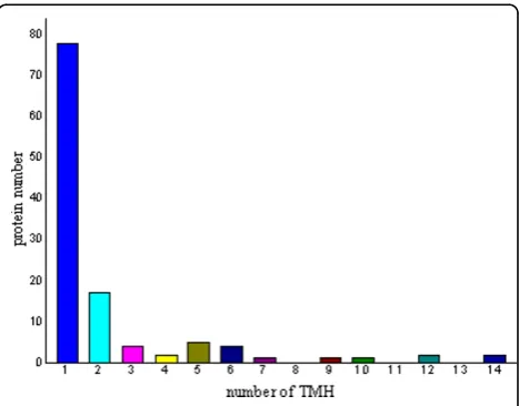
Potential cell wall associated proteins with 1-15 TMHs were assigned using the software TMHMM 2.0 program
against theM. avium subsp.paratuberculosis K10 pro-tein sequence database (excluding the possible signal sequences). In our study, 120 proteins (38.83%) were identified to have at least 1 transmembrane domain. The predicted TMH numbers of these proteins ranged from 1 to 14, 18 proteins contained two TMHs and 25 proteins (8.09%) with three or more TMHs. The profile of TMH in cell wall proteins of M. aviumsubsp.
para-tuberculosis K10 is very similar to previous reports
about TMH in M. tuberculosiscell wall proteome [15]. The distribution of these TMHs is shown in Fig. 1. Among the 309 cell wall proteins identified, it is very interesting to find that there are 157 designated as cyto-plasmic, 85 proteins have an unassigned location and 67 proteins are designated as cell wall related when ana-lyzed by PSORTb location predictions.
Molecular mass and pIdistributions of the identified cellwall proteins
The theoreticalMrdistribution of the identified cell wall proteins ranged from 2.92 kDa to 683.12 kDa. More-over, proteins between Mr10 and 50 kDa were in the majority, representing approximately 58.25% (180 out of 309) of all the identified cell wall proteins. Detailed dis-tributions are shown in Fig. 2. The theoretical pIscores of the identified cell wall proteins ranged from 3.77 to 12.31. Detailed distributions are shown in Fig. 3. There are 39 proteins with pIscores over 10 and 15 proteins with Mr over 100 kDa. Taking GRAVY value into account, there will be at least 39 proteins beyond the general 2-DE separation limits. Additionally, there are 49 proteins with predicted signal peptide in the 309 identified cell wall proteins (Fig. 4A).
Analysis of functional groups in identified cell wall protein
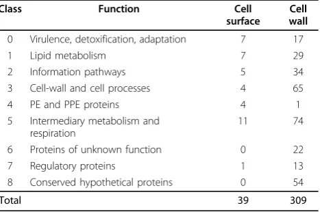
Based on the Pasteur Institute functional classification tree http://genolist.pasteur.fr/TubercuList/, 309 identified proteins were distributed across eight of these functional groups (See table 1 for details). Most of the identified proteins were involved in intermediary metabolism and respiration (functional class 5, 23.95%), cell wall and cell process (21.04%) and conserved hypothetical proteins (17.48%). 62.47% of proteins were involved in the three major functional categories above. Many unexpected pro-teins such as the ribosomal propro-teins were found to be cell wall associated, which were also found in cell wall by pre-vious research [7,15]. It is probable that these proteins interact tightly with the cell wall and join in cell envelop processes and would be potential significance in vaccine studies. Overlap between cytosolic, membrane and cell wall proteins in large scale proteomic studies is not uncommon. Additional studies are necessary to investi-gate the proteins with multiple cellular locations.
The fatty acid components are the most energetically expensive molecules to produce, and thus the regulation of fatty acid production is very tightly controlled to match the growth rate of cells. Mycolic acids are major and specific long-chain fatty acids of the cell envelope of several important human pathogens such as M.
avium subsp. paratuberculosis, M. tuberculosis, M.
leprae, and Corynebacterium diphtheriae. Their
bio-synthesis is essential for mycobacterial growth and represents an attractive target for developing new anti-tuberculous drugs. In this study, 19 proteins related to lipid metabolism were identified as cell wall associated proteins, which include CmaA1(Mycolic acid synthase), CmaA2, FadE25_2, fadD32, fadA_1, FadB_1, fadD12_1, FadE3_2, FadD6, FadE24, FadE23, FadD29, fadA2, FadE20_3, Pks13, DesA1, DesA2, DesA3_2, fabG.
Figure 2 The distribution of molecular mass (Mr) of the
identifiedMycobacterium aviumsubsp.paratuberculosisK10 cell wall proteome.
Figure 3The distribution of isoelectric points(PIvalues) of the identifiedMycobacterium aviumsubsp.paratuberculosisK10 cell wall proteome.
(A) (B)
With signalP 50% Without signalP
50% With signalP
6%
Without signalP 94%
Figure 4The distribution of proteins with asignalpeptide (SP)in (A)Mycobacterium aviumsubsp.paratuberculosisK10 cell wall proteome; (B)Mycobacterium aviumsubsp.paratuberculosis
K10 cell surface-exposed proteome.
Table 1 Functional classification of the identified
Mycobacterium aviumsubsp.paratuberculosisK10 cell wall proteins according to Tubercurolist.
Class Function Cell
surface
Cell wall
0 Virulence, detoxification, adaptation 7 17
1 Lipid metabolism 7 29
2 Information pathways 5 34
3 Cell-wall and cell processes 4 65
4 PE and PPE proteins 4 1
5 Intermediary metabolism and respiration
11 74
6 Proteins of unknown function 0 22
7 Regulatory proteins 1 13
8 Conserved hypothetical proteins 0 54
It is known for many bacterial species that there are tens of proteins required for cell division, for most of which exact functions are still unknown. In this study, the proteins related to cell division, ftsH, ftsZ, ftsX, ftsE, Wag31 (a homologue of the cell division protein DivIVA), PknA/PknB were identified as cell wall related proteins in this study.
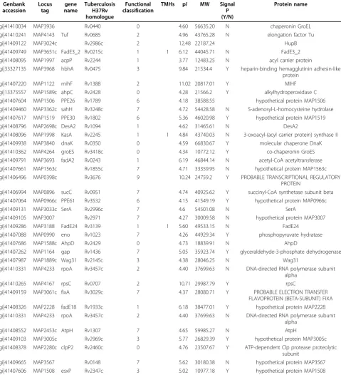
Surface exposed proteins
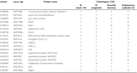
The integrity of the cells after trypsin treatment was confirmed by microscopy (live/dead staining) and culti-vation methods, results of which confirmed the integrity of the cells (data not shown). Peptides released into the supernatant were collected to be fully digested with trypsin for 12~14 hrs, then concentrated and analyzed by LC-MS/MS. A total of 38 cell surface exposed pro-teins were successfully identified (as seen in table. 2). The predicted TMH numbers of these proteins ranged from 1 to 3, and 19% of which contained at least two TMHs. The distribution of these TMHs is listed in Fig. 5. 50% of the identified proteins have signal peptides (Fig 4B). As seen from Fig. 6, 18 proteins of 38 found surface-exposed proteins overlapped with the cell wall proteins, which include 3-oxoacyl-(acyl carrier protein) synthase II, acetyl-CoA acetyltransferase, acyl carrier protein, AhpD, AtpH, chaperonin GroEL, DesA2, DNA-directed RNA polymerase subunit alpha, elongation fac-tor Tu, FadE24, FadE3_2, FixB, hypothetical protein MAP1563c, hypothetical protein MAP3007, hypothetical protein MAP3567, S-adenosyl-L-homocysteine hydro-lase, SerA and Wag31. As seen from table. 3, among the 18 proteins that were identified as both the cell wall and cell surface proteins, there are two proteins (acyl carrier protein and S-adenosyl-L-homocysteine hydrolase) which are not found in the environmentalM. smegma-tis, five proteins (acyl carrier protein, AtpH, DesA2, hypothetical protein MAP1563c and hypothetical pro-tein MAP3567) which are not found inNocardia farci-nica, a pathogenic member of the Actinomycetes, and nine proteins (acyl carrier protein, AhpD, AtpH, DesA2, FadE24, hypothetical protein MAP1563c, hypothetical protein MAP3007, hypothetical protein MAP3567 and Wag31) which are not found in Streptomyces coelicolor A3, a soil-dwelling member of the Actinomycetes.
Discussion
In this study, cell wall proteins were first separated by SDS-PAGE according to their molecular weight followed by in-gel digested with trypsin into complex peptide mixture, and then the mixture was analyzed directly by LC-MS/MS. Subsequently, protein identifications were determined by database searching software [16]. Our experiments led to the identification of a much wider range of proteins in cell wall fraction than those
identified using the conventional 2-DE based method [17] and can therefore be used as a comprehensive reference profile forMycobacterium spp. cell wall teomic studies. Additionally, the surface exposed pro-teome was identified by an enzymatic shaving technique. Two interesting observations result from the cell wall profile. Firstly, there is a discrepancy between the identi-fied surface exposed proteins and the complete cell wall proteome. This is likely due to the loose association of these proteins with the cell wall which makes them prone to detachment. Indeed, some surface proteins are assumed to be attached to the cell wall in a non-cova-lent way and have been reported to be lost during mild standard manipulations [18,19]. Secondly, some proteins are not expected to be localized in the cell wall based on their annotated function. Till now, it is still unclear how proteins such as GroEL and elongation factor TU, leaving the bacterial cell, are retained on the cell surface and whether they have an additional function when associated with the cell wall different from their known function inside the bacterial cell. EF-Tu indeed was identified as a cell wall related protein in this study and has already been identified as cell wall protein in other studies [7]. It was found that only a small percentage of the proteins identified were classified as membrane bound by PSORTb in this study. The existing methods of subcellular localization have been developed for pro-karyotic proteins mainly for bacterial proteins like PSORTb, PSLpred, CELLO, LOCtree, P-classifier, Gpos-ploc, GNBSL [20,21]. Not any method could correctly predict all proteins location. One of the challenges in subcellular localization is to predict location of proteins having multiplelocation [22]. It was reported that PSORTb version 2.0 correctly predicted 88% cytoplas-mic, 81% integral membrane and 80% secretory proteins. PSORTb predicted only 18% membrane-attached into cytoplasmic membrane proteins and rest of them as unknown proteins [23].
In this study, one PPE protein was identified in the cell wall fraction and four PPE proteins were identified in the cell surfaced exposed proteome. The names PE and PPE are derived from the motifs Pro-Glu and Pro-Pro-Glu, respectively, found in conserved domains near the N ter-mini of these proteins. The PE and PPE gene families are highly expanded in the pathogenic members of this genus but show a conspicuous paucity in the nonpathogenic spe-cies. Although no precise function is known for any mem-ber of these families, memmem-bers of the PE and PPE families have been linked to virulence [24,25] or have at least been shown to influence interactions with other cells [26].
the cell division protein DivIVA), PknA/PknB were identified as cell wall related proteins in this study. The divIVA gene, which for the most part is confined to gram-positive bacteria, was first identified in Bacillus
subtilis. Cells with a mutation in this gene have a
reduced septation frequency and undergo aberrant polar division, leading to the formation of anucleate minicells
[19,22,25]. A divIVA gene is also present in Strepto-myces coelicolor [27] and in other actinomycetes, like
Mycobacterium tuberculosis, where Wag31 (antigen 84)
is proposed to be involved in cell shape maintenance [28]. FtsZ is a bacterial cytoskeletal protein that is essen-tial for cell division many prokaryotes [29]. It has been shown to be a bacterial homolog of eukaryotic tubulin,
Table 2 Cell surface proteins identified by trypsin-shaving
Genbank accession
Locus tag
gene name
Tuberculosis H37Rv homologue
Functional classification
TMHs pI MW Signal P (Y/N)
Protein name
gi|41410034 MAP3936 Rv0440 0 4.60 56635.20 N chaperonin GroEL
gi|41410241 MAP4143 Tuf Rv0685 2 4.96 43765.28 N elongation factor Tu
gi|41409122 MAP3024c Rv2986c 2 12.48 22187.24 HupB
gi|41409749 MAP3651c FadE3_2 Rv0215c 1 1 6.12 44045.71 N FadE3_2
gi|41408095 MAP1997 acpP Rv2244 1 3.77 12483.25 N acyl carrier protein
gi|33327135 MAP3968 hbhA Rv0475 3 9.84 21534.4 Y heparin-binding hemagglutinin adhesin-like protein
gi|41407220 MAP1122 mihF Rv1388 2 11.02 20817.01 Y MIHF
gi|13375557 MAP1589c ahpC Rv2428 0 4.28 21566.2 Y alkylhydroperoxidase C gi|41407604 MAP1506 PPE26 Rv1789 6 4.18 38588.55 hypothetical protein MAP1506 gi|41409460 MAP3362c sahH Rv3248c 7 4.72 54428.58 N S-adenosyl-L-homocysteine hydrolase gi|41407617 MAP1519 PPE30 Rv1802 6 5.36 46020.98 Y hypothetical protein MAP1519
gi|41408796 MAP2698c DesA2 Rv1094 1 4.62 31465.61 N DesA2
gi|41408096 MAP1998 KasA Rv2245 1 1 4.84 43740.03 N 3-oxoacyl-(acyl carrier protein) synthase II gi|41409938 MAP3840 dnaK Rv0350 0 4.59 66830.67 Y molecular chaperone DnaK gi|41410362 MAP4264 groES Rv3418c 0 4.34 10772.12 Y co-chaperonin GroES gi|41409791 MAP3693 fadA2 Rv0243 1 6.19 46844.14 N acetyl-CoA acetyltransferase gi|41407661 MAP1563c Rv1855c 7 4.71 33359.95 N hypothetical protein MAP1563c gi|41406496 MAP0398c Rv3676 9 10.24 24759.2 Y PROBABLE TRANSCRIPTIONAL REGULATORY
PROTEIN
gi|41406994 MAP0896 sucC Rv0951 7 4.74 40925.62 Y succinyl-CoA synthetase subunit beta gi|41407064 MAP0966c PPE61 Rv3532 6 4.15 41549.19 Y hypothetical protein MAP0966c
gi|41409131 MAP3033c SerA Rv2996c 7 4.6 54501.08 N SerA
gi|41409105 MAP3007 Rv2971 7 4.27 30009.58 N hypothetical protein MAP3007
gi|41409286 MAP3188 FadE24 Rv3139 1 1 5.60 49533.15 N FadE24
gi|41407088 MAP0990 eno Rv1023 7 4.26 44929.34 Y phosphopyruvate hydratase
gi|41407686 MAP1588c AhpD Rv2429 0 4.73 18839.91 N AhpD
gi|41407262 MAP1164 gap Rv1436 7 5.05 35923.74 Y glyceraldehyde-3-phosphate dehydrogenase
gi|41407987 MAP1889c Wag31 Rv2145c 3 4.38 28046.25 N Wag31
gi|41410331 MAP4233 rpoA Rv3457c 2 4.40 37699.63 N DNA-directed RNA polymerase subunit alpha
gi|41410265 MAP4167 rpsC Rv0707 2 10.71 29987.79 Y rpsC
gi|41409159 MAP3061c fixA Rv3029c 7 4.37 28080.71 Y PROBABLE ELECTRON TRANSFER FLAVOPROTEIN (BETA-SUBUNIT) FIXA gi|41408326 MAP2228 fadE18 Rv1933c 1 6.18 38477.01 Y hypothetical protein MAP2228 gi|41410331 MAP4233 rpoA Rv3457c 2 4.40 37699.63 N DNA-directed RNA polymerase subunit
alpha
gi|41408552 MAP2453c AtpH Rv1307 7 4.65 59985.27 N AtpH
gi|41409103 MAP3005c Rv2969c 3 5.77 26829.39 Y hypothetical protein MAP3005c gi|41408378 MAP2280c clpP2 Rv2460c 0 4.76 23507.67 Y ATP-dependent Clp protease proteolytic
subunit
based both on a low sequence identity and a striking structural similarity [30]. It appears to act at the earliest step in septation and is required through the final step of cytokinesis [31]. FtsE, in association with the integral membrane protein FtsX, is involved in the assembly of potassium ion transport proteins, both of which being relevant to the tubercle bacillus. Recently FtsE and FtsX have been found to localize to the septal ring inE. coli, with the localization requiring the cell division proteins FtsZ, FtsA, and ZipA but not FtsK, FtsQ, FtsL, and FtsI proteins, suggestive of a role for FtsEX in cell division. The receptor-like protein kinase PknB is encoded by the distal gene in a highly conserved operon, present in all actinobacteria, that may control cell shape and cell divi-sion. Genes coding for a PknB-like protein kinase are also found in many more distantly related gram-positive bacteria. It was demonstrated that the Ser/Thr protein kinase PknB is essential for sustaining mycobacterial growth and support the development of protein kinase inhibitors as new potential antituberculosis drugs [32].
The fatty acid components are the most energetically expensive molecules to produce, and thus the regulation of fatty acid production is very tightly controlled to match the growth rate of cells. Mycolic acids are major and speci-fic long-chain fatty acids of the cell envelope of several important human pathogens such as Mycobacterium
1 TMH 11%
2 TMHs 8%
3 TMHs 11% No TMH 70%
Figure 5 Transmembrane helices (TMH) in the identified surface exposed proteins ofMycobacterium avium subsp.
paratuberculosisK10.
Figure 6Venn diagram showing the overlap between the identified cell wall and cell surface exposed proteins.
Table 3 Orthologues of theM. aviumsubsp.paratuberculosisproteins that were identified as both cell surface and cell wall associated in related organisms
accession Locus tag Protein name Orthologues (Y/N)
M. avium 104
M. smegmatis
Nocardia farcinica
Streptomyces coelicolor A3 gi|41408096 MAP1998 3-oxoacyl-(acyl carrier protein) synthase II Y Y Y Y
gi|41409791 MAP3693 acetyl-CoA acetyltransferase Y Y Y Y
gi|41408095 MAP1997 acyl carrier protein Y N N N
gi|41407686 MAP1588c AhpD Y Y Y N
gi|41408552 MAP2453c AtpH Y Y N N
gi|41410034 MAP3936 chaperonin GroEL Y Y Y Y
gi|41408796 MAP2698c DesA2 Y Y N N
gi|41410331 MAP4233 DNA-directed RNA polymerase subunit alpha Y Y Y Y
gi|41410241 MAP4143 elongation factor Tu Y Y Y Y
gi|41409286 MAP3188 FadE24 Y Y Y N
gi|41409749 MAP3651c FadE3_2 Y Y Y Y
gi|41409159 MAP3061c FixB Y Y Y Y
gi|41407661 MAP1563c hypothetical protein MAP1563c Y Y N N
gi|41409105 MAP3007 hypothetical protein MAP3007 Y Y Y N
gi|41409665 MAP3567 hypothetical protein MAP3567 Y Y N N
gi|41409460 MAP3362c S-adenosyl-L-homocysteine hydrolase Y N Y Y
gi|41409131 MAP3033c SerA Y Y Y Y
aviumsubsp.paratuberculosis,Mycobacterium tuberculo-sis,M. leprae, andCorynebacterium diphtheriae. Their biosynthesis is essential for mycobacterial growth and represents an attractive target for developing new antitu-berculous drugs. In this study, 19 proteins proteins related to lipid metabolism were identified as cell wall associated proteins, which include CmaA1(Mycolic acid synthase), CmaA2, FadE25_2, fadD32, fadA_1, FadB_1, fadD12_1, FadE3_2, FadD6, FadE24, FadE23, FadD29, fadA2, FadE20_3, Pks13, DesA1, DesA2, DesA3_2, fabG.
CmaA1 is aciscyclopropanesynthetase which produces a distalciscyclopropane ring in the alpha mycolate of
M. smegmatis[33]. cmaA2is the transcyclopropane
synthetase for both the methoxy and ketomycolates. pks13 gene encodes condensase, the enzyme that per-forms the final condensation step of mycolic acid bio-synthesis and is flanked by two genes, fadD32 and accD4, both of which have been indicated to play a role in the activation of the substrates of the conden-sase [34]. DesA1 is homologous to the plant stearoyl-ACP desaturase which introduce the first double bond in the saturated fatty acids, C16 and C18, the products of fatty acid biosynthesis. These fatty acids are then incorporated in the membrane glycerolipids, cuticular lipids and oilseeds of plants [35]. Involvement of these proteins in mycolic acid synthesis has been suggested based on sequence annotations [36] and structural characterization. However, experimental evidence regarding their functional roles are not presently avail-able. FadE3_2 and FadE25_2 are enzymes involved in electron transport with acyl-CoA dehydrogenase activ-ity. Such enzymes act at the first dehydrogenase step of the b-oxidation of fatty acids. A study of protein expression of M. avium engulfed by macrophages found that FadE2, a protein with 98% protein domain similarity to FadE3_2, was up-regulated [37]. It appears that these proteins are important in the utilisation of fatty acids as a carbon source and that they may have a direct correlation to mycobacterial replication, particu-larly within host macrophages.
A total of 18 identified proteins, HspR, DnaJ, DnaK, KatG, LprG, HtrA, PhoR, PMM, PepA, MmpL3, sdhA, ClpB, hbhA, HBHA, Tuf, groES, manB, DesA3_2, can be considered putative virulence factors as they have previously been suggested to play some role in virulence [38-52].
Conclusions
We have obtained a comprehensive picture of the M.
avium subsp. paratuberculosis K10 cell wall protein
repertoire, with an additional insight in the portion of these proteins that are cell surface exposed. With 309 distinct proteins identified, this study represents the first proteomic analysis of cell wall proteins of M. avium
subsp.paratuberculosisK10. To our knowledge, this is also the first report of a SDS-PAGE-LC-MS/MS based proteomic approach, supported with cell surface enzy-matic digestion, to localize proteins in the mycobacterial cell wall. Many of the cell wall-associated proteins found in this study are involved in cell division, lipid metabo-lism or are putative virulence factors. Therefore, they should be considered as new potential antigens for vac-cine development to prevent M. avium subsp. paratu-berculosisK10 infection.
Additional file 1: summarization of the identified cell wall proteins ofM. aviumsubsp.paratuberculosis. The data provided summarization of the identified cell wall proteins ofM. aviumsubsp.paratuberculosis.
Acknowledgements
This work was financially supported by a grant of the Crohn’s and Colitis Foundation of Canada.
Authors’contributions
ZH carried out the experiments, participated in the data analysis and drafted the manuscript. JDB conceived of the study, and participated in its design and coordination. All authors read and approved the final manuscript.
Competing interests
The authors declare that they have no competing interests.
Received: 24 September 2009 Accepted: 8 April 2010 Published: 8 April 2010
References
1. Wu CW, Schmoller SK, Bannantine JP, Eckstein TM, Inamine JM, Livesey M, Albrecht R, Talaat AM:A novel cell wall lipopeptide is important for biofilm formation and pathogenicity ofMycobacterium aviumssp. paratuberculosis.Microb Pathog2009,46(4):222-30.
2. Uzoigwe JC, Khaitsa ML, Gibbs PS:Mycobacterium aviumssp. paratuberculosisas a cause of Crohn’s disease.Epidemiol Infect2007,
135(7):1057-68.
3. Rowe MT, Grant IR:Mycobacterium aviumssp.paratuberculosisand its potential survival tactics.Lett Appl Microbio2006,42:305-311. 4. Lindahl G, Stalhammar-Carlemalm M, Areschoug T:Surface proteins of
Streptococcus agalactiaeand related proteins in other bacterial pathogens.Clin Microbiol Rev2005,18:102-127.
5. Lin J, Huang S, Zhang Q:Outer membrane proteins: key players for bacterial adaptation in host niches.Microbes Infect2002,4:325-331. 6. Niemann HH, Schubert WD, Heinz DW:Adhesins and invasins of
pathogenic bacteria: a structural view.Microbes Infect2004,6:101-112. 7. Rodríguez-Ortega MJ, Norais N, Bensi G, Liberatori S, Capo S, Mora M,
Scarselli M, Doro F, Ferrari G, Garaguso I, Maggi T, Neumann A, Covre A, Telford JL, Grandi G:Characterization and identification of vaccine candidate proteins through analysis of the group AStreptococcus surface proteome.Nat Biotechnol2006,24(2):191-197.
8. Muskens J, van Zijderveld F, Eger A, Bakker D:Evaluation of the long-term immune response in cattle after vaccination against paratuberculosis in two Dutch dairy herds.Vet Microbiol2002,86(3):269-78.
9. Newton V, McKenna SL, De Buck J:Presence of PPE proteins in Mycobacterium aviumsubsp.paratuberculosisisolates and their immunogenicity in cattle.Vet Microbiol2009,135(3-4):394-400. 10. Rezwan M, Lanéelle MA, Sander P, Daffé M:Breaking down the wall:
fractionation of mycobacteria.J Microbiol Methods2007,68(1):32-9. 11. Shevchenko A, Tomas H, Havlis J, Olsen JV, Mann M:In-gel digestion for
mass spectrometric characterization of proteins and proteomes.Nat
Protoc2006,1(6):2856-60.
Streptococcus pyogenes, with a potential application to other Gram-positive bacteria.Proteomics2008,9(1):61-73.
13. Wei C, Yang J, Zhu J, Zhang X, Leng W, Wang J, Xue Y, Sun L, Li W, Wang J, Jin Q:Comprehensive proteomic analysis ofShigella flexneri2a membrane proteins.J Proteome Res2006,5(8):1860-5.
14. Krogh A, Larsson B, von Heijne G, Sonnhammer EL:Predicting transmembrane protein topology with a hiddenMarkov model: application to complete genomes.J Mol Biol2001,305:567-580. 15. Mawuenyega KG, Forst CV, Dobos KM, Belisle JT, Chen J, Bradbury EM,
Bradbury ARM, Chen X:Mycobacterium tuberculosisfunctional network analysis by global subcellular protein profiling.Mol Biol Cell2005,
16:396-404.
16. Wu CC, Yates JR III:The application of mass spectrometry to membrane proteomics.Nat Biotechnol2003,21:262-267.
17. Ebanks RO, Chisholm K, McKinnon S, Whiteway M, Pinto DM:Proteomic analysis ofCandida albicansyeast and hyphal cell wall and associated proteins.Proteomics2006,6(7):2147-56.
18. Kocincova D, Sonden B, Mendonca-Lima L, Gicquel B, Reyrat JM:The Erp protein is anchored at the surface by a carboxy-terminal hydrophobic domain and is important for cell-wall structure inMycobacterium smegmatis.Fems Microbiol Lett2004,231:191-196.
19. Lichtinger T, Burkovski A, Niederweis M, Kramer R, Benz R:Biochemical and biophysical characterization of the cell wall porin ofCorynebacterium glutamicum:The channel is formed by a low molecular mass polypeptide.Biochemistry1998,37:15024-15032.
20. Guo J, Lin Y, Liu X:GNBSL: a new integrative system to predict the subcellular location for Gram-negative bacteria proteins.Proteomics2006,
6:5099-5105.
21. Gardy JL, Laird MR, Chen F, Rey S, Walsh CJ, Ester M, Brinkman FS:PSORTb v.2.0: expanded prediction of bacterial protein subcellular localization and insights gained from comparative proteome analysis.Bioinformatics
2005,21:617-623.
22. Chou KC, Shen HB:Euk-mPLoc: a fusion classifier for largescale eukaryotic protein subcellular location prediction by incorporating multiple sites.Journal of Proteome Research2007,6:1728-1734. 23. Rashid M, Saha S, Raghava GPS:Support Vector Machine-based method
for predicting subcellular localization of mycobacterial proteins using evolutionary information and motifs.BMC Bioinformatics2007,8:337. 24. Li YJ, Miltner E, Wu M, Petrofsky M, Bermudez LE:AMycobacterium avium
PPE gene is associated with the ability of the bacterium to grow in macrophages and virulence in mice.Cell Microbiol2005,7:539-548. 25. Ramakrishnan L, Federspiel NA, Falkow S:Granuloma-specific expression
ofMycobacteriumvirulence proteins from the glycine-rich PE-PGRS family.Science2000,288:1436-1439.
26. Brennan MJ, Delogu G, Chen YP, Bardarov S, Kriakov J, Alavi M, Jacobs WR:
Evidence that mycobacterial PE_PGRS proteins are cell surface constituents that influence interactions with other cells.Infect Immun
2001,69:7326-7333.
27. Stewart GR, Newton SM, Wilkinson KA, Humphreys IR, Murphy HN, Robertson BD, Wilkinson RJ, Young DB:The stress-responsive chaperone alpha-crystallin 2 is required for pathogenesis ofMycobacterium tuberculosis.Mol Microbiol2005,55:1127-1137.
28. Stewart GR, Robertson BD, Young DB:Tuberculosis: a problem with persistence.Nat Rev Microbiol2003,1:97-105.
29. Bramhill D:Bacterial cell division.Annu Rev Cell Dev Biol1997,13:395-424. 30. Erickson HP:Atomic structures of tubulin and FtsZ.Trends Cell Biol1998,
8:133-137.
31. Ma X, Ehrhardt DW, Margolin W:Colocalization of cell division proteins FtsZ and FtsA to cytoskeletal structures in livingEscherichia colicells by using the green fluorescent protein.Proc Natl Acad Sci USA1996,
93:12998-13003.
32. Fernandez P, Saint-Joanis B, Barilone N, Jackson M, Gicquel B, Cole ST, Alzari PM:The Ser/Thr protein kinase PknB is essential for sustaining mycobacterial growth.J Bacteriol2006,188(22):7778-84.
33. Yuan Y, Lee RE, Besra GS, Belisle JT, Barry CE:3rd Identification of a gene involved in the biosynthesis of cyclopropanated mycolic acids in Mycobacterium tuberculosis.Proc Natl Acad Sci USA1995,92(14):6630-6634. 34. Portevin D, de Sousa-D’Auria C, Montrozier H, Houssin C, Stella A,
Lanéelle MA, Bardou F, Guilhot C, Daffé M:The acyl-AMP ligase FadD32 and AccD4-containing acyl-CoA carboxylase are required for the synthesis of mycolic acids and essential for mycobacterial growth:
identification of the carboxylation product and determination of the acyl-CoA carboxylase components.J Biol Chem2005,280(10):8862-74. 35. Ohlrogge J, Browse J:Lipid biosynthesis.Plant Cell1995,7:957-970. 36. Raman K, Rajagopalan P, Chandra N:Flux balance analysis of mycolic acid
pathway: targets for anti-tubercular drugs.PLoS Comput Biol2005,1:e46. 37. Brunori L, Giannoni F, Bini L, Liberatori S, Frota C, Jenner P, Thoresen OF,
Orefici G, Fattorini L:Induction ofMycobacterium aviumproteins upon infection of human macrophages.Proteomics2004,4:3078-3083. 38. Grandvalet C, de Crecy-Lagard V, Mazodier P:The ClpB ATPase of
Streptomyces albusG belongs to the HspR heat shock regulon.Mol
Microbiol1999,31:521-532.
39. Gamer J, Multhaup G, Tomoyasu T, McCarty JS, Rudiger S, Schonfeld HJ, Schirra C, Bujard H, Bukau B:A cycle of binding and release of the DnaK, DnaJ and GrpE chaperones regulates activity of theEscherichia coliheat shock transcription factor sigma32.EMBO J1996,153:607-617. 40. Ng VH, Cox JS, Sousa AO, MacMicking JD, McKinney JD:Role of LprG
catalase-peroxidase in mycobacterial pathogenesis: countering the phagocyte oxidative burst.Mol Microbiol2004,52(5):1291-302. 41. Gehring AJ, Dobos KM, Belisle JT, Harding CV, Boom WH:Mycobacterium
tuberculosisLprG (Rv1411c): a novel TLR-2 ligand that inhibits human macrophage class II MHC antigen processing.J Immunol2004,
173(4):2660-8.
42. Stack HM, Sleator RD, Bowers M, Hill C, Gahan CG:Role for HtrA in stress induction and virulence potential inListeria monocytogenes.Appl Environ
Microbiol2005,71(8):4241-7.
43. Gonzalo Asensio J, Maia C, Ferrer NL, Barilone N, Laval F, Soto CY, Winter N, Daffé M, Gicquel B, Martín C, Jackson M:The virulence-associated two-component PhoP-PhoR system controls the biosynthesis of polyketide-derived lipids inMycobacterium tuberculosis.J Biol Chem2006,
281(3):1313-6.
44. Kim SH, Ahn SH, Lee JH, Lee EM, Kim NH, Park KJ, Kong IS:Genetic analysis of phosphomannomutase/phosphoglucomutase fromVibrio furnissiiand characterization of its role in virulence.Arch Microbio2003,180(4):240-50. 45. Hauser AR, Kang PJ, Engel JN:PepA, a secreted protein ofPseudomonas
aeruginosa, is necessary for cytotoxicity and virulence.Mol Microbiol
1998,27(4):807-18.
46. Domenech P, Reed MB, Barry CE:Contribution of theMycobacterium tuberculosisMmpL protein family to virulence and drug resistance.Infect
Immun2005,73(6):3492-501.
47. Laguna RK, Creasey EA, Li Z, Valtz N, Isberg RR:A Legionella pneumophila-translocated substrate that is required for growth within macrophages and protection from host cell death.Proc Natl Acad Sci USA2006,
103:18745-18750.
48. Chastanet A, Derre I, Nair S, Msadek T:clpB, a novel member of the Listeria monocytogenesCtsR regulon, is involved in virulence but not in general stress tolerance.J Bacteriol2004,186(4):1165-74.
49. Pethe K, Alonso S, Biet F, Delogu G, Brennan MJ, Locht C, Menozzi FD:The heparin-binding haemagglutinin ofM. tuberculosisis required for extrapulmonary dissemination.Nature2001,412(6843):190-4. 50. Locht C, Hougardy J-M, Rouanet C, Place S, Mascart F:Heparin-binding
hemagglutinin, from an extrapulmonary dissemination factor to a powerful diagnostic and protective antigen against tuberculosis.
Tuberculosis2006,86:303-309.
51. Kunert A, Losse J, Gruszin C, Hühn M, Kaendler K, Mikkat S, Volke D, Hoffmann R, Jokiranta TS, Seeberger H, Moellmann U, Hellwage J, Zipfel PF:
Immune evasion of the human pathogenPseudomonas aeruginosa: elongation factor Tuf is a factor H and plasminogen binding protein.J
Immunol2007,179(5):2979-88.
52. Lin YF, Wu MS, Chang CC, Lin SW, Lin JT, Sun YJ, Chen DS, Chow LP:
Comparative immunoproteomics of identification and characterization of virulence factors fromHelicobacter pylorirelated to gastric cancer.Mol
Cell Proteomics2006,5(8):1484-96.
doi:10.1186/1477-5956-8-21