RESEARCH
Construction and development of an
auto-regulatory gene expression system
in
Bacillus subtilis
Chengran Guan
1, Wenjing Cui
1*, Jintao Cheng
1, Li Zhou
1, Junling Guo
1, Xu Hu
1, Guoping Xiao
2and Zhemin Zhou
1*Abstract
Background: Bacillus subtilis is an all-important Gram-positive bacterium of valuable biotechnological utility that has been widely used to over-produce industrially and pharmaceutically relevant proteins. There are a variety of expression systems in terms of types of transcriptional patterns, among which the auto-inducible and growth-phase-dependent promoters are gaining increasing favor due to their inducer-independent feature, allowing for the potential to industrially scale-up. To expand the applicability of the auto-inducible expression system, a novel auto-regulatory expression system coupled with cell density was constructed and developed in B. subtilis using the quorum-sensing related promoter srfA (PsrfA).
Results: The promoter of the srf operon was used to construct an expression plasmid with the green fluorescent pro-tein (GFP) downstream of PsrfA. The expression displayed a cell-density-dependent pattern in that GFP had a fairly low expression level at the early exponential stage and was highly expressed at the late exponential as well as the station-ary stages. Moreover, the recombinant system had a similar expression pattern in wild-type B. subtilis 168, WB600, and WB800, as well as in B. subtilis 168 derivative strain 1681, with the complete deletion of PsrfA, indicating the excellent compatibility of this system. Noticeably, the expression strength of PsrfA was enhanced by optimizing the −10 and −35 core sequence by substituting both sequences with consensus sequences. Importantly, the expression pattern was successfully developed in an auto-regulatory cell-density coupling system by the simple addition of glucose in which GFP could not be strongly expressed until glucose was depleted, resulting in a greater amount of the GFP product and increased cell density. The expression system was eventually tested by the successful over-production of aminopeptidase to a desired level.
Conclusion: The auto-regulatory cell density coupling system that is mediated by PsrfA is a novel expression system that has an expression pattern that is split between cell-growth and over-expression, leading to an increase in cell density and elevating the overall expression levels of heterologously expressed proteins. The broad applicability of this system and inducer-free expression property in B. subtilis facilitate the industrial scale-up and medical applications for the over-production of a variety of desired proteins.
Keywords: Bacillus subtilis, Protein expression system, Promoter, Quorum sensing, Auto-inducible, Auto-regulatory, Over-production, srf operon
© 2015 Guan et al. This article is distributed under the terms of the Creative Commons Attribution 4.0 International License (http://creativecommons.org/licenses/by/4.0/), which permits unrestricted use, distribution, and reproduction in any medium, provided you give appropriate credit to the original author(s) and the source, provide a link to the Creative Commons license, and indicate if changes were made. The Creative Commons Public Domain Dedication waiver (http://creativecommons.org/ publicdomain/zero/1.0/) applies to the data made available in this article, unless otherwise stated.
Open Access
*Correspondence: [email protected]; [email protected]
1 School of Biotechnology, Key Laboratory of Industrial Biotechnology
(Ministry of Education), Jiangnan University, 1800 Lihu Road, Wuxi, Jiangsu 214122, China
Page 2 of 15 Guan et al. Microb Cell Fact (2015) 14:150
Background
Bacillus subtilis, a rod-shaped Gram-positive soil bacte-rium, has been regarded to be a “generally recognized as safe” (GRAS) microbe that can naturally secrete numer-ous extracellular proteins. These properties make this bacterium to be one of the most popular hosts for natu-rally producing a variety of enzymes and over-expressing a large number of pharmaceutical and industrial recom-binant proteins of interest, such as amylase [1–4], lipase [5], alkaline PGL [6], and so on. The major advantages of B. subtilis compared to other protein expression hosts are that the host has high-cell-density growth and can secrete proteins directly into the cultural medium, which greatly simplifies the following steps for purification and preparation.
The most commonly used expression systems in B. subtilis are inducible expression systems that contain inducer-specific promoters, such as the T7 promoter; grac promoter; spac promoter induced by
isopropyl-β-d-thiogalactopyranoside (IPTG); xylA promoter
driven by xylose; SacB promoter induced by sucrose; and α-amylase promoter transcribed in response to
starch [4, 7, 8]. Although a variety of homologous and heterologous proteins have been successfully over-pro-duced in B. subtilis under the control of these induc-ible promoters, a noticeable basal expression level was observed from these expression systems. Moreover, the high cost of inducers that need to be added to the medium during fermentation at a large scale would limit the industrial utilization of these protein expres-sion systems. Recently, two strictly inducible systems, the subtilin-regulated expression (SURE) system and the LiaRS-controlled expression system (LIKE), which rely on the regulation of the promoter for the spa-box and the cell envelope stress-responsive liaI promoter, respectively, have been developed for scientific and industrial applications. Both of these protein expres-sion systems are strictly controlled by the addition of lantibiotic subtilin and bacitracin, respectively, after the exponential phase of cell growth, preventing the leakage of transcription under non-inducing condi-tions [9, 10].
To avoid the addition of any inducers, promoters under the control of a variety of environmental stresses, such as heat shock, salt, acid, and ethanol stresses, as well as oxygen or glucose starvation [11–13], have also been used to construct systems for the expression of a variety of proteins in B. subtilis, none of which, could be used to over-produce industrially and pharmaceutically utilized proteins and enzymes because of the low and non-per-sistent expression levels of these systems as well as the difficulty of controlling protein expression. Very recently,
an artificial auto-inducible system using a growth-phase promoter (cry3Aa promoter, transcribed during the sta-tionary phase) and another expression system using the nutrient starvation response promoter (pst promoter, responded to phosphate starvation) [14, 15] were devel-oped and characterized. Importantly, a self-inducible system for the reliable and low-cost over-production of recombinant proteins has been developed by using the manP-encoding phosphotransferase system (PTS), which is under the control of the relatively strong and strict promoter manP; manP is transcribed during a glucose-limited process in the engineered B. subtilis strain TQ356 [16]. The self-controlled system is suitable for high-cell-density fermentation because cell growth and over-expression are discrete at different stages.
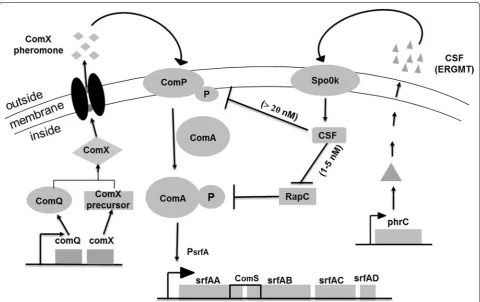
Bacterial quorum sensing (QS) systems are cell-den-sity-dependent regulatory networks that coordinate bac-terial behavioral changes from single cellular organisms at low cell densities to multicellular organisms when their population density reaches a threshold level. The two peptides, ComX and CSF, mediate the quorum sensing control of competence and sporulation in B. subtilis. The accumulation of a high concentration of peptides in the late-growth phase triggers a series of gene transcription events downstream of the ComA-ComP two-component system in the cell [17], among which the srf operon is stimulated via a complex-regulation phosphorylated cas-cade (Fig. 1) [18]. As a monitor of cell density, the ComX pheromone is expressed, transported and accumulated in the broth along with cell growth. When the cells reach a certain density, the ComX pheromone reaches the obligatory concentration to activate the signal transduc-tion system, which is composed of the two-component regulatory proteins ComP and ComA, and finally, phos-phorylated ComA binds to the promoter (PsrfA) of the srf operon to activate the transcription of corresponding gene expression [19–21], leading to temporally control-lable transcription.
Results and discussion
Expression pattern of GFP under the control of PsrfA in B. subtilis 168
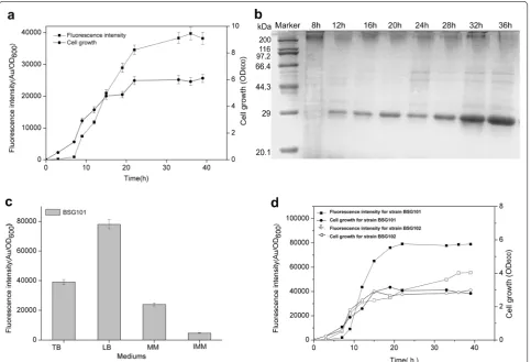
In recent years, several types of expression systems that are free of inducers via a variety of transcriptional patterns have been developed in B. subtilis or other Gram-positive hosts to over-express industrially and pharmaceutically used proteins [13, 15, 16]. To expand the application of self-inducible or auto-inducible gene expression systems in B. subtilis, a novel recombinant plasmid, pBSG03, har-boring a cell-density-responsive promoter, PsrfA, upstream of the reporter protein GFP was constructed, permitting the monitoring of gene expression in the hosts. To char-acterize the expression pattern and strength for PsrfA, pBSG03 was transformed into B. subtilis 168 and then cultivated in Terrific Broth (TB) medium for 40 h, during which the culture was sampled periodically to examine the expression level of GFP. The results demonstrate that the expression level of GFP was considerably low from the lag phase to the early exponential phase. Subsequently,
the expression level increased sharply when cell growth entered the late exponential phase. Thereafter, the expres-sion level increased gradually throughout the station-ary phase until the relative fluorescence unit reached the vertex (Fig. 2a). In parallel, the intracellular proteins that were sampled in a time course were determined by SDS-PAGE analysis. The data indicated that the expression of GFP quantitatively was accumulated 9 h after fermenta-tion, before which there were few GFP proteins that were scarcely detected on the PAGE (Fig. 2b). These results indicate that the transcription of PsrfA occurs in the late growth phase, whereas the activity of the promoter is inhibited in the early stage of growth, permitting cell-density-dependent expression. The cell-density-depend-ent transcription of PsrfA is subjected to simple regulated mechanism of the ComA-ComP phosphorylation cascade compared to that of the recently reported gene expression system, by which the PmanP is involved in a rather com-plex regulatory network, making it necessary to delete the manP gene in the operon to achieve self-inducible Fig. 1 The schematic model for the regulation of the transcription of the srf operon network involved in two extracellular signaling peptide-medi-ated quorum sensing in B. subtilis. The srf operon was composed of four genes encoding surfactin synthetase with comS embedded between gene
Page 4 of 15 Guan et al. Microb Cell Fact (2015) 14:150
expression properties. Otherwise, the gene expression under PmanP is typically mannose-inducible in wild-type B. subtilis [16].
Meanwhile, the effect of four different culture media (TB medium, MM medium, IMM medium and LB medium) on PsrfA-mediated GFP expression in B. subti-lis 168 (BSG101) was estimated in the following experi-ments. The data of the fluorescence intensity showed that the recombinant strain grown in LB medium achieved the maximal expression level, in which the expression level of GFP was approximately twofold higher than that in TB medium (Fig. 2c).
PHpaII is a strong constitutive promoter, has compa-rable expression strength to that of P43, and has been
used to construct an array of vectors for the over-pro-duction of recombinant proteins [22, 23]. To contrast the transcriptional level of PsrfA with that of PHpaII, plas-mid pBSG04 harboring the PHpaII-GFP expression cas-sette was constructed and transformed into B. subtilis 168, generating BSG102. Over the 40 h of fermentation in LB medium, each culture of the two recombinant strains was sampled periodically to measure the expres-sion level. The data showed that the overall expresexpres-sion of GFP in BSG101 was higher than that of BSG102 by 1.5-fold (Fig. 2d), even though the final biomasses of the two strains were nearly the same. These results indicate that the PsrfA is a potentially strong promoter for gene expres-sion in B. subtilis.
Fig. 2 Identification of the expression level and patterns of GFP in recombinant B. subtilis 168. a The fluorescence intensity of GFP (solid rectangle) and cell growth (solid cycle) under the control of wild-type PsrfA were monitored in recombinant B. subtilis 168 during cultivation in TB medium. b
SDS-PAGE analysis for the expression of GFP in B. subtilis 168 harboring pBSG03 with PsrfA, which was cultivated in TB medium, showed the
expres-sion pattern. Samples were collected periodically at different time points, and total proteins of 30 μg were loaded onto the gel. c The impact of the medium on the expression level of GFP in B. subtilis 168 harboring pBSG03 (BSG101). The expression level of GFP under the control of PsrfA
was determined by the total fluorescence intensity at OD600 (denoted as Au/OD600). The recombinant strain was cultivated in TB, MM, IMM and LB
medium. All of the data were collected in triplicate and were presented as the mean ± SD. d Comparison of the expression patterns and levels controlled by PsfrA and PHpaII. B. subtilis 168 harboring pBSG03 (BSG101) and pBSG04 containing the PHpaII promoter (BSG102) were cultivated in LB
medium for 40 h. The fluorescence intensity and cell growth for each of the recombinant strains were examined by periodic sampling throughout cultivation. The cell density of BSG101 (solid cycle) and BSG102 (open cycle) was monitored by measuring the OD600. The fluorescence intensity of
Expression pattern of GFP in markerless deletion mutant and the protease‑deficient mutants
Based on the organization of the srf operon, the native PsrfA would also be activated, resulting in the expression of genes in the operon. To ascertain whether the back-ground activation of the srf operon occurred simultane-ously with heterologous gene expression, the markerless deletion mutant BSG1681 was constructed by completely deleting the full-length of PsrfA on the chromosome.
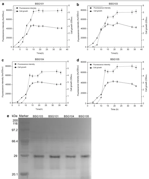
To compare the transcriptional features and expres-sion levels of PsrfA in BSG1681 with the other three types of hosts (wild-type 168, B. subtilis WB600 and WB800), plasmid pBSG03 was introduced into the above hosts, generating BSG103, BSG101, BSG104, and BSG105, respectively. The levels of fluorescence intensity were monitored in the four recombinant strains throughout culturing. As expected, the transcriptional feature of PsrfA in BSG103 was similar to that in BSG101, BSG104 and BSG105, giving a transcription pattern of cell-density-dependent auto-regulation. Noticeably, the final fluo-rescence intensity in BSG101, BSG103 and BSG105 was similar, but slightly higher than that in BSG104 (Fig. 3a– d). These results suggest that PsrfA has a broad transcrip-tional compatibility with the cell-density-dependent pattern in various hosts. The final expression levels of GFP in the four different hosts were analyzed by SDS-PAGE, and the data were consistent with those of the fluorescence intensity (Fig. 3e).
Enhancement of the expression level by engineering the consensus sequence within the srfA promoter
Although the cell-density-dependent expression sys-tem was characterized as highly transcribed and broadly compatible in various B. subtilis hosts, the duration of exponential expression under the control of the wild-type promoter was short, scarcely 10 h (Fig. 3a–d). The exponential expression phase in the other constructed auto-regulation systems starts at approximately the mid-exponential growth phase and terminated during the beginning of the stationary phase [24]. In addition, the activity of the wild-type promoter (e.g., cry3Aa), which behaves in a cell-density-dependent manner, usually dis-allows the over-production of a large amount of heter-ologous proteins to the desired level. Thus, engineering the promoters for higher capacity is the major demand for augmenting the over-production of heterologous pro-teins [9].
As one of the most efficient and prevalent strategies, promoter engineering has been widely used to tune the gene expression level, both for high levels of protein expression and tunable gene expression in synthetic biol-ogy [25, 26]. The major essential elements in prokary-otic promoters that mainly determine their overall
transcriptional activity are the −10 and −35 consensuses motifs, while other factors, such as the gene context, could also influence transcriptional activity [27]. This feature indicates that the two motifs are potential targets to be engineered within the promoter sequence. In this study, to enhance the activity of PsrfA, the sequence of the −10 and −35 motifs in PsrfA was compared with the
σA-dependent consensuses in B. subtilis. The alignment
showed that the two motifs in PsrfA were in poor homol-ogy to the consensus. Therefore, the consensus −10 (TATAAT) and −35 (TTGACT) motifs of σA-dependent
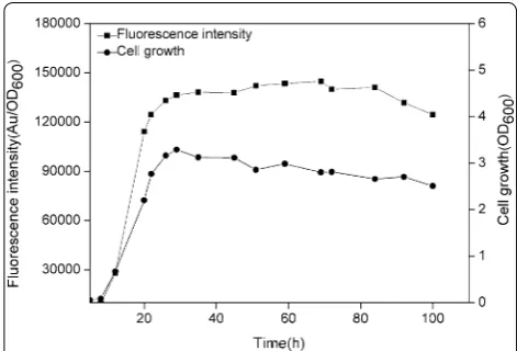
promoters substituted for the −10 (TAAACT) and −35 (GTGATA) motifs of the PsrfA in plasmid pBSG03, result-ing in plasmid pBSG05. After transformresult-ing pBSG05 into B. subtilis 168 (generating BSG106), the cell growth and fluorescence intensity were monitored throughout culturing. After 20 h of culture, the fluorescence inten-sity peaked, and thereafter, the maximal level did not decrease until 80 h of culture. The maximal expression level under the control of engineered PsrfA was approxi-mately twofold stronger than that of the wild-type pro-moter (Fig. 4), demonstrating that engineering the poorly conserved −10 and −35 motifs within the promoters lead to fold increases of the transcriptional level. Despite engineering the non-inducible promoters, a set of other promoters, including inducible promoters, has been engineered to achieve a stronger over-production level through this strategy [15, 28].
Development of the cell‑density‑dependent expression system to an auto‑regulatory system
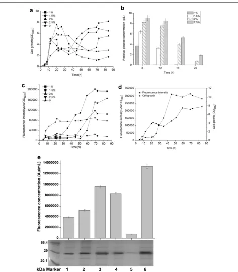
The aforementioned expression system displayed a cell-density-dependent expression pattern. However, the cell density did not achieve a higher level than expected. According to the data referring to the expression pat-tern, we proposed that the initiation of a high expression level overlapped the rapid growth stage, leading to com-petition for energy and chemicals. Therefore, to achieve a high cell density and the desired over-production, expression initiation should be retarded until the cell density reaches a high level, avoiding mutual intervention between the two biological events. Nutrients that delayed expression while promoting cell growth were regarded as prospective candidates. A previous study revealed that the addition of glucose to the growth medium inhibits the expression of many genes, including srf operon [29].
Page 6 of 15 Guan et al. Microb Cell Fact (2015) 14:150
Fig. 3 Over-expression of GFP using the plasmid pBSG03 in B. subtilis 168 (a), BSG1681 (b), B. subtilis WB600 (c), and B. subtilis WB800 (d) was compared by determining the cell density and relative fluorescence intensity (Au/OD600). The final expression level of GFP in the different four
the final cell-density in the BSG101 strain; the addition of 1.5 % glucose led to the highest cell density, which was nearly threefold higher than that of the glucose-free culture (Fig. 5a). The 1.5 % glucose in the medium was depleted after 16 h, at which time the 2 and 2.5 % glu-cose remained in the medium (Fig. 5b). GFP expression sharply increased in the 1, 1.5, and 2 % glucose groups when glucose was depleted, before which GFP was sig-nificantly inhibited (Fig. 5c). Among the four groups, the highest expression level of GFP was acquired by the addition of 1.5 % glucose. The highest expression level was more than 2.5-fold higher than that of glucose-free cultivation (Fig. 5c), indicating that the addition of glu-cose not only augment the expression level of heterolo-gous genes but also give the system an auto-regulatory expression pattern. Interestingly, the addition of 2.5 % glucose completely inhibited expression throughout cul-tivation (Fig. 5c). Then, the expression of GFP in BSG106 was examined to determine the effect of 1.5 % glucose on the expression level of GFP under the engineered PsrfA (mutP
srfA). As expected, the expression of GFP initiated after the 1.5 % glucose was depleted and maintained at the maximal level from 40 h until the end of cultivation (Fig. 5d). The maximal expression level under the control of mutP
srfA was approximately fourfold higher than that of the wild-type promoter PsrfA without adding of 1.5 % glucose (Fig. 2d). Moreover, the total amount of GFP was determined by measuring the average fluorescence intensity (as calculated by arbitrary units per milliliter of culture) and by SDS-PAGE analysis. The highest flu-orescence intensity and accumulative GFP protein were obtained under the control of mutP
srfA with the addition of 1.5 % glucose (Fig. 5e).
Taken together, the cell-density-dependent system that is mediated by PsrfA could be contrived to be an auto-reg-ulatory device by the regauto-reg-ulatory effect of glucose repres-sion. This simple method eventually achieves a high cell density without a complex gene deletion or modification of the genome in the host. Regarding other developed auto-inducible expression systems, certain genes that are involved in the regulatory network should be disrupted or deleted to make an auto-inducible expression system while preventing native gene expression from interfering with the heterologous transcription that is mediated by the used promoters. Generally, B. subtilis under the con-trol of PmanP, which is intrinsically a mannose-induced system, has been successfully fabricated to an auto-inducible expression by the deletion of the manP gene in the PTS pathway [16].
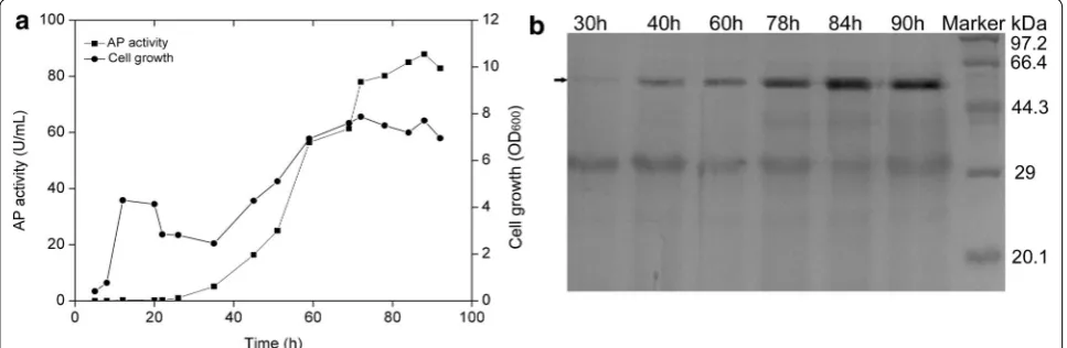
Over‑production of AP using the auto‑regulatory gene expression system
To estimate the suitability of the system for the over-pro-duction of the heterologous protein, AP from B. subtilis Zj016, plasmid pBSG06 was constructed under the con-trol of promoter mutP
srfA. A previous report demonstrated the expression level of AP in B. subtilis WB600 under the control of PHpaII. However, the expression and secretion levels were incomparable to those of other proteins [22,
30, 31]. Plasmid pBSG06 was introduced into B. subtilis 168, generating recombinant strain BSG107. The culture procedure was similar to that for the auto-regulatory expression mentioned above. The cell growth, AP activ-ity, and protein over-production were measured through-out cultivation. As expected, AP expression displayed the auto-regulatory pattern coupled with a cell-density-dependent manner. The highest activity of AP that was obtained was 87.89 U/mL, and the cell-density at OD600 achieved a level of approximately 7.87 (Fig. 6a). The expression property also be verified at the protein level by SDS-PAGE analysis (Fig. 6b). Those results indicate that mutP
srfA-mediated system could be used to express heterologous proteins in B. subtilis.
Conclusions
In this study, a novel auto-regulatory gene expression sys-tem coupled to cell density in B. subtilis was constructed and developed using the srfA promoter that is associ-ated with triggering quorum sensing. Using the expres-sion system, expresexpres-sion was separated by the cell growth phase and high expression phase, during which the addition of glucose not only augmented the two-phase expression profile and enabled auto-regulation but also increased the final cell density. In addition, the expres-sion strength was remarkably enhanced by engineering the −10 and −35 region via substitution to the highly Fig. 4 The expression level of GFP controlled by the mutant PsrfA
(mutP
srfA) in B. subtilis 168. The recombinant strain harboring pBSG05
Page 8 of 15 Guan et al. Microb Cell Fact (2015) 14:150
Fig. 5 The effect of different concentrations of glucose on the PsrfA- and mutPsrfA-controlled expression of GFP. a The cell density profiles for BSG101
harboring wild-type PsrfA in LB medium with the addition of 1 % (solid rectangle), 1.5 % (solid cycle), 2 % (solid triangle), and 2.5 % (solid inverse triangle)
glucose. Medium without the addition of glucose was used as the control group (solid pentangle). b Glucose consumption profiles at 8, 12, 16, and 20 h with different initial concentrations. c The fluorescence intensity profiles that were detected in BSG101 with different concentrations of glucose corresponded to those in a. d The fluorescence intensity and the cell density that were detected in recombinant strain BSG106 that harbored
mutP
srfA in which the intrinsic sequences of −10 and −35 were replaced by the corresponding conserved sequences for σA-dependent promoters.
e Comparison of the highest fluorescence intensity and the corresponding GFP expression level with or without the addition of glucose in B. subtilis
168 under the control of wild-type PsrfA and mutPsrfA. Sample 1 represents the BSG101 without the addition of glucose, which was designated as
the control. Samples 2–5 represent strain BSG101 with the addition of 1, 1.5, 2, and 2.5 % glucose, respectively. Sample 6 represents strain BSG106 (harboring mutP
conserved sequences. Furthermore, aminopeptidase was successfully over-produced as the test protein using the newly constructed gene expression system, achieving a 1.7-fold over-expression level compared to that under the control of the generally regarded strong promoter PHpaII [30]. These features of the auto-regulatory gene expres-sion system give the system great potential for the over-production of enormous high-value-added proteins at the industrial scale.
Methods
Strains, plasmids and growth conditions
The bacterial strains and plasmids that were used in this study are listed in Table 1. E. coli JM109 was used as a host for gene cloning. B. subtilis 168 and engineered B. subtilis BSG1681 were used for the gene expres-sion and gene integration experiments. The media used in this study were glucose minimal medium (MM) [32], improved minimal medium (IMM, adding 0.02 % Casamino acids to MM medium), Luria–Bertani medium (LB), and TB. All of the strains were cultured in the appropriate medium with aeration at 37 °C. When appro-priate, B. subtilis growth media was supplemented with kanamycin (5 μg/mL) and E. coli growth media with
ampicillin (10 μg/mL). The cell density was determined
by measuring the OD600 with a UV-1800/PC spectro-photometer (Shanghai, MAPADA Instrument Co., Ltd., China).
Recombinant DNA techniques
Plasmid construction was performed in E. coli, and extraction was processed following the standard proce-dure as previously described [33]. Recombinant plasmids were transformed into B. subtilis as previously described
[30]. Enzymes were obtained from TOYOBO (Osaka, Japan), TaKaRa (Dalian, China), or New England Biolabs (Beijing, China) and were used according to the manu-factures’ protocols. The primers used in this study are listed in Table 2. PCR was performed using KOD DNA polymerase (Osaka, Japan). All of the recombinant plas-mids that were constructed in this work were confirmed by DNA sequencing (Shanghai Sangon Biotech Co., Ltd., China).
Construction of plasmids and mutant strains with markerless gene deletion
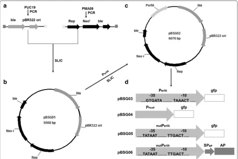
Plasmid construction was carried out using the Sequence and Ligation Independent Cloning (SLIC) method, with few modifications [34]. The primers that were used to amplify the donor sequence were designed to have 30-bp flanking regions that are homologous to the recep-tor plasmid insertion regions, and the receprecep-tor plasmid was linearized by inverse PCR. The main procedures used for construction are shown in Fig. 7. The plasmids constructed in this study were E. coli–B. subtilis shuttle vectors.
To construct pBSG01, Fragment 1, which is 2.2 kb and comprises the ampicillin resistance gene (bla) and repli-cation region (pBR322 ori) and was flanked by the cor-responding homology to Fragment 2, was amplified from pUC19 with primers P1 and P2. Concomitantly, Frag-ment 2 carrying the replication protein (Rep), kanamy-cin resistance gene (neor), and bleomycin resistance gene (ble), which was flanked by the corresponding homology of Fragment 1, was amplified from plasmid pMA09 using primers P3 and P4. These two fragments were mixed at the molar ratio of 1:2 prior to treatment with T4 DNA polymerase. After a 2.5-min treatment, the mixture was Fig. 6 Over-production of aminopeptidase (AP) using the constructed auto-regulatory gene expression system. Plasmid pBSG06 harboring the signal peptide (SPap) and AP downstream of the mutPsrfA was introduced into B. subtilis 168. The recombinant strain BSG107 was then cultured in
Page 10 of 15 Guan et al. Microb Cell Fact (2015) 14:150
incubated on ice for 10 min, allowing for the two frag-ments to be annealed, after which the mixture was transformed to E. coli JM109 competent cells [35]. The resulting plasmid was termed pBSG01.
To introduce promoter PsrfA into the plasmid pBSG01, the 0.6-kb PsrfA flanked by a 30-bp homology sequence upstream and downstream of the inserted position in pBSG01 was amplified from the chromosome of B. sub-tilis 168 using primers P5 and P6. Plasmid pBSG01 was then linearized using inverse PCR with primers P7 and P8. The two fragments were joined according to the steps described above. The resulting plasmid was termed pBSG02. To insert GFP downstream of PsrfA in pBSG02, the gfp gene was amplified from pBS1154 with primers P9 and P10, flanked by the 30-bp homology sequence corre-sponding to the upstream and downstream sequences at the inserted position. Meanwhile, pBSG02 was linearized by inverse PCR with primers P11 and P2. These two frag-ments were joined by the SLIC method mentioned above, generating plasmid pBSG03 for expression in B. subtilis. To construct the comparative plasmid pBSG04 under the
control of promoter HpaII (PHpaII), PHpaII was inserted into pBSG03 by replacing PsrfA using mega-primer PCR with a whole plasmid protocol using primers P7 and P12 [36]. Plasmid pBSG05, harboring the mutant PsrfA, was constructed with primers P13 and P14 using pBSG03 as the template through inverse PCR. Then, the endo-nuclease DpnI was used to cut the template plasmid. To express aminopeptidase (AP) under the control of PsrfA, plasmid pMA05 was used as the template to amplify AP with primers P15 and P16, and pBSG02 was linearized with primers P11 and P2. Plasmid pBSG06 was obtained by processing these two fragments in the same manner as for pBSG02. All of the plasmid constructs were verified by DNA sequencing.
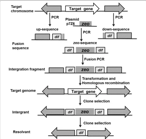
The markerless deletion of genes on the chromosome of B. subtilis 168 was performed as previously described, with some modification [37, 38]. The sequences of the B. subtilisdif site (dif B. subtilis:
ACTTCCTAGAATATATAT-TATGTAAACT) were used in this study.
To delete the promoter region in the srf operon from the B. subtilis 168 chromosome, two tripartite fragments Table 1 Strains and plasmids used in this study
amutP
srfA: the −10 region (TAAACT) and −35 region (GTGATA) of the PsrfA was replaced by the consensus −10 region (TATAAT) and −35 region (TTGACT) of σA
-dependent promoters
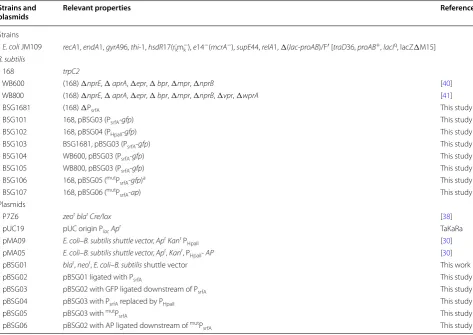
Strains and
plasmids Relevant properties References
Strains
E. coli JM109 recA1, endA1, gyrA96, thi-1, hsdR17(rk-m k
−), e14−(mcrA−), supE44, relA1, Δ(lac-proAB)/F′ [traD36, proAB+, lacIq, lacZΔM15]
B. subtilis
168 trpC2
WB600 (168) ΔnprE, ΔaprA, Δepr, Δbpr, Δmpr, ΔnprB [40]
WB800 (168) ΔnprE, ΔaprA, Δepr, Δbpr, Δmpr, ΔnprB, Δvpr, ΔwprA [41]
BSG1681 (168) ΔPsrfA This study
BSG101 168, pBSG03 (PsrfA-gfp) This study
BSG102 168, pBSG04 (PHpaII-gfp) This study
BSG103 BSG1681, pBSG03 (PsrfA-gfp) This study
BSG104 WB600, pBSG03 (PsrfA-gfp) This study
BSG105 WB800, pBSG03 (PsrfA-gfp) This study
BSG106 168, pBSG05 (mutP
srfA-gfp)a This study
BSG107 168, pBSG06 (mutP
srfA-ap) This study
Plasmids
P7Z6 zeorblarCre/lox [38]
pUC19 pUC origin PlacApr TaKaRa
pMA09 E. coli–B. subtilis shuttle vector, AprKanr P
HpaII [30]
pMA05 E. coli–B. subtilis shuttle vector, Apr, Kanr, P
HpaII- AP [30]
pBSG01 blar, neor, E. coli–B. subtilis shuttle vector This work
pBSG02 pBSG01 ligated with PsrfA This study
pBSG03 pBSG02 with GFP ligated downstream of PsrfA This study
pBSG04 pBSG03 with PsrfA replaced by PHpaII This study
pBSG05 pBSG03 with mutP
srfA This study
pBSG06 pBSG02 with AP ligated downstream of mutP
(Fig. 8) were amplified from the B. subtilis genome using the primers that are listed in Table 2. The specific pro-cedure is described as follows. The up-sequence, which consisted of the approximately 1-kb homologous frag-ment upstream of PsrfA, the dif B. subtilis site, and 6-bp
homologous region to the 5′ end of the gene zeo, was amplified by PCR with primers P17 and P18. Similarly, the down-sequence, which consisted of a 5-bp homol-ogous fragment to the 3′ end of the gene zeo, the difB.
subtilis, and the approximately 1-kb homologous fragment
downstream of the gene PsrfA, was amplified with prim-ers P19 and P20. Next, the zeocin resistance gene zeo was amplified from plasmid p7Z6 using primers P21 and P22, flanked by a 5-bp homology to the sequence in the chromosome followed by the 28-bp dif sequence at the 5′ terminus and the 28-bp dif sequence followed by a 5-bp homology to the chromosome. These three fragments were assembled by fusion PCR. The resulting 2.5-kb frag-ment was then sequenced and transformed into the B. subtilis 168 competent cells. The integrants were selected on LB agar medium containing 25 μg/mL zeocin. The
positive clones were selected before inoculating the LB broth in the absence of antibiotics and were incubated for approximately 36 h to produce zeocin-sensitive recombinant clones. Then, the culture was identified by
replica agar plating with and without the selective anti-biotic. Finally, the clones that grew on the agar plate in the absence of antibiotics were selected in liquid medium for identification by PCR using primers P17 and P20. The positive mutant strain that was deficient in PsrfA was named BSG1681 (B. subtilis 168: ΔPsrfA).
GFP fluorescence assay
A single colony of the appropriate B. subtilis strain picked on LB agar plates was inoculated in 5 mL of LB liquid medium and was cultured overnight for more than 12 h to serve as a preculture. Then, 0.6 mL of preculture was transferred into 250-mL shaking flasks that were loaded with 30 mL of LB liquid medium to cultivate for 3 days, during which the culture medium was sampled every 2 h. After cultivation, the cells were harvested by centrifuga-tion, washed by PSB buffer (50 mM Tris–HCl, 100 mM NaCl, pH 7.5) for three times, and suspended in an appropriate diluted ratio. Then, a final volume of 200 μL
of the cell suspension was transferred to each well, and the fluorescence value (expressed as Au) was recorded.
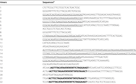
The fluorescent activity of GFP was monitored via fluo-rescence spectroscopy on a black flat 96-well plate using a Synergy™ H4 multimode microplate reader (BioTek Instruments, Inc., USA). The excitation wavelength was Table 2 Oligonucleotides used in this study
a Homologous sequences were underlined, and dif
B. subtilis sequence was shown in bold
Primers Sequencesa
P1 CTCTTCCGCTTCCTCGCTCACTGACTCGC
P2 GCGGTATTTTCTCCTTACGCATCTGTGCGG
P3 GCGAGTCAGTGAGCGAGGAAGCGGAAGAGTAGAAGAAGCTTGGAGACAAGGTAAAGG
P4 CCGCACAGATGCGTAAGGAGAAAATACCGCCATATGTAAATCGCTCCTTTTTAGGTGGCAC
P5 CCCCCTTTGCTGAGGTGGCAGAGGGCAGGTATCGACAAAAATGTCATGAA
P6 CCGCACAGATGCGTAAGGAGAAAATACCGCCATTGTCATACCTCCCCTAATCTTTATAAG
P7 ACCTGCCCTCTGCCACCTCAGC
P8 GCGGTATTTTCTCCTTACGCATC
P9 CTTATAAAGATTAGGGGAGGTATGACAATGATGAGTAAAGGAGAAGAACTTTTCACTGGAG
P10 CCGCACAGATGCGTAAGGAGAAAATACCGCTTATTTGTATAGTTCATCCATG
P11 CATTGTCATACCTCCCCTAATC
P12 ATGAGTAAAGGAGAAGAACT
P13 ACTTTTCACCCATTTTTCGGTTGACAAAACATTTTTTTCATTTATAATGAACGGTAGAAAGATAAAAAATATTGAAA
P14 TTTTATCTTTCTACCGTTCATTATAAATGAAAAAAATGTTTTGTCAACCGAAAAATGGGTGAAAAGTTTCATGCGGG
P15 CTTATAAAGATTAGGGGAGGTATGACAATGATGAAAAAGCTTTTGACTGTC
P16 CCGCACAGATGCGTAAGGAGAAAATACCGCTTATTTGATATCTTCAAAAATG
P17 CGCAGATGTAGTCAACACCGAGTGCGTC
P18 ATCAAGAGTTTACATAATATATATTCTAGGAAGTATCAATCAATTCCATATAGCCTTTCCC
P19 ACTGAACTTCCTAGAATATATATTATGTAAACTATGGAAATAACTTTTTACCCTTTAACGG
P20 ATCGTTGATTAGGAGATTATACGG
P21 TTGATACTTCCTAGAATATATATTATGTAAACTCTTGATATGGCTTTTTATATGTG
Page 12 of 15 Guan et al. Microb Cell Fact (2015) 14:150
495/9 nm, and the emission wavelength was 525/9 nm with gain 80. The determination of the GFP expression was calculated with the fluorescence intensity divided by the OD600 that was previously measured [39].
The relative fluorescence intensities which reflected the expression levels were calculated according to the method described by Toymentseva et al. [10]. The rela-tive fluorescence intensity (Au/OD600) of three parallel samples of the recombinant B. subtilis strains harbor-ing empty vector pBSG01 were averaged and subtracted from that of the recombinant strains harboring plasmids with GFP at the consistency time of cultivation. Growth was monitored by measuring the absorbance at 600 nm. The data were averaged from three independent samples of the same time points.
Heterologous expression of aminopeptidase and enzyme activity analysis
A fresh overnight culture of the recombinant B. subti-lis strain BSG107 harboring pBSG06 under the control of mutant PsrfA (mutPsrfA) was inoculated into 250-mL shaking flasks that were loaded with 30 mL of LB liquid medium, the optical density of which was adjusted to OD600 0.2. Then, the culture was cultivated at 37 °C with rigorous shaking for 2 days, and sampling was imple-mented throughout the fermentation process with an interval of 2 h. Thereafter, crude AP in the fermenta-tion supernatant was obtained by removing recombi-nant B. subtilis cells via centrifugation (5 min, 10,000×g). AP activity was determined via a previously described method [23]. One unit of activity was defined as the Fig. 7 The schematic diagram of the construction of the E. coli–B. subtilis recombinant shuttle plasmids. a Fragment 1, harboring the ampicillin resistance gene (bla) and replication origin (pBR322 ori), was amplified from pUC19. Fragment 2, containing the replication protein (Rep), kanamycin resistance gene (neor), and bleomycin resistance gene (ble), was similarly obtained from pMA09 by PCR. The terminus of each of the two fragments
was flanked by a 30-bp homology corresponding to each other. b These two fragments were fused using the Sequence and Ligation Independ-ent Cloning (SLIC) method [35], with some modifications, and yielding Plasmid pBSG01. c Construction of pBSG02 with the insertion of promoter PsrfA into pBSG01. d GFP was inserted downstream of the PsrfA, generating expression plasmid pBSG03. To compare the transcriptional strength,
promoter PHpaII was substituted for PsrfA, generating plasmid pBSG04. To engineer PsfrA, the −10 (TAAACT) and −35 (GTGATA) motifs in PsrfA were
amount of enzyme that released 1 µmol L−1p -nitroani-line min−1 at 37 °C (ε
405 nm = 9.98 L mmol−1 cm−1). The results are the averages of triplicate assays.
Protein analysis and SDS‑PAGE
After cultivation for appropriate recombinant B. subti-lis, the cells were harvested by centrifugation. The pel-leted cells were then re-suspended in PBS buffer prior
to disruption by ultra-sonication on ice. The crude cell extracts were separated by centrifugation, the superna-tant was analyzed by SDS-PAGE according to the stand-ard procedure, and the gels were subsequently stained by Coomassie brilliant blue R250 staining. The concentra-tion of the total proteins was determined by the Bradford assay and equal amounts of total protein were loaded onto SDS-PAGE gels.
Fig. 8 The sketch displays the procedure for the deletion of the target gene on a chromosome using the dif/Xer recombination system. The shaded regions represented homology between the integration cassette and the genes flanking the target gene. zeo, the zeocin resistance gene zeo. The front (up-sequence) and back (down-sequence) regions flanking the target gene to be deleted were amplified from the chromosome of B. subtilis
Page 14 of 15 Guan et al. Microb Cell Fact (2015) 14:150
Abbreviations
PsrfA: the promoter of the srf operon; PTS: phosphotransferase system; QS:
quorum sensing; AP: aminopeptidase; GFP: green fluorescent protein; SLIC: sequence and ligation independent cloning; bla: ampicillin resistance gene; pBR322 ori: replication region; Rep: replication protein; neor: kanamycin
resist-ance gene; ble: bleomycin resistance gene; PHpaII: promoter of HpaII; SPap: the
signal peptide of aminopeptidase; mutP
srfA: the mutant of promoter PsrfA.
Authors’ contributions
CG constructed the plasmids, the variant strain, accomplished the determina-tion of the recombinant expression system, analyzed the data and wrote the manuscript. WC conceived the idea, designed this study, analyzed the data and wrote the manuscript. ZL, JC, JG, XH were participated in the determi-nation of expression pattern using GFP reporter gene. GX participated in the experiment of expressing aminopeptidase. ZZ conceived of the study, participated in its design, and coordination. All authors read and approved the final manuscript.
Author details
1 School of Biotechnology, Key Laboratory of Industrial Biotechnology
(Minis-try of Education), Jiangnan University, 1800 Lihu Road, Wuxi, Jiangsu 214122, China. 2 Wuxi Biortus Bioscience Co., Ltd, Wuxi, Jiangsu 214122, China.
Acknowledgements
The investigation was supported by a Project Funded by the Priority Academic Program Development of Jiangsu Higher Education Institutions,the 111 Pro-ject (No. 111-2-06),and the Jiangsu province “Collaborative Innovation Center for Advanced Industrial Fermentation” industry development program, the National High Technology Research and Development Program of China (863 Program, 2014AA021304), the Fundamental Research Funds for the Central Universities (JUSRP51411B), the National Natural Science Foundation of China (31400058), the Natural Science Foundation of Jiangsu Province (BK20130139), and the Public Project for Key Laboratory of Industrial Biotechnology, Ministry of Education (KLIB-KF201306).
Compliance with ethical guidelines
Competing interests
The authors declare that they have no competing interests.
Received: 17 June 2015 Accepted: 12 September 2015
References
1. Chen J, Gai Y, Fu G, Zhou W, Zhang D, Wen J. Enhanced extracellular production of alpha-amylase in Bacillus subtilis by optimization of regula-tory elements and over-expression of PrsA lipoprotein. Biotechnol Lett. 2015;37(4):899–906.
2. Rabbani M, Mirmohammad Sadeghi H, Moazen F, Rahimi M, Salehi G. Cloning and expression of randomly mutated Bacillus subtilis alpha-amyl-ase genes in HB101. Biotechnol Res Int. 2011. doi:10.4061/2011/305956. 3. Yang H, Liu L, Li J, Du G, Chen J. Heterologous expression, biochemical
characterization, and overproduction of alkaline alpha-amylase from
Bacillus alcalophilus in Bacillus subtilis. Microb Cell Fact. 2011;10:77. 4. Ying Q, Zhang C, Guo F, Wang S, Bie X, Lu F, et al. Secreted expression of
a hyperthermophilic alpha-amylase gene from Thermococcus sp. HJ21 in
Bacillus subtilis. J Mol Microbiol Biotechnol. 2012;22(6):392–8.
5. Lu Y, Lin Q, Wang J, Wu Y, Bao W, Lv F, et al. Overexpression and characteri-zation in Bacillus subtilis of a positionally nonspecific lipase from Proteus vulgaris. J Ind Microbiol Biotechnol. 2010;37(9):919–25.
6. Zhang J, Kang Z, Ling Z, Cao W, Liu L, Wang M, et al. High-level extracel-lular production of alkaline polygalacturonate lyase in Bacillus subtilis with optimized regulatory elements. Bioresour Technol. 2013;146:543–8. 7. Chen PT, Shaw JF, Chao YP, David Ho TH, Yu SM. Construction of
chromosomally located T7 expression system for production of heterologous secreted proteins in Bacillus subtilis. J Agric Food Chem. 2010;58(9):5392–9.
8. Wieland KP, Wieland B, Gotz F. A promoter-screening plasmid and xylose-inducible, glucose-repressible expression vectors for Staphylococcus carnosus. Gene. 1995;158(1):91–6.
9. Bongers RS, Veening JW, Van Wieringen M, Kuipers OP, Kleerebezem M. Development and characterization of a subtilin-regulated expression system in Bacillus subtilis: strict control of gene expression by addition of subtilin. Appl Environ Microbiol. 2005;71(12):8818–24.
10. Toymentseva AA, Schrecke K, Sharipova MR, Mascher T. The LIKE system, a novel protein expression toolbox for Bacillus subtilis based on the liaI
promoter. Microbial Cell Factories. 2012;11:143.
11. Bhavsar AP, Zhao X, Brown ED. Development and characterization of a xylose-dependent system for expression of cloned genes in Bacillus subti-lis: conditional complementation of a teichoic acid mutant. Appl Environ Microbiol. 2001;67(1):403–10.
12. Phan TT, Nguyen HD, Schumann W. Novel plasmid-based expression vectors for intra- and extracellular production of recombinant proteins in
Bacillus subtilis. Protein Expr Purif. 2006;46(2):189–95.
13. Le Thuy AT, Schumann W. A novel cold-inducible expression system for
Bacillus subtilis. Protein Expr Purif. 2007;53(2):264–9.
14. Qi Y, Kobayashi Y, Hulett FM. The pst operon of Bacillus subtilis has a phosphate-regulated promoter and is involved in phosphate transport but not in regulation of the pho regulon. J Bacteriol. 1997;179(8):2534–9. 15. Lee SJ, Pan JG, Park SH, Choi SK. Development of a stationary
phase-specific autoinducible expression system in Bacillus subtilis. J Biotechnol. 2010;149(1–2):16–20.
16. Wenzel M, Muller A, Siemann-Herzberg M, Altenbuchner J. Self-inducible
Bacillus subtilis expression system for reliable and inexpensive protein production by high-cell-density fermentation. Appl Environ Microbiol. 2011;77(18):6419–25.
17. Stefanic P, Decorosi F, Viti C, Petito J, Cohan FM, Mandic-Mulec I. The quo-rum sensing diversity within and between ecotypes of Bacillus subtilis. Environ Microbiol. 2012;14(6):1378–89.
18. Cosby WM, Vollenbroich D, Lee OH, Zuber P. Altered srf expression in
Bacillus subtilis resulting from changes in culture pH is dependent on the Spo0K oligopeptide permease and the ComQX system of extracellular control. J Bacteriol. 1998;180(6):1438–45.
19. Roggiani M, Dubnau D. ComA, a phosphorylated response regulator protein of Bacillus subtilis, binds to the promoter region of srfA. J Bacteriol. 1993;175(10):3182–7.
20. Schneider KB, Palmer TM, Grossman AD. Characterization of comQ and comX, two genes required for production of ComX pheromone in Bacil-lus subtilis. J Bacteriol. 2002;184(2):410–9.
21. Yazgan Karatas A, Çetin S, Özcengiz G. The effects of insertional muta-tions in comQ, comP, srfA, spo0H, spo0A and abrB genes on bacilysin biosynthesis in Bacillus subtilis. Biochimica et Biophysica Acta (BBA). 2003;1626(1–3):51–6.
22. Liu SL, Du K. Enhanced expression of an endoglucanase in Bacillus subtilis
by using the sucrose-inducible sacB promoter and improved properties of the recombinant enzyme. Protein Expr Purif. 2012;83(2):164–8. 23. Gao X, Liu Z, Cui W, Zhou L, Tian Y, Zhou Z. Enhanced thermal stability
and hydrolytic ability of Bacillus subtilis aminopeptidase by removing the thermal sensitive domain in the non-catalytic region. PLoS One. 2014;9(3):e92357.
24. Kleerebezem M, Bongers R, Rutten G, de Vos WM, Kuipers OP. Autoregula-tion of subtilin biosynthesis in Bacillus subtilis: the role of the spa-box in subtilin-responsive promoters. Peptides. 2004;25(9):1415–24. 25. Blazeck J, Alper HS. Promoter engineering: recent advances in
con-trolling transcription at the most fundamental level. Biotechnol J. 2013;8(1):46–58.
26. Dehli T, Solem C, Jensen PR. Tunable promoters in synthetic and systems biology. Subcell Biochem. 2012;64:181–201.
27. Leavitt JM, Alper HS. Advances and current limitations in transcript-level control of gene expression. Curr Opin Biotechnol. 2015;34C:98–104. 28. Phan TT, Nguyen HD, Schumann W. Development of a strong intracellular
expression system for Bacillus subtilis by optimizing promoter elements. J Biotechnol. 2012;157(1):167–72.
30. Gao X, Cui W, Tian Y, Zhou Z. Over-expression, secretion, biochemical characterisation, and structure analysis of Bacillus subtilis aminopeptidase. J Sci Food Agric. 2013;93(11):2810–5.
31. Kang HK, Jang JH, Shim JH, Park JT, Kim YW, Park KH. Efficient constitutive expression of thermostable 4-alpha-glucanotransferase in Bacillus subtilis
using dual promoters. World J Microbiol Biotechnol. 2010;26(10):1915–8. 32. Spizizen J. Transformation of biochemically deficient strains of
Bacillus Subtilis by deoxyribonucleate. Proc Natl Acad Sci USA. 1958;44(10):1072–8.
33. Guan C, Cui W, He X, Hu X, Xu J, Du G, et al. Construction and develop-ment of a novel expression system of Streptomyces. Protein Expr Purif. 2015;113:17–22.
34. Li MZ, Elledge SJ. Harnessing homologous recombination in vitro to generate recombinant DNA via SLIC. Nat Methods. 2007;4(3):251–6. 35. Jeong JY, Yim HS, Ryu JY, Lee HS, Lee JH, Seen DS, et al. One-step
sequence- and ligation-independent cloning as a rapid and versatile cloning method for functional genomics studies. Appl Environ Microbiol. 2012;78(15):5440–3.
36. Miyazaki K, Takenouchi M. Creating random mutagenesis libraries using megaprimer PCR of whole plasmid. Biotechniques. 2002;33(5):1033–4 (1036–s8).
37. Bloor AE, Cranenburgh RM. An efficient method of selectable marker gene excision by Xer recombination for gene replacement in bacterial chromosomes. Appl Environ Microbiol. 2006;72(4):2520–5.
38. Yan X, Yu HJ, Hong Q, Li SP. Cre/lox system and PCR-based genome engi-neering in Bacillus subtilis. Appl Environ Microbiol. 2008;74(17):5556–62. 39. Botella E, Fogg M, Jules M, Piersma S, Doherty G, Hansen A, et al.
pBaSys-Bioll: an integrative plasmid generating gfp transcriptional fusions for high-throughput analysis of gene expression in Bacillus subtilis. Microbiol-ogy. 2010;156:1600–8.
40. Wu XC, Lee W, Tran L, Wong SL. Engineering a Bacillus subtilis expression-secretion system with a strain deficient in six extracellular proteases. J Bacteriol. 1991;173(16):4952–8.
41. Wu SC, Yeung JC, Duan Y, Ye R, Szarka SJ, Habibi HR, et al. Functional production and characterization of a fibrin-specific single-chain antibody fragment from Bacillus subtilis: effects of molecular chaperones and a wall-bound protease on antibody fragment production. Appl Environ Microbiol. 2002;68(7):3261–9.
Submit your next manuscript to BioMed Central and take full advantage of:
• Convenient online submission
• Thorough peer review
• No space constraints or color figure charges
• Immediate publication on acceptance
• Inclusion in PubMed, CAS, Scopus and Google Scholar
• Research which is freely available for redistribution