R E S E A R C H
Open Access
Markers of increased atherosclerotic risk in
patients with chronic kidney disease: a
preliminary study
Anna Gluba-Brzózka
1,6*, Marta Michalska-Kasiczak
2, Beata Franczyk
1, Marek Nocu
ń
3, Peter P. Toth
4,5,
Maciej Banach
2,6and Jacek Rysz
1,6Abstract
Background:The prevalence of chronic kidney disease is rising continuously. Cardiovascular disease is among leading causes of death and premature mortality of patients with chronic kidney disease. Even the earliest stages of chronic kidney disease are associated with higher risk of subsequent coronary heart disease. The aim of this study was to determine markers of increased risk of atherosclerosis in CKD.
Methods:The study group consisted of a total of 80 patients (20 patients with stage I/II CKD, 20 with stage III CKD, 20 stage IV CKD and 20 stage V/dialysis) and 24 healthy volunteers. Levels of proteins (osteoprotegerin,
osteopontin, osteocalcin, matrixγ-carboxyglutamic acid protein, fetuin A, MMP-2, MMP-9, TIMP-1, TIMP-2) and biochemical parameters were measured to analyse their influence on atherosclerosis risk in CKD patients. Cardiac echocardiography was performed to assess structural integrity and function, presence of left ventricular hypertrophy and systolic and diastolic function dysfunction.
Results:This study shows that the prevalence of ventricular hypertrophy (95.3 %) and diastolic dysfunction (93.2 %) in CKD patients is high. Also E/E’ratio was significantly higher (13.6 ± 4.4,p= 0.001), tricuspid insufficiency (27.3 in CKD I/II vs. 71.4 in CKD V,p= 0.016), contractile dysfunction (33.3 in CKD I/II vs. 78.9 in CKD V,p= 0.040), mitral valve calcification (0 in CKD I/II vs. 28.6 in CKD V,p= 0.044) and aortic valve calcification (0 in CKD I/II vs. 61.9 in CKD V,p= 0.0008) were significantly more frequent in patients with CKD stage V/dialysis than in other groups. Only MMP-2, MMP-2/TIMP-2 ratio and TIMP-1 differed significantly between groups.
Conclusions:This study shows high prevalence of ventricular hypertrophy and diastolic dysfunction in CKD patients. Contractile dysfunction, mitral and aortic valve calcification in HD patients were significantly more frequent than in patients with other CKD stages. Significantly increased levels of MMP-2, MMP-2/TIMP-2 ratio and lower TIMP-1 suggests that these factors may be involved in the pathogenesis of atherosclerosis in CKD patients.
Background
Chronic kidney disease (CKD) is defined by KDIGO in Clinical Practice Guideline for the Evaluation and Man-agement of Chronic Kidney Disease published in 2013 as abnormalities of kidney structure or function, present for over 3 months, with implications for health and CKD is classified based on cause, GFR category, and al-buminuria category (CGA) [1]. The prevalence of
chronic kidney disease is rising continuously. According to National Kidney Foundation (NKF) KDOQI guide-lines, chronic kidney disease, irrespective of diagnosis, is associated with increased risk of cardiovascular disease (CAD), including coronary heart disease, cerebrovascular disease, peripheral vascular disease, and heart failure, due to both ‘traditional’ (defined in the Framingham Heart Study) and ‘chronic kidney disease related’ CAD risk factors, and, thus, these patients have risk on par with the highest CAD risk group [2]. The prevalence of uraemia-related (non-traditional) factors increases along with the decline in kidney function. Cardiovascular dis-ease is one of the leading causes of death and premature
* Correspondence:aniagluba@yahoo.pl
1Department of Nephrology, Hypertension and Family Medicine, WAM
University Hospital of Lodz, Poland,Żeromskiego 113, 90-549Łódź, Poland
6Healthy Aging Research Center, Medical University of Lodz, Lodz, Poland
Full list of author information is available at the end of the article
mortality of patients with chronic kidney disease. Ac-cording to recent studies, even the earliest stages of chronic kidney disease are associated with higher risk of subsequent coronary heart disease [3, 4]. It has been suggested that the assessment of CKD-associated CAD risk factors together with conventional risk factors should be performed in order to improve the prediction of coronary heart disease risk [2]. Moreover, patients with manifestations of cardiovascular disease should be screened for evidence of kidney disease [3, 5, 6]. The re-duction in risk factors seems to be effective in lowering cardiovascular morbidity and mortality in patients with CKD [2]. According to the report of NKF Task Force on Cardiovascular Disease in Chronic Renal Disease, the mortality due to CVD was 10 to 30 times higher in dia-lysis patients than in the general population despite stratification for sex, race, and the presence of diabetes [7]. CVD mortality in dialysis patients remained ~5-fold higher than in the general population after stratification for age [8]. In patients with CKD the prevalence of ar-teriosclerosis (remodelling of large arteries) and cardio-myopathy is higher than in general population [9]. A high prevalence of a proinflammatory state, endothelial dysfunction, hypertension, and dyslipidemia associated with renal disease may explain the acceleration of ath-erosclerosis with a high prevalence of coronary ischemia and CV events in CKD. However, the exact mechanisms of atherosclerotic and arteriosclerotic changes in the set-ting of CKD formation are not yet fully characterized.
Aim
The aim of this study was to determine markers of in-creased risk of atherosclerosis in CKD.
Methods
The study group consisted of a total of 80 patients (20 patients with stage I/II CKD, 20 with stage III CKD, 20 stage IV CKD and 20 stage V/dialysis) hospitalized in the Department of Nephrology, Hypertension and Family Medicine. The control group consisted of 24 vol-unteers without CKD, recruited among patients hospital-ized due to causes other than CAD, tumours or diabetes mellitus. All persons involved in this study signed informed a consent form before the collection of blood samples. The purpose and methodology of this study was approved by the Bioethics Committee of the Med-ical University of Lodz (no. RNN/79/12/KB). Total cholesterol, low-density lipoprotein (LDL), high-density lipoprotein (HDL), triglyceride (TG), albuminuria, serum calcium and phosphate, Fe, total iron-binding capacity (TIBC), C-reactive protein (CRP), alkaline phosphatase activity, creatinine, urea, uric acid, total protein, the level of fibrinogen and D-dimer were also determined. In addition, cardiac echocardiography was performed to
assess structural integrity and function, the presence of left ventricular hypertrophy and systolic and diastolic function dysfunction. Contractility disorder was diag-nosed when ejection fraction (EF) is below 44 %. The E/ A is defined as a ratio of the early (E) to late (A) ven-tricular filling velocities, while E/E’is a ratio of early fill-ing (E) and early diastolic mitral annular velocity (E’) [10]. The levels of studied proteins and biochemical markers were analysed in blood of all people involved in the study. The study excluded patients with diagnosed cancer and advanced cardiovascular disease. In the present study, concentrations of proteins involved in the processes of vessel wall calcification and bone metabolism disorders (osteoprotegerin [TECOMedical, no. 8034], osteopontin [RayBiotech, ELH-OPN-001], osteocalcin [TECOMedical, no. 8002], matrix γ -carboxyglutamic acid protein (MGP) [USCN Life Science, E91477Hu], fetuin A [TECOMedical, no. KT-800]) and vascular remodelling (MMP-2, MMP-9, TIMP-1, TIMP-2 [Raybiotech: MMP2-001, ELH-MMP9-001, ELH-TIMP1-001, ELH-TIMP2-001]) were measured in order to analyse their influence on athero-sclerosis risk in CKD patients. Levels of these proteins were determined by the ELISA method according to the manufacturer’s instructions. Estimated glomerular filtra-tion rate (GFR-MDRD) was calculated using the Modifi-cation of Diet in Renal Disease:
GFR (ml/min/1.73 m2) = 186 x (Creat/88.4)−
1.154
x (Age)−0.203x (0.742 if female) x (1.210 if black), and the classification into CKD stage confirmed using the CKD-EPI equation:
GFR ¼ 141 x min Sð cr=κor 1Þαx max Sð cr=κor 1Þ−1:209
x 0:993Agex 1ð :018 if femaleÞ x 1ð :159 if blackÞ;
where:
Scr- serum creatinine (mg/dL),κ- 0.7 for females and
0.9 for males, α is -0.329 for females and -0.411 for males [74].
Creatinine level was measured with enzymatic method.
This work was funded by Iuventus Plus 2010 grant no. IP2010009870 from the Polish Ministry of Science and Higher Education.
Statistical analysis
homogeneity of variance. Standard Student t test was used for the comparison of data showing no departures from normality and for multiple comparisons (more than two groups) one-way ANOVA with post hoc Scheffe tests was used. If at least one of the aforemen-tioned criteria is not met, non-parametric Mann– Whit-ney U test and detailed or non-parametric analysis of variance (Kruskal-Wallis test) with post hoc Connover-Inman tests was used, respectively. The χ2 test of inde-pendence was used for the analysis of discontinuous var-iables. The analysis of logistic regression was used for the analysis of relationship between the occurrence of cardiovascular disorders and CKD, age, sex, and the con-centration of selected proteins. All the echocardio-graphic images were analysed by the single investigator and repeated in order to assess the intra-observer vari-ability. Intra-observer variability of echocardiographic parameters was determined on the basis of the intra-class correlation coefficient (ICC) with 95 % CI. A value ofp< 0.05 was considered significant. Calculations were made with the use of statistical R program [11].
Results
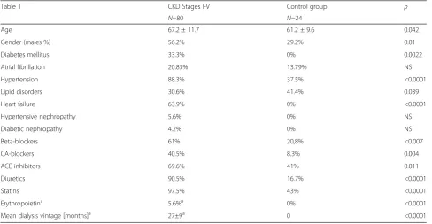
Eighty patients in the study group with the average age of 67.2 ± 11.7 and 24 patients in control group with the age of 61.2 ± 9.6 (P= 0.042) were involved in the study. There were 45 men and 35 women (56.2 % and 43.8 %, respectively) in the study group and 7 men and 17 women (29.2 % and 70.8 %) in the control group (P =
0.01). Hypertension occurred significantly more often in the study group (88.3 % vs. 37.5 %,p< 0.0001).
The analysis of biochemical parameters in both groups revealed in patients with CKD stages I-V statistically lower concentration of Na+ (137.9 ± 3.4 vs. 140.1 ± 2.8,
P = 0.006), haemoglobin level (11.7 ± 1.9 vs. 12.8 ± 1.4,
P <0.01), iron (12.1 ± 5.6 vs. 18.1 ± 7.0,P= 0.001), and higher level of hsCRP (14.7 ± 30.7 vs. 3.9 ± 6.6, P < 0.005), inorganic phosphate (1.36 ± 0.44 vs. 1.14 ± 0.15,
P= 0.001), and triglycerides (1.88 ± 1.07 vs. 1.46 ± 0.62,
P = 0.02), in comparison with the control group. More-over, in the study group, levels of markers of renal func-tion, such as urea (14.0 ± 8.4 vs. 5.5 ± 1.9, P <0.0001), creatinine (276.2 ± 217.4 vs. 80.1 ± 11.3,P <0.0001) and uric acid (386.7 ± 135.9 vs. 271.7 ± 53.0,P<0.0001) were also significantly increased. In patients with chronic kid-ney disease, GFR-MDRD was significantly lower in com-parison to the control group (35.0 ± 24.5 vs. 86.4 ± 16.4). The prevalence of comorbidities and frequency of used drugs differed significantly between control and study groups. Baseline characteristics of enrolled pa-tients is summarized in Table 1.
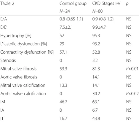
Echocardiographic examination results are summa-rized in Table 2.
All the echocardiographic measurements were per-formed by the same person. The intra-observer variabil-ity by ICC (interclass correlation coefficient) varied from 0.82 to 0.96. Echocardiographic examination revealed significant differences only in the occurrence of mitral valve fibrosis and aortic valve calcification between the
Table 1Baseline characteristics of enrolled patients
Table 1 CKD Stages I-V Control group p
N=80 N=24
Age 67.2 ± 11.7 61.2 ± 9.6 0.042
Gender (males %) 56.2% 29.2% 0.01
Diabetes mellitus 33.3% 0% 0.0022
Atrial fibrillation 20.83% 13.79% NS
Hypertension 88.3% 37.5% <0.0001
Lipid disorders 30.6% 41.4% 0.039
Heart failure 63.9% 0% <0.0001
Hypertensive nephropathy 5.6% 0% NS
Diabetic nephropathy 4.2% 0% NS
Beta-blockers 61% 20,8% <0.007
CA-blockers 40.5% 8.3% 0.004
ACE inhibitors 69.6% 41% 0.011
Diuretics 90.5% 16.7% <0.0001
Statins 97.5% 43% <0.0001
Erythropoietina 5.6%a 0% <0.0001
Mean dialysis vintage [months]a 27±9a 0 <0.0001
a
study and control groups. More significant differences in echocardiographic results were observed when each CKD stage was analysed separately. The results of this analysis are presented below, in Table 3.
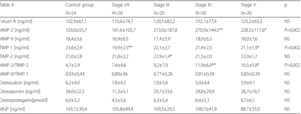
This analysis revealed significant differences in E/E’, presence of contractility disorders, occurrence of mitral and aortic valve calcifications and tricuspid insufficiency. Our analysis of the relationship between selected pro-teins and CKD stage are demonstrated in Table 4.
The analysis of the concentrations of proteins associ-ated with bone metabolism (fetuin A, osteocalcin, osteo-pontin, osteoprotegerin and MPG) revealed no statistically significant differences between the control group and patients with chronic renal failure. It was ob-served that the concentration of osteocalcin was highest
in patients with stage I/II CKD and gradually decreased to its lowest value in patients with stage V/dialysis. Simi-lar proportional decreases through CKD stages were ob-served with osteocalcin and MPG. Osteoprotegerin concentration was lowest in subjects with stage I/II CKD and gradually increased to reach its highest value in patients with stage V/dialysis. However, these trends were not statistically significant which may be related to the small size of each group.
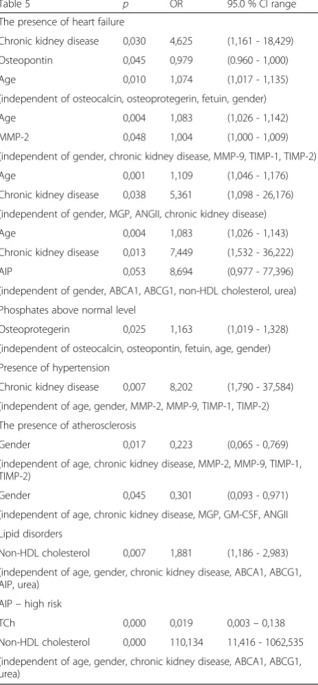
Statistically higher levels of MMP-2 in patients with chronic kidney disease are observed as compared to the control group (p = 0.002) were observed in this study (Table 4). The lowest concentration of MMP-2 was seen in patients with CKD stage I/II and the highest in pa-tients with stage IV and in those on dialysis. Significant differences in serum concentrations of metalloproteinase inhibitor TIMP-1 (p= 0.002) and MMP-2/TIMP-2 ratio were also observed. Statistically significant results of multivariable analysis are presented in Table 5.
In this analysis, the presence of heart failure was asso-ciated with the presence of chronic kidney disease, the level of osteopontin, age, MMP-2 and AIP and this rela-tionship was independent of osteocalcin, osteoproteg-erin, fetuin, gender, MMP-9, TIMP-1, TIMP-2, MGP, ANGII, ABCA1, ABCG1, non-HDL cholesterol and urea).
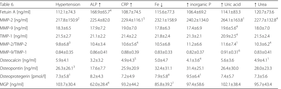
There was also relationship between phosphates above normal level and osteoprotegerin. Moreover, the pres-ence of hypertension was associated with chronic kidney disease, the presence of atherosclerosis associated only with gender, while lipid disorders with non-HDL choles-terol, AIP (high risk) and total cholesterol. An analysis of the relationship between protein concentrations and various biochemical markers was also performed. Statis-tically significant results of this analysis are presented in Table 6.
Table 2The summary of echocardiographic examination results
Table 2 Control group CKD Stages I-V p
N=24 N=80
E/A 0.8 (0.65-1.1) 0.9 (0.8-1.2) NS
E/E’ 7.5±2.1 9.9±4.7 NS
Hypertrophy [%] 52 95.3 NS
Diastolic dysfunction [%] 29 93.2 NS
Contractility dysfunction [%] 57.1 52.8 NS
Stenosis 0 3.2 NS
Mitral valve fibrosis 53.3 81.3 P<0.01
Aortic valve fibrosis 0 14.1 NS
Mitral valve calcification 13.3 14.1 NS
Aortic valve calcification 0 30.2 P<0.02
IM 46.7 63.1 NS
IA 0 6.7 NS
IT 16.7 43.8 NS
Abbreviations used in Table 1:IMmitral insufficiency,IAatrial insufficiency,IT
tricuspid insufficiency,NSnot significant
Table 3The summary of echocardiographic examination results of patients divided into CKD stage groups
Table 3. Stage I/II Stage III Stage IV Stage V p
N=20 N=20 N=20 N=20
E/A 0.8 (0.8-1.1) 0.8 (0.7-0.95) 0.9 (0.8-1.2) 0.9 (0.8-1.3) NS
E/E’ 7.4±2.0 8.1±3.9 7.1±3.5 13.6±4.41,3,6 P=0.001
Diastolic dysfunction [%] 81.8 93.3 100.0 94.7 NS
Contractility disorders [%] 33.3 42.9 36.4 78.91,5 P=0.040
Mitral valve fibrosis [%] 81.8 80.0 70.6 90.5 NS
Aortic valve fibrosis [%] 9.1 20.0 23.5 4.8 NS
Mitral valve calcifications [%] 0 0 17.6 28.61 P=0.044
Aortic valve calcifications [%] 0 13.3 23.5 61.92,5 P=0.0008
IM [%] 54.5 50.0 58.8 81.0 NS
IA [%] 10.0 12.5 0 4.8 NS
IT [%] 27.3 25.0 37.5 71.41 P=0.016
1
Discussion
This preliminary study analysed the possible markers of atherosclerotic and calcification processes occurring in CKD patients and possible novel mechanisms of in-creased cardiovascular risk in this group of patients.
The results of biochemical parameters analysis (signifi-cantly lower levels of Na+, haemoglobin and iron, as well as higher levels of CRP, PO4−and triglycerides in patients
with CKD stages I-V in comparison with the control group) are consistent with results of other studies [12, 13] and are associated with kidney damage. Also, signifi-cantly higher concentrations of urea, creatinine and uric acid in patients with CKD stages I-V are not surprising, since these are established markers of kidney function.
This study shows that the prevalence of ventricular hypertrophy (95.3 %) and diastolic dysfunction (93.2 %) in CKD patients is high. According to the literature, left ventricular hypertrophy appears in approximately 40 % of patients with chronic renal insufficiency, and is even more frequent (75 %) at the onset of ESRD [14, 15]. Pro-gressive left ventricular enlargement is considered as the most typical morphological pattern of dialysis patients and it is a crucial prognostic factor for cardiovascular mortality in ESRD patients [16, 17]. Diastolic dysfunc-tion, which is frequent in chronic kidney disease (CKD) patients, accounts for 40 %-66 % of cardiovascular com-plications [18]. However, there is still a controversy con-cerning which parameter (E/A, E’, E/E’) is of better predictive and prognostic value for the diagnosis of dia-stolic dysfunction and the assessment of its clinical out-comes [19]. It has been suggested that the use of multiple echocardiographic indices to diagnose and to grade diastolic dysfunction seems to be the best solution. This analysis revealed that in patients with CKD stage V/dialysis the E/E’ratio was significantly higher than in other groups (13.6 ± 4.4, p = 0.001) and that in this
group of patients tricuspid insufficiency was significantly more prevalent (p = 0.016). The results of the de Bie et al. [20] study confirm that diastolic dysfunction is highly prevalent among dialysis patients but they imply that its prevalence in this group of patients might be underestimated using conventional measurements. The diagnosis of LV diastolic dysfunction has been demon-strated to provide independent, prognostic value for long-term mortality and cardiovascular death in patients with end-stage renal disease [21]. As shown by Han et al. [19], the increase in E/E’(E/E’> 15) and left atrium (LA) volume index (LAVI > 32 mL/m2) are significant risk factors for CV events in incident dialysis patients with preserved LV systolic function.
Vascular calcification (VC) within the media and in-tima layers of arteries contributes considerably to the greater mortality of patients with chronic kidney disease [22, 23]. This pathological calcification seems to be asso-ciated with an elevated serum calcium phosphate and with differentiation of vascular or mesenchymal cells into osteoblast-like cells [24]. Changes of the mitral ring, which extend towards valve leaflets, are particularly fre-quent [25, 26]. In this study mitral valve calcification in CKD patients was observed, but it was only slightly more frequent than in the control group. However, pa-tients with CKD were more likely to have aortic valve calcification (study group 30.2 % vs. 0 % control group,
p< 0.02). In another study, the joint prevalence of mitral or aortic valve calcification was 31 % in pre-dialysis pa-tients, 50 % in dialysis patients and 12 % in control group (p= 0.001) [27]. In this study, contractile dysfunc-tion, mitral valve calcification and aortic valve calcifica-tion in HD patients were significantly more frequent than in other groups of patients suffering from chronic kidney disease (p= 0.040;p= 0.044;p= 0.0008, respect-ively), which is consistent with results obtained in other Table 4The results of analysis of selected proteins concentrations
Table 4 Control group Stage I/II Stage III Stage IV Stage V p
N=24 N=20 N=20 N=20 N=20
Fetuin A [ng/ml] 102,9±61,1 110,6±74,7 120,1±82,2 102,1±77,4 125,2±63,3 NS
MMP-2 [ng/ml] 103,0±55,7 161,6±105,7 213,0±187,8 270,9±144,5** 228,5±117,6* P=0,002
MMP-9 [ng/ml] 18,4±5,6 16,9±6,5 17,4±5,9 18,0±6,5 18,0±7,6 NS
TIMP-1 [ng/ml] 23,6±2,9 19,9±2,5** 22,1±2,7 21,4±2,5 21,1±1,9* P=0,002
TIMP-2 [ng/ml] 21,0±2,8 21,8±2,2 22,9±1,4* 21,5±2,0 22,0±1,7 NS
MMP-2/TIMP-2 4,7±2,9 7,4±4,6 9,2±7,9 11,9±6,9** 10,5±5,8* P=0,002
MMP-9/TIMP-1 0,93±0,43 0,80±36 0,77±0,26 0,81±0,39 0,83±0,39 NS
Osteocalcin [ng/ml] 6,2±4,0 7,8±4,5 7,0±3,8 5,0±4,4 3,9±4,1 NS
Osteopontin [ng/ml] 38,0±22,5 11,3±5,1 29,7±33,6 29,8±29,9 28,7±19,7 NS
Osteoprotegerin[pmol/l] 6,0±5,3 4,5±5,6 6,3±5,4 6,4±5,1 8,7±6,1 NS
MGP [ng/ml] 103,7±30,4 105,8±44,4 103,5±29,5 100,7±41,9 88,7±55,0 NS
studies [27]. Some studies demonstrated that the severity of vascular and valvular calcification in haemodialysis patients is associated with the incidence of cardiovascu-lar complications and predicts cardiovascucardiovascu-lar mortality [25, 28]. Due to the fact that high frequency of cardio-vascular disease cannot be explained only by the
influence of traditional risk factors including smoking, hypertension, diabetes, disturbed lipid metabolism and aging there is a need to look for new mechanisms in-volved in its pathogenesis [22, 29, 30].
According to studies, in comparison to non-uremic serum, uremic serum increases the mineralization of vascular smooth muscle cells (VSMCs) and up-regulates the expression of Cbfa1/Runx2 and osteopontin (OPN), regardless of the serum P2concentration [29, 31]. Bone-associated proteins such as fetuin A, osteoprotegerin (OPG), osteopontin (OPN) and MGP have been demon-strated to be expressed in atherosclerotic plaques and to participate in its calcification, while exogenous osteocal-cin was shown to inhibit the process of calcification [32]. The level of osteocalcin (which is a non-collagenous, vitamin K-dependent protein produced by osteoblasts) is considered to be a non-invasive marker of osteoblast ac-tivity and bone formation [33]. In this study, no statisti-cally significant differences in osteocalcin concentration between the control group and patients with chronic kidney disease were observed. However, it was found that the concentration of osteocalcin was highest in pa-tients with stage I/II CKD and gradually decreased to reach its lowest value in patients with stage V/dialysis. Levels of osteocalcin in patients with CKD stage I-II and III were higher than in patients with higher CKD stages. Similar results were obtained in the study of Delmas et al. [34] who observed elevated levels of osteocalcin in patients with mild or moderate renal impairment. Ac-cording to them, such results reflect the enhanced bone metabolism rather than decreased renal filtration.
Vitamin K-dependent MGP (matrix Gla-protein) is an-other important inhibitor of vascular calcification, which directly inhibits calcium precipitation and crystallization in the vessel wall and also plays a role in maintaining a normal phenotype of VSMCs and in preventing their differentiation into osteoblasts [29]. Although this study failed to reveal significant differences in the concentra-tion of this protein between CKD patients and healthy volunteers, it was noted that MGP concentration de-creased with worsening kidney function, which is con-sistent with other studies demonstrating significantly lower serum levels of uncarboxylated MGP (ucMGP) in dialyzed adult compared to healthy controls [35–37]. However, Schurgers et al. [36] observed that plasma levels of the inactive, dephosphorylated, uncarboxylated MGP (dp-ucMGP) levels increased progressively in the setting of CKD. Moreover, they reported an independent association between higher dp-ucMGP levels and aortic calcification as well as a limited relationship to overall mortality risk in CKD patients [38]. Osteoprotegerin de-ficiency is associated with vascular calcification through the inhibition of osteoclast differentiation and the modu-lation of bone resorption [29, 39]. Serum concentrations Table 5Multivariate analysis of obtained results
Table 5 p OR 95.0 % CI range
The presence of heart failure
Chronic kidney disease 0,030 4,625 (1,161 - 18,429)
Osteopontin 0,045 0,979 (0.960 - 1,000)
Age 0,010 1,074 (1,017 - 1,135)
(independent of osteocalcin, osteoprotegerin, fetuin, gender)
Age 0,004 1,083 (1,026 - 1,142)
MMP-2 0,048 1,004 (1,000 - 1,009)
(independent of gender, chronic kidney disease, MMP-9, TIMP-1, TIMP-2)
Age 0,001 1,109 (1,046 - 1,176)
Chronic kidney disease 0,038 5,361 (1,098 - 26,176)
(independent of gender, MGP, ANGII, chronic kidney disease)
Age 0,004 1,083 (1,026 - 1,143)
Chronic kidney disease 0,013 7,449 (1,532 - 36,222)
AIP 0,053 8,694 (0,977 - 77,396)
(independent of gender, ABCA1, ABCG1, non-HDL cholesterol, urea)
Phosphates above normal level
Osteoprotegerin 0,025 1,163 (1,019 - 1,328)
(independent of osteocalcin, osteopontin, fetuin, age, gender)
Presence of hypertension
Chronic kidney disease 0,007 8,202 (1,790 - 37,584)
(independent of age, gender, MMP-2, MMP-9, TIMP-1, TIMP-2)
The presence of atherosclerosis
Gender 0,017 0,223 (0,065 - 0,769)
(independent of age, chronic kidney disease, MMP-2, MMP-9, TIMP-1, TIMP-2)
Gender 0,045 0,301 (0,093 - 0,971)
(independent of age, chronic kidney disease, MGP, GM-CSF, ANGII
Lipid disorders
Non-HDL cholesterol 0,007 1,881 (1,186 - 2,983)
(independent of age, gender, chronic kidney disease, ABCA1, ABCG1, AIP, urea)
AIP–high risk
TCh 0,000 0,019 0,003–0,138
Non-HDL cholesterol 0,000 110,134 11,416 - 1062,535
(independent of age, gender, chronic kidney disease, ABCA1, ABCG1, urea)
Abbreviations:MMP-2matrix metalloproteinase 2,MMP-9matrix metalloproteinase 9,TIMP-1 & TIMP-2tissue inhibitor of metalloproteinases-1&2,MGPmatrix Gla protein,AngIIangiotensin II,GM-CSF granulocyte-macrophage colony-stimulating factor,AIPatherogenic index of plasma,TCh
total cholesterol,ABCA1ATP binding cassette subfamily A member 1,ABCG1
of osteoprotegerin seems to be a useful biomarker for early diagnosis of chronic kidney disease-mineral and bone disorder (CKD-MBD) [40]. In this study, osteopro-tegerin concentrations in the control and study groups did not differ significantly. Osteoprotegerin levels were lowest in subjects with I/II stage CKD and gradually in-creased to reach its highest values in patients with stage V/dialysis. Morena et al. [41] also observed that a de-cline in renal function was associated with a significant increase in OPG. Omland et al. [42] demonstrated that raised levels of circulating OPG in patients with chronic kidney disease are associated with both aortic calcifica-tion and increased mortality. Moreover, in a study by Nascimento et al. [43], elevated OPG levels independ-ently correlated with all-cause mortality and atheroscler-osis assessed on the basis of increased IMT. However, it is still not known whether the increased levels of OPG levels reflects a protective, counter-regulatory effect or is associated with inflammatory processes which underlies the development of atherosclerosis [44, 45].
No significant differences in the levels of osteopontin and fetuin A between the control and study group were seen in this study. Fetuin A is a calcification inhibitor and reduced serum levels of this protein are associated with increased cardiovascular mortality in dialysis pa-tients [24]. Westenfeld et al. [24] demonstrated that the co-existence of CKD, atherosclerotic vascular damage, hyperphosphatemia and fetuin-A deficiency is associated with significant increases in vascular calcification, almost exclusively intimal calcification of atheromatous lesions. Moreover, fetuin A deficiency in HD patients was found to be a predictor of inflammation-related cardiovascular and all-cause mortality, respectively [46, 47].
Osteopontin (OPN) has been recently identified as a component of human atherosclerotic plaque (in symp-tomatic carotid atherosclerosis [48] and in calcified cor-onary plaques [49]) implying a role for this protein in atherogenesis [50]. OPN protein was found to be
abundant at calcification sites in human atherosclerotic plaques [49] and to be associated with carotid plaque vulnerability [51, 52], the presence and extent of coron-ary artery disease [50] in non-renal adult patients and myocardial remodeling, which might further influence ventricular function [53]. Barreto et al. [50] demon-strated elevated plasma OPN levels also in patients with chronic kidney disease, even at early stages, in compari-son to healthy volunteers. They also reported that the positive association between plasma osteopontin level and clinical outcomes of CKD patients depended on their inflammatory status [50]. The lack of association between fetuin A and osteopontin in this study may be due to the relatively small sample size.
We also analysed the concentration of two matrix me-talloproteinases (MMPs) and their inhibitors (TIMPs). Matrix metalloproteinases are endopeptidases respon-sible for the tissue remodeling and degradation of the extracellular matrix (ECM). The analysed MMPs−2 and
−9 degrade type IV collagen, which is the main struc-tural component of basement membrane [54]. Metallo-proteinases are involved in atherogenesis and over-expression of MMP-2 and −9 has been observed within plaques [54, 55]. MMPs are able to damage fibrous cap of an atherosclerotic plaque thus making it unstable [56]. Matrix metalloproteinases (MMPs) production from macrophages could be enhanced by interferon (IFN)-γ from Th1 lymphocytes. IL-33 within IL-33/ST2 signaling pathway lowers serum levels of IFN-γand pre-vents MMPs activation, retarding extracellular matrix destruction and plaque rupture [57]. Elevated serum levels of MMP-9 has been observed during the acute phase of myocardial infarction [58] with its maximum concentrations in the culprit coronary artery rather than systemic circulation [59]. In patients with non-ST seg-ment elevation myocardial infarction (NSTEMI) the lower serum levels of IL-33 negatively correlated with MMP-9 (r =−0.461,p< 0.05) levels [56, 60]. Moreover, Table 6The relationship between selected protein concentration and other parameters
Table 6. Hypertension ALP↑ CRP↑ Fe↓ ↑inorganic P ↑Uric acid ↑Urea
Fetuin A [ng/ml] 112.1±74.3 168.9±65.74 108.7±74.5 115.6±77.3 106.4±69.2 114.1±83.3 120.7±73.6
MMP-2 [ng/ml] 217.8±150.92 225.4±82.0 229.4±116.15 232.1±158.9 240.2±134.0 264.1±163.81 227.7±132.84
MMP-9 [ng/ml] 18.3±6.5 17.9±7.2 19.0±7.0 17.8±6.3 17.4±6.9 19.6±5.64 18.0±7.0
TIMP-1 [ng/ml] 21.5±2.7 21.1±2.2 21.4±2.2 21.8±2.4 21.3±2.1 20.9±2.54 21.5±2.4
MMP-2/TIMP-2 9.8±6.83 10.4±3.4 10.6±5.64 10.5±6.8 11.2±6.6 11.6±7.41 10.3±6.24
MMP-9/TIMP-1 0.84±0.35 0.86±0.41 0.88±0.39 0.83±0.33 0.82±0.37 0.91±0.316 0.83±0.41
Osteocalcin [ng/ml] 5.9±4.1 3.2±3.2 4.9±4.35 5.0±4.7 4.1±3.64 5.6±3.6 4.9±4.11
Osteopontin [ng/ml] 26.3±26.13 17.6±7.7 25.9±20.9 32.4±31.1 31.4±25.1 26.4±30.0 28.0±23.3
Osteoprotegerin [pmol/l] 7.3±5.81 8.2±4.3 7.2±4.9 7.9±5.84 9.5±6.41 7.4±5.7 7.3±5.6
MGP [ng/ml] 103.7±30.4 62.0±28.44 93.2±44.2 85.8±39.21 97.4±58.6 102.1±38.4 95.7±43.4
1
p<0.01;2
p<0.0001;3
p=0.065;4
p<0.05;5
p<0.07;6
it has been suggested that elevated levels of MMP-2 and decreased concentration of MMP-9 are associated with the development of chronic kidney disease [61]. This study revealed significantly higher levels of MMP-2 in patients with chronic kidney disease in comparison to the control group. The lowest concentration of MMP-2 was seen in patients with CKD stage I/II and the highest in patients with stage IV and in those on dialysis. Our results are in accordance with the study of Pawlak et al. [62] who observed increased serum MMP-2 and also−9 in HD patients with a history of cardiovascular disease in comparison to patients without such history and con-trol group. Chen et al. [63] demonstrated the role of MMP-2 and MMP-9 in arterial calcification. Moreover, they observed increased expression of MMP-2 and MMP-9 in the aorta of rats with progressive CKD as well as elevated serum activity of MMP-2. The over-expression of these two metalloproteinases was accom-panied by the increased expression of transcription fac-tor RUNX-2, which is thought to play an important role in the osteochondrocytic differentiation of VSMC and further in calcification [64].
Significant differences in concentration were also ob-served in metalloproteinase inhibitor TIMP-1. The high-est concentration was observed in the control group, and the lowest in the group of patients with stage I/II CKD and in all CKD patients’ levels of TIMP-1 was lower than in the control group. Similarly to the results obtained by Musiał et al. [65] in the study of children with CKD, in this analysis, serum TIMP-1 concentra-tions increased in the late stages (II, IV) of renal failure which might be an anti-fibrotic response to extracellular matrix accumulation [66]. Some studies demonstrated that abundant TIMP-1 expression in the kidneys posi-tively correlated with the extent of fibrosis [67–69]. However, in this study in all patients with CKD, TIMP-1 concentration was lower than in control group. Statisti-cally significant results were found also for the MMP-2/ TIMP-2 ratio, with the lowest values in the control group and the highest in patients with stage IV chronic kidney disease as well as in patients with stage V CKD and on dialysis. It has been suggested that CKD-associated MMP/TIMP imbalance disrupts the integrity of the extracellular matrix and leads to tissue remodel-ing, cells damage and matrix accumulation and further to atherosclerosis, renal fibrosis and enhanced cell mi-gration to sites of inflammation [70]. Also, Rysz et al. [71] observed increased MMP-2/TIMP-2 ratio in HD patients compared with patients with CKD and controls. In contrast, in the study of Musiał et al. [70], MMP-2/ TIMP-2 ratio was higher in CKD stages 2–3 vs. controls and thus they suggested that disturbances in MMP/ TIMP balance are noticeable in early CKD, but as chronic kidney disease progresses it becomes corrected
and stabilized. The discrepancies between studies results may be explained by differences in ethnicity of analysed populations, age, and CKD aetiology. The results of matrix metalloproteinases and their inhibitors analysis can be treated with caution due to the fact that their concentration may be influenced by used medications. According to Tayebjee MH [72] circulating MMP-9 levels are decreased while circulating TIMP-1 levels are increased after antihypertensive treatment. Moreover, it has been shown that nitroglycerin increases the expres-sion and the activity of MMP-2, MMP-7 and MMP-9, and reduces TIMP-1 levels [73]. Medications such as calcium channel blockers (amlodipine, diltiazem), angio-tensin II and angioangio-tensin converting enzyme (ACE) in-hibitors affects the activity of MMPs, not affecting its expression [74–76].
Multivariable analysis of comorbidities and protein concentrations demonstrated that the presence of heart failure was associated with the presence of chronic kid-ney disease. Also in the study of Heywood et al. [77] there was a relationship between the prevalence of cor-onary artery disease and worsening kidney function. However, due to the fact that heart failure and CKD share common risk factors it is often difficult to assess whether CKD in heart failure is prevalent or incident CKD, or rather a manifestation of cardio-renal syndrome [78, 79]. This multivariable analysis also revealed associ-ation between heart failure and osteopontin level. Also López et al. [80] found that plasma OPN was abnormally increased in patients with HF of hypertensive origin. Moreover, multivariable analysis including demographic, clinical and biochemical parameters indicated that osteo-pontin could be an independent predictor of death (haz-ard ratio 2.3, 95 % confidence interval 1.4 to 3.5, P < 0.001) and that it might be useful as a novel prognostic biomarker in patients with chronic heart failure [81].
concentration in rapidly progressing atherosclerosis in ESRD. AIP was also suggested to be a subclinical athero-sclerosis marker [84].
Moreover, in our multivariate analysis there was also relationship between phosphates (Pi) above normal level and osteoprotegerin (OPG). The study of paediatric pa-tients with chronic kidney disease [85] provided plaus-ible explanation for the association observed in our study. Siomou et al. [85] demonstrated a positive correl-ation between OPG levels and fibroblast growth factor-23 (FGF-factor-23) levels which was not independent of serum Pi concentrations, which as they suggested may indicates possible compensatory reaction of OPG synthesis in re-sponse to increased Pi levels. In case of elevated serum phosphate levels, FGF-23 is secreted from the bone and it acts on the kidney to induce phosphaturia in order to maintain phosphate homeostasis [85].
Additionally, in our study the presence of hypertension was associated with chronic kidney disease, while lipid disorders with non-HDL cholesterol, AIP (high risk) and total cholesterol.
Both associations are not a new finding. It is com-monly known that the relationship between hyperten-sion and CKD is of cyclic nature. On the one hand, uncontrolled hypertension is an important risk factor for the development of CKD and is the second leading cause of ESRD [5], but on the other hand chronic kidney dis-ease is one of the most common causes of secondary hypertension with prevalence increasing progressively with the severity of CKD [86]. Also the relationship be-tween lipid disorders and non-HDL cholesterol, total cholesterol and AIP (high risk) is not surprising. Numer-ous studies indicate abnormalities in lipid metabolism in patients with all stages of chronic kidney disease (CKD) [87–89]. These abnormalities refer to all lipoprotein classes and depend on the degree of renal impairment, the aetiology of primary disease and dialysis method [90]. In CKD and dialysis patients, hypertriglyceridemia seems to be the most common form of dyslipidemia. [91]. All lipid abnormalities observed in chronic kidney disease including also diminished serum apoA-1 and high-density lipoprotein (HDL) concentrations, defective HDL maturation and its impaired antioxidant, anti-inflammatory and reverse cholesterol transport proper-ties as well as compromised clearance of very low-density lipoprotein and chylomicrons in addition to oxi-dative stress are associated with increased risk of athero-sclerosis in this group of patients [92]. Thus, it is not surprising that high risk AIP was observed in CKD pa-tients in this study. Since AIP, as it was mentioned above, is the logarithm of plasma triglycerides to high-density lipoprotein cholesterol, its relationship with lipid disorders is not surprising. Moreover, it should be kept in mind that the size of LDL-c particles (and perhaps
HDL particles) may be more important than their concentration.
We also analysed the relationship between protein concentration and various biochemical markers. An as-sociation between higher fetuin A concentration and in-creased level of alkaline phosphatase (ALP) was noted. We did not find any study observing a similar correl-ation. Serum ALP is a marker of bone turnover used to monitor the metabolic bone disease associated with renal insufficiency [93]. Experimental studies revealed that alkaline phosphatase might promote vascular calcifi-cation [94]. A high level of fetuin A coexisting with in-creased concentration of ALP may act as a defence mechanism against calcification. However, fetuin-A-mediated inhibition is overwhelmed in CKD and espe-cially in CKD/HD [95]. Our study also revealed an asso-ciations between lower levels of osteocalcin and both elevated serum inorganic P and increased levels of urea as well as between higher levels of osteoprotegerin and increased concentrations of inorganic P. In patients with CKD, it is well established that hyperphosphatemia is as-sociated with the development of vascular calcification [27, 62, 96]. In the past, vascular calcification induced by high serum phosphate was explained by simply exceed-ing (Ca2-P2) solubility, resulting in the precipitation of
calcium phosphate. However, recent studies have dem-onstrated that high extracellular phosphate levels induce the transformation of VSMCs into osteoblast-like cells, which suggests that vascular calcification is an active process. Moreover, elevated extracellular phosphate levels are associated with the induction of Cbfa1/Runx2, a specific transcription factor for osteoblastic differenti-ation and the increase in bone-associated proteins such as osteocalcin, osteopontin and alkaline phosphatase (ALP) [27, 97, 98].
positively correlated with MMP-9 and MMP-9/TIMP-1 ratio in haemodialysis patients and patients with CKD [69].
Conclusions
This study shows that the prevalence of ventricular hypertrophy and diastolic dysfunction in CKD patients is high. Moreover, in patients with CKD stage V/dialysis the E/E’ ratio was significantly higher than in other groups and tricuspid insufficiency was significantly more prevalent. Additionally, contractile dysfunction, mitral valve calcification and aortic valve calcification in HD patients were significantly more frequent than in other groups of patients suffering from chronic kidney disease. In this study, significantly increased levels of MMP-2, MMP-2/TIMP-2 ratio and lower levels of TIMP-1 were observed, suggesting that these factors may be involved in the pathogenesis of atherosclerosis in patients with CKD. Analysis of the levels of proteins associated with bone metabolism did not show statistically significant differences in the level of the analysed proteins between the healthy group and patients with chronic renal failure. Lack of significant correlations between bone-associated proteins could be due to the fairly small size of groups. In patients with CKD hypertrophy and calcification of the aortic valve were observed more frequently, which may suggest the reasons for increased cardiovascular risk in CKD patients.
Limitations
Our study has some limitations. The number of partici-pants included to the study is relatively small (80 pa-tients with CKD and 24 healthy volunteers) due to the fact that it was a preliminary study. In this study, there may be a selection bias toward patients with associated disorders that might influence laboratory results due to the fact that patients for both control and study group were recruited among hospitalized persons. Study and control groups differ in age due to the difficulty to find healthy people aged 60–70 years to match study group. There are also differences in other demographic data such as sex, diabetes mellitus and hypertension between groups. Another limitation of this study is its cross-sectional design.
Competing interest
The authors declare that they have no competing interests.
Authors’contributions
AGB designed this study, enrolled patients, carried out immunoassays, prepared database and wrote the article, MMK enrolled patients, carried out immunoassays, BF performed echocardiographic examination, MN was responsible for statistical analysis of obtained data, PT corrected language of this article, MB and JR participated in the design of the study. All authors read and approved the final manuscript.
Acknowledgement
This work was funded by Iuventus Plus 2010 grant no. IP2010009870 from the Polish Ministry of Science and Higher Education.
Three of authors are (partially) supported by the Healthy Ageing Research Centre project (REGPOT-2012-2013-1, 7FP).
Author details
1
Department of Nephrology, Hypertension and Family Medicine, WAM University Hospital of Lodz, Poland,Żeromskiego 113, 90-549Łódź, Poland.
2Department of Hypertension, Medical University of Lodz, Poland,
Żeromskiego 113, 90-549Łódź, Poland.3Nofer Institute of Occupational
Medicine, Lodz, Poland,Św. Teresy od Dzieciątka Jezus 8, 91-348Łódź, Poland.4Preventive Cardiology, CGH Medical Center, Sterling, IL, USA.5The
Johns Hopkins Ciccarone Center for the Prevention of Heart Disease, Baltimore, MD, USA.6Healthy Aging Research Center, Medical University of
Lodz, Lodz, Poland.
Received: 7 October 2015 Accepted: 27 January 2016
References
1. Kidney Disease: Improving Global Outcomes (KDIGO) CKD Work Group. KDIGO 2012 clinical practice guideline for the evaluation and management of chronic kidney disease. Kidney Int Suppl. 2013;3:1–150.
2. National Kidney Foundation. K/DOQI Clinical Practice Guidelines for Chronic Kidney Disease: Evaluation, Classification and Stratification. Am J Kidney Dis. 2002;39 suppl 1:S1–266.
3. Di Angelantonio E, Chowdhury R, Sarwar N, Aspelund T, Danesh J, Gudnason V. Chronic kidney disease and risk of major cardiovascular disease and non-vascular mortality: prospective population based cohort study. BMJ. 2010;341:c4986.
4. Franczyk-Skóra B, Gluba-Brzózka A, Wranicz JK, Banach M, Olszewski R, Rysz J. Sudden cardiac death in CKD patients. Int Urol Nephrol. 2015; 47(6):971–82.
5. Sarnak MJ, Levey AS, Schoolwerth AC, Coresh J, Culleton B, Hamm LL, et al. Kidney disease as a risk factor for development of cardiovascular disease: a statement from the American Heart Association Councils on Kidney in Cardiovascular Disease, High Blood Pressure Research, Clinical Cardiology, and Epidemiology and Prevention. Circulation. 2003;108(17): 2154–69.
6. Brosius III FC, Hostetter TH, Kelepouris E, Mitsnefes MM, Moe SM, Moore MA, et al. Detection of chronic kidney disease in patients with or at increased risk of cardiovascular disease: a science advisory from the American Heart Association Kidney and Cardiovascular Disease Council; the Councils on High Blood Pressure Research, Cardiovascular Disease in the Young, and Epidemiology and Prevention; and the Quality of Care and Outcomes Research Interdisciplinary Working Group: developed in collaboration with the National Kidney Foundation. Circulation. 2006;114:1083–7.
7. Levey AS, Beto JA, Coronado BE, et al. Controlling the epidemic of cardiovascular disease in chronic renal disease: what do we know? What do we need to learn? Where do we go from here? National Kidney Foundation Task Force on Cardiovascular Disease. Am J Kidney Dis. 1998;32:853–906. 8. Foley RN, Parfrey PS, Sarnak MJ. Clinical epidemiology of cardiovascular
disease in chronic renal disease. Am J Kidney Dis. 1998;32:S112–9. 9. London GM, Marchais SJ, Guerin AP, et al. Arterial structure and function in
end-stage renal disease. Nephrol Dial Transplant. 2002;17:1713–24. 10. Franczyk-Skóra B, Gluba A, Olszewski R, Banach M, Rysz J. Heart function
disturbances in chronic kidney disease - echocardiographic indices. Arch Med Sci. 2014;10(6):1109–16.
11. R Development Core Team. R: a language and environment for statistical computing. Wieden, Austria: R Foundation for Statistical Computing; 2011. 12. Martínez-Castelao A, Górriz JL, Portolés JM, De Alvaro F, Cases A, Luño J,
et al. Baseline characteristics of patients with chronic kidney disease stage 3 and stage 4 in Spain: the MERENA observational cohort study. BMC Nephrol. 2011;12:53.
13. Incidence, prevalence, patient characteristics, & modality. http://www.usrds. org/2012/pdf/v2_ch1_12.pdf
15. Foley RN, Parfrey PS, Harnett JD, Kent GM, Martin CJ, Murray DC, et al. Clinical and echocardiographic disease in patients starting end-stage renal disease therapy. Kidney Int. 1995;47:186–92.
16. Foley RN, Parfrey PS, Harnett JD, Kent GM, Murray DC, Barre PE. The long-term evolution of uremic cardiomyopathy. Kidney Int. 1998;54:1720–5. 17. Middleton RJ, Parfrey PS, Foley RN. Left ventricular hypertrophy in the renal
patient. JASN. 2001;12(5):1079–84.
18. Arodiwe EB, Ulasi II, Ijoma CK, Ike SO. Left ventricular diastolic function in a predialysis patient population. West Afr J Med. 2010;29(4):225–9. 19. Han JH, Han JS, Kim EJ, Doh FM, Koo HM, Kim CH, et al. Diastolic
dysfunction is an independent predictor of cardiovascular events in incident dialysis patients with preserved systolic function. PLoS One. 2015;10(3), e0118694.
20. de Bie MK, Ajmone Marsan N, Gaasbeek A, Bax JJ, Groeneveld M, Gabreels BA, et al. Left ventricular diastolic dysfunction in dialysis patients assessed by novel speckle tracking strain rate analysis: prevalence and determinants. Int J Nephrol. 2012;2012:963504.
21. Wang AYM, Wang M, Lam CWK, Chan IHS, Zhang Y, Sanderson JE. Left ventricular filling pressure by Doppler echocardiography in patients with end-stage renal disease. Hypertension. 2008;52(1):107–14.
22. London GM, Guerin AP, Marchais SJ, Metivier F, Pannier B, Adda H. Arterial media calcification in end-stage renal disease: Impact on all-cause and cardiovascular mortality. Nephrol Dial Transplant. 2003;18:1731–40. 23. Lehto S, Niskanen L, Suhonen M, Ronnemaa T, Laakso M. Medial artery
calcification. A neglected harbinger of cardiovascular complications in non-insulin-dependent diabetes mellitus. Arterioscler Thromb Vasc Biol. 1996;16: 978–83.
24. Westenfeld R, Schäfer C, Krüger T, Haarmann C, Schurgers LJ, Reutelingsperger C, et al. Fetuin-A protects against atherosclerotic calcification in CKD. J Am Soc Nephrol. 2009;20(6):1264–74. 25. Janicka L, Czekajska-Chebab E, Duma D, et al. The study of several risk
factors of calcification in coronary vessels in patients treated with peritoneal dialysis. Pol Arch Med Wewn. 2006;4:14–20. In Polish.
26. Relterowa M, Moe SM. Vascular calcification in dialysis patients:
Pathogenesis and consequences. Am J Kidney Dis. 2003;41(3 Suppl 1):S96–9. 27. Leskinen Y, Paana T, Saha H, Groundstroem K, Lehtimäki T, Kilpinen S, et al.
Valvular calcification and its relationship to atherosclerosis in chronic kidney disease. J Heart Valve Dis. 2009;18(4):429–38.
28. Brenner BM. Remission of renal disease: recounting the challenge, acquiring the goal. J Clin Invest. 2002;110:1753–8.
29. Mizobuchi M, Towler D, Slatopolsky E. Vascular calcification: the killer of patients with chronic kidney disease. J Am Soc Nephrol. 2009;20:1453–64. 30. Wang AY, Wang M, Woo J, Lam CW, Li PK, Lui SF, et al. Cardiac valve
calcification as an important predictor for all-cause mortality and cardiovascular mortality in long-term peritoneal dialysis patients: A prospective study. J Am Soc Nephrol. 2003;14:159–68.
31. Moe SM, Duan D, Doehle BP, O’Neill KD, Chen NX. Uremia induces the osteoblast differentiation factor Cbfa1 in human blood vessels. Kidney Int. 2003;63:1003–11.
32. Wada T, McKee MD, Steitz S, et al. Calcification of vascular smooth muscle cell cultures: inhibition by osteopontin. Circ Res. 1999;84:166–78. 33. Price PA, Parthemore JG, Deftos LJ. New biochemical marker for bone
metabolism. Measurement by radioimmunoassay of bone GLA protein in the plasma of normal subjects and patients with bone disease. J Clin Invest. 1980;66:878–83.
34. Delmas PD, Wilson DM, Mann KG, et al. Effect of renal function on plasma levels of bone Gla-protein. J Clin Endocrinol Metab. 1983;57:1028–30. 35. Hermans MM, Vermeer C, Kooman JP, Brandenburg V, Ketteler M, Gladziwa U, et al. Undercarboxylated matrix GLA protein levels are decreased in dialysis patients and related to parameters of calcium-phosphate metabolism and aortic augmentation index. Blood Purif. 2007;25:395–401.
36. Shroff RC, Shah V, Hiorns MP, Schoppet M, Hofbauer LC, Hawa G, et al. The circulating calcification inhibitors, fetuin-A and osteoprotegerin, but not matrix Gla protein, are associated with vascular stiffness and calcification in children on dialysis. Nephrol Dial Transplant. 2008;23:3263–71.
37. Schurgers LJ, Cranenburg EC, Vermeer C. Matrix Gla-protein: The calcification inhibitor in need of vitamin K. Thromb Haemost. 2008; 100:593–603.
38. Schurgers LJ, Barreto DV, Barreto FC, Liabeuf S, Renard C, Magdeleyns EJ, et al. The circulating inactive form of matrix gla protein is a surrogate
marker for vascular calcification in chronic kidney disease: a preliminary report. Clin J Am Soc Nephrol. 2010;5(4):568–75.
39. Jono S, McKee MD, Murry CE, Shioi A, Morii HO, et al. Phosphate regulation of vascular smooth muscle cell calcification. Circ Res. 2000;87(7):E10–7. 40. Jiang JQ, Lin S, Xu PC, Zheng ZF, Jia JY. Serum osteoprotegerin
measurement for early diagnosis of chronic kidney disease-mineral and bone disorder. Nephrology (Carlton). 2011;16(6):588–94.
41. Morena M, Jaussent I, Dupuy AM, Bargnoux AS, Kuster N, Chenine L, et al. Osteoprotegerin and sclerostin in chronic kidney disease prior to dialysis: potential partners in vascular calcifications. Nephrol Dial Transplant. 2015 [Epub ahead of print]
42. Omland T, Drazner MH, Ueland T, Abedin M, Murphy SA, Aukrust P, et al. Plasma osteoprotegerin levels in the general population: relation to indices of left ventricular structure and function. Hypertension. 2007;49:1392–8. 43. Nascimento MM, Hayashi SY, Riella MC, Lindholm B. Elevated levels of
plasma osteoprotegerin are associated with all-cause mortality risk and atherosclerosis in patients with stages 3 to 5 chronic kidney disease. Braz J Med Biol Res. 2014 [Epub ahead of print]
44. Anand DV, Lim E, Darko D, Bassett P, Hopkins D, Lipkin D, et al. Determinants of progression of coronary artery calcification in type 2 diabetes role of glycemic control and inflammatory/vascular calcification markers. J Am Coll Cardiol. 2007;50:2218–25.
45. Beaussier H, Masson I, Collin C, Bozec E, Laloux B, Calvet D, et al. Carotid plaque, arterial stiffness gradient, and remodeling in hypertension. Hypertension. 2008;52:729–36.
46. Ketteler M, Bongartz P, Westenfeld R, Wildberger JE, Mahnken AH, Bohm R, et al. Association of low fetuin-A (AHSG) concentrations in serum with cardiovascular mortality in patients on dialysis: A cross-sectional study. Lancet. 2003;361:827–33.
47. Hermans MM, Brandenburg V, Ketteler M, Kooman JP, van der Sande FM, Boeschoten EW, et al. Association of serum fetuin-A levels with mortality in dialysis patients. Kidney Int. 2007;72:202–7.
48. Golledge J, McCann M, Mangan S, Lam A, Karan M. Osteoprotegerin and osteopontin are expressed at high concentrations within symptomatic carotid atherosclerosis. Stroke. 2004;35:1636–41.
49. Fitzpatrick LA, Severson A, Edwards WD, Ingram RT. Diffuse calcification in human coronary arteries. Association of osteopontin with atherosclerosis. J Clin Invest. 1994;94:1597–604.
50. Barreto DV, Lenglet A, Liabeuf S, Kretschmer A, Barreto FC, Nollet A, et al. Prognostic implication of plasma osteopontin levels in patients with chronic kidney disease. Nephron Clin Pract. 2011;117(4):c363–72.
51. Kadoglou NP, Gerasimidis T, Golemati S, Kapelouzou A, Karayannacos PE, Liapis CD. The relationship between serum levels of vascular calcification inhibitors and carotid plaque vulnerability. J Vasc Surg. 2008;47:55–62.
52. Ohmori R, Momiyama Y, Taniguchi H, Takahashi R, Kusuhara M, Nakamura H, et al. Plasma osteopontin levels are associated with the presence and extent of coronary artery disease. Atherosclerosis. 2003;170:333–7. 53. Singh M, Foster CR, Dalal S, Singh K. Osteopontin: role in extracellular matrix
deposition and myocardial remodeling post-MI. J Mol Cell Cardiol. 2010;48: 538–43.
54. Sand JM, Larsen L, Hogaboam C, Martinez F, Han M, Røssel Larsen M, et al. MMP mediated degradation of type IV collagen alpha 1 and alpha 3 chains reflects basement membrane remodeling in experimental and clinical fibrosis–validation of two novel biomarker assays. PloS one. 2013;8(12): e84934.
55. Pasterkamp G, Schoneveld AH, Hijnen DJ, de Kleijn DPV, Teepen H, van der Wal AC, et al. Atherosclerotic arterial remodeling and the localization of macrophages and matrix metalloproteases 1, 2 and 9 in the human coronary artery. Atherosclerosis. 2000;150:245–53.
56. Ciccone MM, Cortese F, Gesualdo M, Riccardi R, Di Nunzio D, Moncelli M, et al. Molecules. 2013;18(12):15314–28.
57. Sweet MJ, Leung BP, Kang D, Sogaard M, Schulz K, Trajkovic V, et al. A novel pathway regulating lipopolysaccharide-induced shock by ST2/T1 via inhibition of Toll-like receptor 4 expression. J Immunol. 2001;166:6633–9. 58. Inokubo Y, Hanada H, Ishizaka H, Fukushi T, Kamada T, Okumura K. Plasma
levels of matrix metalloproteinase-9 and tissue inhibitor of
metalloproteinase-1 are increased in the coronary circulation in patients with acute coronary syndrome. Am Heart J. 2001;141:211–7.
artery in patients with acute myocardial infarction: Clinical evidence from distal protection. Circ J. 2005;69:1180–5.
60. Guzel S, Serin O, Guzel EC, Buyuk B, Yılmaz G, Güvenen G. Interleukin-33, Matrix 9, and tissue inhibitor of matrix metalloproteinase-1 in myocardial infarction. Korean J Intern Med. 20metalloproteinase-13;28:metalloproteinase-165–73.
61. Chang H-R, Yang S-F, Li M-L, Lin C-C, Hsieh Y-S, Lian J-D. Relationship between circulating matrix metalloproteinase-2 and−9 and renal function in patients with chronic kidney disease. Clin Chim Acta. 2006;366:243–8. 62. Pawlak K, Pawlak D, Mysliwiec M. Serum matrix metalloproteinase-2 and increased oxidative stress are associated with carotid atherosclerosis in hemodialyzed patients. Atherosclerosis. 2007;190:199–204.
63. Chen NX, O’Neill KD, Chen X, Kiattisunthorn K, Gattone VH, Moe SM. Activation of Arterial Matrix Metalloproteinases Leads to Vascular Calcification in Chronic Kidney Disease. Am J Nephrol. 2011;34:211–9. 64. Moe SM, Chen NX. Mechanisms of vascular calcification in chronic kidney
disease. J Am Soc Nephrol. 2008;19:213–6.
65. Musial K, Zwolinska D. Pleiotropic functions of TIMP-1 in patients with chronic kidney disease. Cell Mol Life Sci. 2014;71(8):1547–8.
66. Musial K, Zwolinska D. Novel indicators of fibrosis-related complications in children with chronic kidney disease. Clin Chim Acta. 2014;430:15–9. 67. Wang L, Wang J, Wang Y, Fu Q, Lei YH, Nie ZY, et al. Protective effect of
exogenous matrix metalloproteinase-9 on chronic renal failure. Exp Ther Med. 2014;7(2):329–34.
68. Duymelinck C, Dauwe SE, De Greef KE, et al. TIMP-1 gene expression and PAI-1 antigen after unilateral ureteral obstruction in the adult male rat. Kidney Int. 2000;58:1186–201.
69. Hörstrup JH, Gehrmann M, Schneider B, et al. Elevation of serum and urine levels of TIMP-1 and tenascin in patients with renal disease. Nephrol Dial Transplant. 2002;17:1005–13.
70. Musial K, Zwolinska D. Matrix metalloproteinases (MMP-2,9) and their tissue inhibitors (TIMP-1,2) as novel markers of stress response and atherogenesis in children with chronic kidney disease (CKD) on conservative treatment. Cell Stress Chaperones. 2011;16(1):97–103.
71. Rysz J, Banach M, Stolarek RA, Mikhailidis DP, Cialkowska-Rysz A, Pokoca L, et al. Serum metalloproteinases MMP-2, MMP-9 and metalloproteinase tissue inhibitors TIMP-1 and TIMP-2 in patients on hemodialysis. Int Urol Nephrol. 2011;43(2):491–8.
72. Tayebjee MH, Nadar S, Blann AD, Gareth Beevers D, MacFadyen RJ, Lip GY. Matrix metalloproteinase-9 and tissue inhibitor of metalloproteinase-1 in hypertension and their relationship to cardiovascular risk and treatment: a substudy of the Anglo-Scandinavian Cardiac Outcomes Trial (ASCOT). Am J Hypertens. 2004;17(9):764–9.
73. Death AK, Nakhla S, McGrath KC, Martell S, Yue DK, Jessup W, et al. Nitroglycerin upregulates matrix metalloproteinase expression by human macrophages. J Am Coll Cardiol. 2002;39(12):1943–50.
74. Eickelberg O, Roth M, Mussmann R, et al. Calcium channel blockers activate the interleukin-6 gene via the transcription factors NF-IL-6 and NF-KB in primary human vascular smooth muscle cells. Circulation. 1999;99:2276–82. 75. Funck RC, Wilke A, Rupp H, Brilla CG. Regulation and role of myocardial
collagen matrix remodeling in hypertensive heart disease. Adv Exp Med Biol. 1997;432:35–44.
76. Papakonstantinou E, Roth M, Kokkas B, Papadopoulos C, Karakiulakis G. Losartan inhibits the angiotensin II-induced modifications on fibrinloysis and matrix deposition by primary human vascular smooth muscle cells. J Cardiovasc Pharmacol. 2001;38:715–28.
77. Heywood JT, Fonarow GC, Costanzo MR, Mathur VS, Wigneswaran JR, Wynne J. High prevalence of renal dysfunction and its impact on outcome in 118,465 patients hospitalized with acute decompensated heart failure: a report from the ADHERE database. J Card Fail. 2007;13:422–30.
78. Schrier RW. Cardiorenal versus renocardiac syndrome: is there a difference? Nat Clin Pract Nephrol. 2007;3:637.
79. Ahmed A, Campbell RC. Epidemiology of Chronic Kidney Disease in Heart Failure. Heart Fail Clin. 2008;4(4):387–99.
80. López B, González A, Lindner D, Westermann D, Ravassa S, Beaumont J, et al. Osteopontin-mediated myocardial fibrosis in heart failure: a role for lysyl oxidase? Cardiovasc Res. 2013;99(1):111–20.
81. Rosenberg M, Zugck C, Nelles M, Juenger C, Frank D, Remppis A, et al. Osteopontin, a new prognostic biomarker in patients with chronic heart failure. Circ Heart Fail. 2008;1(1):43–9.
82. Papazafiropoulou A, Tentolouris N. Matrix metalloproteinases and cardiovascular diseases. Hippokratia. 2009;13(2):76–82.
83. Dobiasova M, Frohlich J. The plasma parameter log (TG/HDLC) as an atherogenic index: Correlation with lipoprotein particle size and esterification rate in apoB-lipoprotein-depleted plasma (FER (HDL)). Clin Biochem. 2001;34:583–8.
84. Yildiz G, Duman A, Aydin H, Yilmaz A, Hür E, Mağden K, et al. Evaluation of association between atherogenic index of plasma and intima-media thickness of the carotid artery for subclinic atherosclerosis in patients on maintenance hemodialysis. Hemodial Int. 2013;17(3):397–405. 85. Siomou E, Challa A, Printza N, Giapros V, Petropoulou F, Mitsioni A, et al.
Serum osteoprotegerin, RANKL and fibroblast growth factor-23 in children with chronic kidney disease. Pediatr Nephrol. 2011;26(7):1105–14. 86. Tedla FM, Brar A, Browne R, Brown C. Hypertension in Chronic Kidney
Disease: Navigating the Evidence. Int J Hypert. 2011;2011:132405. 87. Appel G. Lipid abnormalities in renal disease. Kidney Int. 1991;39:169. 88. Sentí M, Romero R, Pedro-Botet J, et al. Lipoprotein abnormalities in
hyperlipidemic and normolipidemic men on hemodialysis with chronic renal failure. Kidney Int. 1992;41:1394.
89. Attman PO, Samuelsson O, Alaupovic P. Lipoprotein metabolism and renal failure. Am J Kidney Dis. 1993;21:573.
90. Tsimihodimos V, Mitrogianni Z, Elisaf M. Dyslipidemia Associated with Chronic Kidney Disease. Open Cardiovasc Med J. 2011;5:41–8.
91. Weiner DE, Sarnak MJ. Managing dyslipidemia in chronic kidney disease. J Gen Intern Med. 2004;19:1045.
92. Vaziri ND, Norris K. Lipid disorders and their relevance to outcomes in chronic kidney disease. Blood Purif. 2011;31(1–3):189–96.
93. Kalantar-Zadeh K, Kuwae N, Regidor DL. Survival predictability of time-varying indicators of bone disease in maintenance hemodialysis patients. Kid Int. 2006;70:771–80.
94. Narisawa S, Harmey D, Yadav MC. Novel inhibitors of alkaline phosphatase suppress vascular smooth muscle cell calcification. J Bone Miner Res. 2007; 22:1700–10.
95. Westenfeld R, Schäfer C, Smeets R, Brandenburg VM, Floege J, Ketteler M, et al. Fetuin-A (AHSG) prevents extraosseous calcification induced by uraemia and phosphate challenge in mice. Nephrol Dial Transplant. 2007; 22(6):1537–46.
96. Moe SM, Chen NX. Pathophysiology of vascular calcification in chronic kidney disease. Circ Res. 2004;95:560–7.
97. Chen NX, O’Neill KD, Duan D, Moe SM. Phosphorus and uremic serum up-regulate osteopontin expression in vascular smooth muscle cells. Kidney Int. 2002;62:1724–31.
98. Odenbach J, Wang X, Cooper S, Chow FL, Oka T, Lopaschuk G, et al. MMP-2 mediates angiotensin II-induced hypertension under the transcriptional control of MMP-7 and TACE. Hypertension. 2011;57(1):123–30. 99. Pawlak K, Tankiewicz J, Mysliwiec M, Pawlak D. Systemic levels of MMP2/
TIMP2 and cardiovascular risk in CAPD patients. Nephron Clin Pract. 2010; 115:c251–8.
• We accept pre-submission inquiries
• Our selector tool helps you to find the most relevant journal
• We provide round the clock customer support
• Convenient online submission
• Thorough peer review
• Inclusion in PubMed and all major indexing services
• Maximum visibility for your research
Submit your manuscript at www.biomedcentral.com/submit