R E S E A R C H
Open Access
Disruption of reducing pathways is not essential
for efficient disulfide bond formation in the
cytoplasm of
E. coli
Feras Hatahet, Van Dat Nguyen, Kirsi EH Salo, Lloyd W Ruddock
*Abstract
Background:The formation of native disulfide bonds is a complex and essential post-translational modification for many proteins. The large scale production of these proteins can be difficult and depends on targeting the protein to a compartment in which disulfide bond formation naturally occurs, usually the endoplasmic reticulum of eukaryotes or the periplasm of prokaryotes. It is currently thought to be impossible to produce large amounts of disulfide bond containing protein in the cytoplasm of wild-type bacteria such asE. colidue to the presence of multiple pathways for their reduction.
Results:Here we show that the introduction of Erv1p, a sulfhydryl oxidase and FAD-dependent catalyst of disulfide bond formation found in the inter membrane space of mitochondria, allows the efficient formation of native disulfide bonds in heterologously expressed proteins in the cytoplasm ofE. colieven without the disruption of genes involved in disulfide bond reduction, for exampletrxBand/orgor. Indeed yields of active disulfide bonded proteins were higher in BL21 (DE3) pLysSRARE, anE. colistrain with the reducing pathways intact, than in the commercialΔgorΔtrxBstrain rosetta-gami upon co-expression of Erv1p.
Conclusions:Our results refute the current paradigm in the field that disruption of at least one of the reducing pathways is essential for the efficient production of disulfide bond containing proteins in the cytoplasm ofE. coli
and open up new possibilities for the use ofE. colias a microbial cell factory.
Background
Disulfide bond formation is one of the most common types of protein post-translational modification, with disulfide bonds being found in most outer membrane or secreted proteins. The formation of native disulfide bonds is not trivial and complex pathways have evolved in the three cellular compartments in which catalyzed disulfide bond formation commonly occurs, the endo-plasmic reticulum (ER) of eukaryotes [1], the inter-membrane space of mitochondria [2] and the periplasm of prokaryotes [3]. These pathways include components that catalyze the formation of disulfide bonds and others that catalyze the subsequent rearrangement or isomeri-zation of incorrect or non-native disulfide bonds. Despite the presence of these catalyzed pathways for forming protein disulfides native disulfide bond
formation is often the rate-limiting step in protein fold-ingin vitroand in vivo.
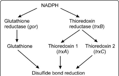
In contrast to the compartments in which catalyzed disulfide bond formation occurs, the environment of the cytoplasm of most prokaryotes has evolved not only lacking components that catalyze formation of disulfide bonds, but also having active systems that result in the reduction of protein disulfide bonds (Figure 1). Due to the presence of these pathways the production of pro-teins that contain disulfide bonds is thought to be impossible in the cytoplasm of most wild-type prokar-yotes such as E. coli [4]. When such proteins are expressed they are unable to attain their native confor-mation and commonly form insoluble aggregates known as inclusion bodies. While such inclusion bodies can be purified and refolded, it would be useful to have a sys-tem for large scale production of disulfide bond contain-ing proteins in the cytoplasm ofE. coli.
* Correspondence: [email protected]
Department of Biochemistry, Linnanmaa Campus, University of Oulu, 90570 Oulu, Finland
To circumvent the problem associated with the pro-duction of disulfide bond formation in the cytoplasm of
E. colia variety of modified strains have been produced [5-9]. These strains, which evolved from seminal studies onE. coli physiology, have a total or partial disruption of one or both of the pathways involved in ensuring that the cytoplasm is reducing. Strains in which both path-ways are disrupted show a significant growth defect con-nected with the reducing pathways being required for other cellular processes e.g. the function of ribonucleo-tide reductase, unless the media is supplemented with a reducing agent [6]. However, this requirement in rich media can be obviated by spontaneous mutations in
aphC [9-11]. The disruption of these two pathways through a knockout of the two NADPH dependent reductases trxB and gor combined with a mutation in
aphCallows for a significant increase in the production of activity of even a complex disulfide bonded protein such as a truncated variant of tissue plasminogen activa-tor [vtPA; 8,12]. The addition of DsbC, a periplasmic disulfide isomerase [13], increased the yields of active vtPA produced a further 20-fold [8]. Such Δgor ΔtrxB
strains with mutations inaphC are available commer-cially, for example origami or rosetta-gami (Novagen) or with DsbC co-expression as the SHuffle system (New England Biolabs). However, the yields of many disulfide bonded proteins from these systems are below that required for commercial production or even for the pro-duction of proteins for academic studies.
WhileΔgorΔtrxBstrains lack the reducing pathways and hence allow thioredoxins to transfer oxidizing equivalents from other metabolic pathways to folding proteins [7], there is no dedicated source or catalyst for disulfide bond formation. This is in contrast to compart-ments in which disulfide bond formation occurs. These
all naturally have catalysts ofde novo disulfide bond for-mation, such as the sulfhydryl oxidases Ero1, found in the ER [14], or Erv1p, found in the mitochondrial inter-membrane space ofS. cerevisiae[15] which can catalyze the reaction:
Dithiol+O2⎯ →⎯ Disulfide+H O2 2
or the transmembrane protein DsbB required for dis-ulfide bond formation in the periplasm [16] which trans-fers electrons to menaquinone or ubiquinone. While all of the catalysts of disulfide bond formation are thought to act via an intermediary protein, protein disulfide iso-merase (PDI) for Ero1 [17], Mia40 for Erv1p [18] and DsbA for DsbB [16], rather than directly on non-native protein substrates, data exists in the literature that Erv1p may be able to function independently of Mia40 [for example 19,20]. In addition, we have observed that Erv1p is able to efficiently oxidatively refold reduced denatured bovine pancreatic trypsin inhibitor to a two disulfide state in the absence of any other factor such as Mia40, glutathione, thioredoxin or PDI (unpublished observations). Hence Erv1p may be a suitable single pro-tein system to catalysede novodisulfide bond formation. To date all reported systems for making disulfide bond containing proteins in the cytoplasm ofE. coliare based on the disruption of one, or more usually both, of the naturally occurring reduction pathways. Here we show that the introduction of the sulfhydryl oxidase Erv1p, an enzyme which can use molecular oxygen to catalyze the oxidation of a dithiol to a disulfide in an FAD-dependent manner, into the cytoplasm of wild-typeE. coliresults in as high or higher levels of produc-tion of active proteins than a ΔgorΔtrxBstrain. Hence our results refute the current paradigm in the field that disruption of at least one of the reducing pathways is essential for the efficient production of disulfide bond containing proteins and opens up new possibilities for their production.
Results
Alkaline phosphatase production
Our initial screen for whether co-expression of the sulf-hydryl oxidase Erv1p could increase the yield of disulfide bond containing proteins was based on E. colialkaline phosphatase (PhoA), a protein which is widely used to examine disulfide bond formation in vivo. PhoA is a hydrolase which naturally folds in the periplasm and contains two sequential disulfide bonds whose formation is essential for activity [21]. While the activity of PhoA has an essential requirement for disulfide bond forma-tion, when expressed in a reducing environment such as the cytoplasm ofE. coli the protein does not form inclu-sion bodies. Hence, expresinclu-sion of the mature form of
PhoA i.e. the protein lacking the periplasmic signal sequence, in the cytoplasm of E. coli resulted in high levels of soluble protein being produced (Figure 2A), but in negligible yields of active protein (Figure 2B and
Table 1), consistent with the reducing environment of the cytoplasm. In contrast, co-expression of Erv1p with PhoA from a polycistronic vector resulted in high yields of active PhoA (Figure 2B and Table 1). Exact
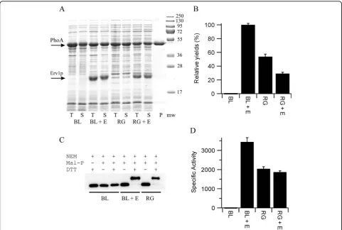
Figure 2Production of PhoA in the cytoplasm ofE. coli. A) SDS-PAGE analysis of the production of PhoA produced in LB media at 30°C. T = totalE. colilysate, S =E. colilysate soluble fraction, P = purified protein from BL21 with co-expression of Erv1p from the NEM-treated lysate and is representative of the quality obtained from all of the samples. Mw = molecular weight markers. BL = BL21 (DE3) pLysSRARE; RG = rosetta-gami; + E = co-expression ofS. cerevisiaeErv1p from a polycistronic vector. The positions of PhoA (upper) and Erv1p (lower) are marked with arrows. B) Relative yields of active PhoA normalized to the system producing the most active protein and shown as percentage mean ± s.d. (n = 4). C) Representative blot from a shift-assay based on alkylation of free thiol groups to examine the disulfide bond status of the PhoA produced. Note that the samples are treated with the thiol-blocking agent N-ethylmaleimide (NEM) before reduction and maleimide based addition of polyethyleneglycol. Hence an increase in apparent molecular weight is consistent with the presence of one or more disulfide bonds in the original sample. The greater the number of disulfide bonds the greater the mass shift. PhoA produced in both theΔgorΔtrxBbackground and in the wild-type background plus co-expression of Erv1p show a homogeneous disulfide bonded protein being produced, however this assay does not determine whether these disulfide bonds are native or not. D) Specific activity (μmole of product formed per minute per mg of protein) of purified PhoA shown as mean ± s.d. (n = 3).
Table 1 Production of PhoA in the cytoplasm ofE. coli
Sample ΔAbsorbance (mAU/min) Final OD of culture Relative yield (%)
PhoA in BL21 (DE3) -0.068 ± 0.053 2.67 ± 0.20 -0.3 ± 0.3
PhoA + Erv1p in BL21 (DE3) 27.6 ± 0.6 1.73 ± 0.01 100 ± 2.1
PhoA in rosetta-gami 16.2 ± 0.6 1.59 ± 0.05 53.9 ± 3.6
PhoA + Erv1p in rosetta-gami 11.0 ± 0.8 1.27 ± 0.03 29.3 ± 1.8
quantification of the fold increase is difficult, given the absence of recombinant PhoA activity when Erv1p is not co-expressed, but it is at least 1000-fold. Further-more, the yield of active PhoA upon co-expression of Erv1p in the E. coli strain BL21(DE3) pLysSRARE, which has the reducing pathways in place, was circa two-fold higher than that produced in the equivalent commercial ΔgorΔtrxBstrain rosetta-gami (Figure 2B) despite similar amounts of protein being produced (Fig-ure 2A). Analysis of the PhoA produced in these strains via gel-shifts after mal-PEG treatment [22] implied that effectively all of the PhoA produced in the cytoplasm of theE. coli strain BL21 upon Erv1p co-expression con-tained disulfide bonds (Figure 2C), despite the protein being produced in a strain with both disulfide reducing pathways intact.
To further characterize the system, PhoA from all four expression conditions was purified by immobilised metal affinity chromatography. This one step purification resulted in highly purified protein (Figure 2A). Reduced PhoA is prone to air oxidation which results in the for-mation of disulfide bonds and a gain in activity. To ensure the specific activity measurements of PhoA were not influenced by this, while allowing analysis of the free thiol content of the purified protein, part of the
E. colilysate was treated with N-ethyl maleimide (NEM) to block free thiol groups and prevent ex vivo disulfide bond formation, while another part was not. Parallel purifications of the two samples from each of the four expression conditions were performed. Despite neither the expression conditions nor the purification procedure being optimised the equivalent of 16 mg/L of PhoA was obtained from theE. colistrain BL21 with co-expression of Erv1p. Activity measurements on the PhoA protein from the NEM-blocked samples showed that the specific activity of the PhoA from theE. coli strain BL21 was circa 1000-fold greater when Erv1p was co-expressed and that this material was circa 1.7-fold more active that the PhoA purified from the rosetta-gami strain (Fig-ure 2D). A similar pattern was observed for the PhoA purified from the non-NEM treated lysate except that the specific activity of the PhoA from the BL21 strain was circa 3-fold greater than that from the non-treated lysate, consistent with ex vivo oxidation. An Elman’s assay of the non-NEM treated purified samples, under denaturing conditions, revealed that the PhoA purified from the BL21 strain had circa 3.1 free thiol groups per protein, while there was no detectable free thiol groups on the protein purified from the same strain with co-expression of Erv1p. Electrospray mass spectrometric analysis of non-treated samples showed that the major component for all four proteins were of the expected mass of PhoA (48285 Da; mass accuracy 0.01%), but additional minor peaks were also observed. For all four
samples an additional peak of +80Da was observed in the mass spectra, consistent with the addition of a phos-phate group. The relative intensity of this peak was around 25% except for the PhoA purified from the BL21 strain in the absence of Erv1p co-expression where it was around 10%. For both of the PhoA samples purified from the BL21 strains an additional peak consistent with gluconoylation of the N-terminal hexa-histdine tag [23] was also observed, with this post-translational mod-ification also being found on around 25% of the total protein. It is unclear to what extent this additional post-translational modification, that we commonly observe for N-terminal hexa-histidine tagged proteins expressed in BL21, affects the activity of PhoA. Both SDS-PAGE and mass spectrometry revealed that the PhoA purified from the BL21 (DE3) strain in the absence of Erv1p co-expression became increasingly degraded upon storage, something not observed for the other samples over the same time scale. This is consistent with the lack of disulfide bonds in this protein.
Phytase production
background upon co-expression of Erv1p and DsbC (Figure 3A and Table 2) i.e. once both a catalyst of dis-ulfide bond formation (Erv1p) and of isomerization (DsbC) were expressed. Again absolute quantification of the increase in yield of active protein is difficult due to the negligible amounts of active protein produced in BL21(DE3) pLysSRARE when Erv1p is not co-expressed. However, the yields of active AppA produced with co-expression of Erv1p and DsbC in BL21(DE3) pLysS-RARE are circa three-fold higher than those produced in theΔgorΔtrxBstrain rosetta-gami with co-expression of DsbC (Figure 3A). Furthermore, analysis of the AppA produced via gel-shift assay showed that the AppA pro-duced by co-expression of Erv1p and DsbC in a wild-typeE. colibackground was comprised only of disulfide containing protein that migrated at the same position in the gel-shift assay, while that produced in the Δgor
ΔtrxB strain with or without co-expression of DsbC resulted in a mixture of species with different numbers of disulfide bonds (Figure 3B) implying incomplete oxidation.
Purification of AppA from these strains is complicated by the very low expression levels. Immobilised metal affinity chromatography from a standard culture did not generate sufficient protein for the concentration of the purified AppA to be determined accurately. However, an enzyme activity assay performed immediately after purification revealed that very significant AppA activity was recovered from the BL21 (DE3) pLysSRARE strain with co-expression of Erv1p and DsbC. Relative quantifi-cation of the proteins purified using tryptophan intrinsic fluorescence showed that the protein purified from BL21(DE3) pLysSRARE had less than 1% of the relative activity of that obtained upon co-expression of Erv1p and DsbC, while that from rosetta-gami with co-expres-sion of DsbC was 31%. These results on the purified proteins mirror the results from the E. coli lysates shown in Figure 3A. More than 99% of the AppA activ-ity was lost from all of the purified samples upon a sin-gle freeze-thaw cycle in the purification elution buffer.
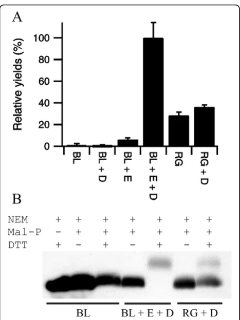
Figure 3 Production of AppA in the cytoplasm ofE. coli. A) Relative yields of active AppA produced in LB media at 30°C, normalized to the system producing the most active protein and shown as percentage mean ± s.d. (n = 4). BL = BL21 (DE3) pLysSRARE; RG = rosetta-gami; + = co-expression from a
polycistronic vector where D = matureE. coliDsbC, E =S. cerevisiae
Erv1p. B) Representative blot from a shift-assay based on alkylation of free thiol groups to examine the disulfide bond status of the AppA produced. While AppA produced upon co-expression of Erv1p in a wild-type background shows a homogeneous disulfide bonded protein being produced, the protein produced in theΔgor
ΔtrxBbackground shows heterogeneity and a lower degree of disulfide bond formation. Note that the molecular weight of the mal-PEG is not homogenous and hence modified proteins, especially those with multiple mal-PEG added, appear as more defuse bands.
Table 2 Production of AppA in the cytoplasm ofE. coli
Sample ΔAbsorbance Final OD of culture Relative yield (%)
AppA in BL21 (DE3) 0.003 ± 0.004 2.60 ± 0.18 1.3 ± 1.3
AppA + DsbC in BL21 (DE3) -0.003 ± 0.004 2.02 ± 0.07 -0.7 ± 1.2
AppA + Erv1p in BL21 (DE3) 0.039 ± 0.009 1.10 ± 0.02 6.2 ± 1.4
AppA + Erv1p + DsbC in BL21 (DE3) 0.420 ± 0.062 1.65 ± 0.01 100 ± 14.2
AppA in rosetta-gami 0.105 ± 0.020 1.91 ± 0.14 28.6 ± 3.0
AppA + DsbC in rosetta-gami 0.123 ± 0.007 2.03 ± 0.01 36.2 ± 1.9
Discussion
The current systems for making disulfide bonded pro-teins in the cytoplasm ofE. coliare based on the disrup-tion of one, or more usually both, of the reducing pathways normally present in this compartment [5-8], with a concomitant mutation in aphC [9-11] which restores growth rates in rich media. While these strains can produce much higher levels of disulfide bonded pro-teins than the wild-type strains the yields and/or quality are often not optimal even when a disulfide isomerase is co-expressed. While thioredoxin is reported to be involved in disulfide bond formation inΔtrxBstrains [7], it is a catalyst of thiol-disulfide exchange and not a cata-lyst ofde novodisulfide bond formation. Hence thiore-doxin becomes oxidised in this system by reducing substrates such as ribonucleotide reductase and transfers the disulfide to folding proteins such as PhoA, such that disulfide bond formation in these strains can potentially be seen as being a“by-product”of other metabolic pro-cesses. In contrast, compartments that naturally produce disulfide bonded proteins have a catalytic system for making disulfide bonds for example a sulfhydryl oxidase which can use molecular oxygen to catalyze the oxidation of a dithiol to a disulfide. The cytoplasm ofE. colilacks such a catalyst. Hence, we thought that the introduction of such a system might significantly increase the yields of active disulphide-bonded protein produced. This turns out to be the case. Indeed the production of disulfide bonded proteins in the cytoplasm ofE. colidoes not require disruption of genes involved in either of the redu-cing pathways. Instead for PhoA and AppA the addition of a catalytic system for the formation of disulfide bonds i.e. co-expression of the sulfhydryl oxidase Erv1p, while not significantly altering the amount of recombinant pro-tein produced, significantly increases the yields of active protein produced. Furthermore, it is more effective than the removal of the reducing pathways for the production of active PhoA and AppA. The expression conditions and purification reported here have not been optimized, but yields of 16 mg/L of culture of purified disulfide bonded protein were obtained from the cytoplasm of E. coli
grown in shake flasks. Furthermore, our preliminary results indicate that such yields are possible for a range of other proteins of academic and commercial interest with multiple disulfide bonds (data not shown).
The ability of co-expression of Erv1p (plus a disulfide isomerase where needed) to enable the production of natively folded disulfide bond containing proteins in the cytoplasm of wild-typeE. colimay seem contradictory with what is known about the environment of this cellular compartment. However, it is compatible with what is known about disulfide bond formation in other compart-ments. Neither the ER nor the inter-membrane space of
mitochondria can be accurately described as an oxidizing environment. While the redox potential of the ER is more oxidizing than that of the cytoplasm it is still a compart-ment in which disulfide bond reduction and isomerization must occur [1] i.e. both oxidative and reducing pathways must co-exist in the same compartment. However, native disulfide bonds in folded proteins are often buried and hence inaccessible to reduction by either glutathione or members of the thioredoxin superfamily. Hence in our system there is probably a kinetic competition between oxidation/folding and reduction with the native disulfide bonded state being stable once formed even in the back-ground of wild-type cytoplasm with both the thioredoxin and glutathione based reducing pathways intact.
The ability of Erv1p to act in the absence of Mia40 may also seem contradictory with what is known about its mechanism of action in the inter-membrane space of mitochondria. It is possible that Erv1p may be acting in our system via an intermediary molecule, such as one of the thioredoxins or glutaredoxins or via a low molecular weight species. However, there is published data that Erv1p is able to act without Mia40 [for example 19,20] and we have preliminaryin vitro data that Erv1p is able to efficiently oxidise dithiols to disulfides in reduced unfolded proteins in the absence of any factors except molecular oxygen. The elucidation of the exact mechan-isms by which Erv1p is acting in the cytoplasm ofE. coli
is not trivial, but this and a more detailed understanding of the other processes that are occurring may help in the optimization of this system for the production of recombinant disulfide bond containing proteins.
Conclusions
By mimicking natural systems, which have both redu-cing and oxidizing pathways in place in compartments where oxidative folding occurs, we are able to generate more efficient production of disulfide bond containing proteins in the cytoplasm while at the same time avoid-ing the problems associated with the disruption of the reducing pathways e.g. the requirement of the reducing pathways for other cellular processes such as the func-tion of ribonucleotide reductase. Furthermore, our results refute the current paradigm in the field that dis-ruption of at least one of the reducing pathways is essential for the efficient production of disulfide bond containing proteins in the cytoplasm ofE. coliand open up new possibilities for the use ofE. coli as a microbial cell factory.
Methods
Vector construction
The genes for mature E. coli alkaline phosphatase (PhoA; Arg22-Lys471), matureE. coli phytase (AppA; Gln23-Leu432) and mature DsbC (Asp21-Lys236) were amplified by PCR using a colony ofE. coliXL1-Blue as a template. The gene for Erv1p (Met1-Glu189) was amplified using a plasmid kindly provided by Prof Tho-mas Lisowsky as a template.
Erv1p and mature DsbC were cloned into pET23a. An alternative cloning strategy for Erv1p using pET23d (which replaces the 5’NdeI restriction site with NcoI) was also used. This adds an extra Glycine between Met1 and Lys2 of Erv1p. Both versions of the protein were used in a variety of co- and pre-expression experiments and no significant differences between the two were observed. Mature PhoA and AppA were cloned into a modified version of pET23a which includes an N-term-inal his-tag in frame with the cloned gene and an addi-tional SpeI site in the multi-cloning site between the EcoRI and SacI sites. The resulting gene products include the sequence MHHHHHHM- prior to the first amino acid of the protein sequence.
Polycistronic vectors were constructed by taking frag-ments encoding the folding factors from the pET23 based constructs which include the ribosome binding site e.g. XbaI/X fragments and ligating them into the SpeI/X cut plasmid encoding the protein of interest (where X is an appropriate restriction site found in the multi-cloning site after SpeI and not found in either gene e.g. XhoI). After a single such ligation this gener-ates a plasmid that contains a single transcription initia-tor/terminator and hence makes a single mRNA, but has two ribosome binding sites and makes two proteins
by co-expression from two translation initiation sites. This ligation results in the loss of the original SpeI site. Transfer of a SpeI site after the second gene into the new vector allows a third gene to be cloned by the same method resulting in a tricistronic vector which makes three proteins from a single mRNA.
All plasmid purification was performed using the QIAprep spin miniprep kit (Qiagen) and all purification from agarose gels was performed using the Gel extrac-tion kit (Qiagen), both according to the manufacturers’ instructions.
All plasmids generated were sequenced to ensure there were no errors in the cloned genes (see Table 3 for plasmid names and details).
Protein expression
For expression in LB media, E. colistrains containing expression vectors were streaked out from glycerol stocks stored at -70°C onto LB agar plates containing suitable antibiotics to allow for selection (100 μg/ml ampicillin for pET23 derivatives, 35μg/ml chloramphe-nicol for pLysS derivatives; with 10 μg/ml tetracycline and 15μg/ml kanamycin for selection of rosetta-gami strains). The next day one colony from these plates were used to inoculate 5 ml of LB media, containing suitable antibiotics (100 μg/ml ampicillin for pET23 derivatives, 35 μg/ml chloramphenicol for pLysS deriva-tives; with 10μg/ml tetracycline and 15μg/ml kanamy-cin for selection of rosetta-gami strains), and grown overnight at 30°C, 200 rpm. This overnight culture was used to seed a 50 ml culture of LB containing suitable antibiotics in a 250 ml conical flask to an optical density of 0.05 at 600 nm (OD600). The addition of FAD to the media is not required for the production of active Erv1p and preliminary experiments suggestion such addition does not increase the yield of active PhoA or AppA or other proteins tested in this system. This culture was grown at 30°C, 200 rpm until the OD600 reached 0.4 at which point protein production was induced by the addition of 0.5 mM IPTG. The cells were then grown for a total of 4 hours post induction at 30°C, 200 rpm and the final OD600 measured. The cells were collected by centrifugation and resuspended to an OD600 equiva-lent of 10 (based on the final OD600 of the culture) in 20 mM sodium phosphate pH 7.4, 20μg/ml DNase, 0.1 mg/ml egg white lysozyme and frozen. Such normaliza-tion allows for easy correcnormaliza-tion for differences in the growth rates of the cultures and allows rapid equal total protein loading of samples for activity assay and SDS-PAGE analysis. Cells were lysed by freeze-thawing. Where appropriate, the insoluble fraction was removed by centrifugation and the soluble fraction removed quickly to a new container. Cell lysates or soluble frac-tions were stored frozen in 1 ml aliquots for further
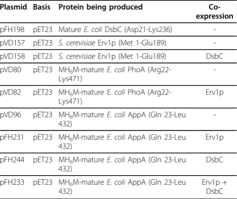
Table 3 Vectors reported in this study
Plasmid Basis Protein being produced Co-expression
pFH198 pET23 MatureE. coliDsbC (Asp21-Lys236) -pVD157 pET23 S. cerevisiaeErv1p (Met 1-Glu189) -pVD158 pET23 S. cerevisiaeErv1p (Met 1-Glu189) DsbC pVD80 pET23 MH6M-matureE. coliPhoA
(Arg22-Lys471)
-pVD82 pET23 MH6M-matureE. coliPhoA
(Arg22-Lys471)
Erv1p
pVD96 pET23 MH6M-matureE. coliAppA (Gln 23-Leu
432)
-pFH231 pET23 MH6M-matureE. coliAppA (Gln 23-Leu
432)
Erv1p
pFH244 pET23 MH6M-matureE. coliAppA (Gln 23-Leu
432)
DsbC
pFH233 pET23 MH6M-matureE. coliAppA (Gln 23-Leu
432)
Erv1p + DsbC
experiments as repeated freeze-thawing clearly influ-enced the results obtained in a protein dependent manner.
Protein analysis from lysates
PhoA activity was measured using a continuous assay at pH 8.0 with 4-nitrophenylphosphate as the substrate. Since PhoA folds in the absence of disulfide bonds and is able to gain activity due to air oxidation post lysis all samples containing PhoA included 100 mM NEM in the lysis buffer to block free thiols and to trap the thiol-disulfide status of the protein during analysis. Similarly 100 mM NEM was added to AppA samples. PhoA activ-ity was determined in 96well micro-titer plates at 37°C. Soluble lysate fractions (from OD600 normalized cul-tures) were diluted 100 fold into 20 mM sodium phos-phate buffer pH 7.4. 20 μl of this was used in a total assay volume of 200 μl formed by adding 50μl of the substrate p-nitrophenyl phosphate solution (0.4% w/v in 1 M Tris pH 8.0) to 130 μl of 1 M Tris pH 8.0. The absorbance was recorded at 410 nm every minute up to 20 minutes and the rate of change in absorbance deter-mined by linear-fit. Yields of active protein were calcu-lated relative to the tested system that generated the most active protein (PhoA with Erv1p co-expression in BL21 (DE3) pLysSRARE) based on the rate of change of absorbance determined in the PhoA assay and the final OD600of the culture (see Table 1 for the values of the individual components).
AppA activity was determined in a similar manner to that of PhoA except that the pH optima of the enzyme and absorbance maxima of the chromogenic product meant that a discontinuous assay had to be performed. In place of the PhoA substrate and Tris solutions, 180μl of p-nitrophenyl phosphate solution (0.4% w/v in 0.25 M Glycine pH 2.5) was used. After 20 min incuba-tion at 37°C the reacincuba-tion was quenched by the addiincuba-tion of 50 μl of 5M NaOH and the absorbance was mea-sured at 410 nm.
The redox state of PhoA and AppA was determined as follows. 10 ml of culture was spun down and resus-pended in 20 mM sodium phosphate pH 7.4, 20 μg/ml DNase, 0.1 mg/ml egg white lysozyme 100 mM NEM to give the equivalent of an OD600of 10 and incubated at 30°C for 5 minutes. 0.5 or 1 ml aliquots were removed for activity measurements (see below) then the samples were frozen at -20C. Cells were then lysed by freeze-thawing. After lysis total protein was precipi-tated with 10% trichloroacetic acid (TCA; final concen-tration) on ice for 10 minutes. The pellet was washed with acetone and then resuspended in 2% SDS, 100 mM Tris pH 7.4 with or without 50 mM DTT for 15 min at 37°C. The proteins were precipitated again with 10% TCA, washed twice with acetone and the
pellet resuspended in alkylation buffer (2% SDS, 100 mM Tris pH 8.0 containing 5 mM malPEG-2000 (NOF Corporation, Japan)) at 37°C for 1 hour. All samples were reduced with 50 mM DTT before load-ing onto 12.5% SDS-PAGE and the PhoA and AppA proteins were detected by western blotting using His-tag antibody (Santa Cruz Biotechnology, USA).
Protein purification
PhoA was expressed as described above and purified by immobilised metal affinity chromatography (IMAC) using TALON superflow metal affinity resin (BD Bios-ciences, USA) with phosphate present in the lysis and purification buffers. Subsequent analysis (see below) revealed that the purified proteins had no PhoA activ-ity and that all of the purified proteins had a mass 80 Da greater than expected, consistent with phosphory-lation. Subsequently, PhoA and AppA were expressed as described above except that the 20 mM sodium phosphate in the lysis buffer was replaced with 50 mM Tris pH 8.0. 100 mM NEM was added to part of the resuspended cell culture, while no NEM was added to the remainder. After cell lysis and removal of the cell debris by centrifugation all samples were puri-fied by IMAC using 400 μl of TALON resin with no phosphate buffer present at any time. Specifically, the resin was washed with 3 mls of water and then equili-brated with 3 mls of equilibration buffer (50 mM Tris, 300 mM sodium chloride, 10 mM immidazole, pH 8.0) before the clarified E. colilysate was loaded. The column was then washed with 5 mls of equilibration buffer before being washed with 3 × 5 mls of wash buffer (50 mM Tris, 300 mM sodium chloride, 20 mM immidazole, pH 8.0), before elution with 2 × 400μl of elution buffer (50 mM Tris, 300 mM sodium chloride, 250 mM immidazole, pH 8.0). The purified PhoA con-centration was determined spectrophotometrically at 280 nm using a calculated molar absorption coeffi-cient (32900 M-1cm-1). The concentration of purified AppA was too low to determine spectrophotometri-cally or by Bradford assay, but relative concentrations were estimated by fluorescence measurements performed on a Perkin-Elmer LS50B spectrometer at 25°C, excitation 280 nm, emission 320-400 nm and slit widths 5 nm.
Analysis of purified proteins
Elman’s assay for free thiol content were performed at room temperature under denaturing conditions in 50 mM Tris buffer, 2 M Guanidine hydrochloride (pH 8.0) using 0.073 mg/ml Elman’s reagent. The change in absorbance at 412 nm was monitored after 15 minutes and the free thiol content calculated using a molar extinction coefficient of 13600 M-1cm-1.
Prior to mass spectrometric analysis the protein sam-ples were desalted using pepClean™C-18 spin columns (Pierce, Rockford, IL, USA) according to manufacturer’s instructions. Molecular masses were measured with a Q-Tof-2 electrospray ionisation mass spectrometer (Micromass, UK) using positive ionisation.
Acknowledgements
This work was supported by the Academy of Finland, Sigrid Juselius Foundation, TULI programme of the Finnish Funding Agency for Technology and Innovation and the University of Oulu. We thank Prof Thomas Lisowsky for kindly providing reagents and Dr Ulrich Bergmann for assistance with the mass spectrometry.
Authors’contributions
FH and VDN participated in the design of the research. FH, VDN and KEHS performed the research. LWR conceived and coordinated the study, participated in its design, performed the research and wrote the manuscript. All authors read and approved the final manuscript.
Competing interests
A patent application has been filed.
Received: 16 April 2010 Accepted: 13 September 2010 Published: 13 September 2010
References
1. Hatahet F, Ruddock LW:Protein disulfide isomerase: a critical evaluation of its function in disulfide bond formation.Antioxid Redox Signal2009,
11:2807-2850.
2. Herrmann JM, Köhl R:Catch me if you can! Oxidative protein trapping in the intermembrane space of mitochondria.J Cell Biol2007,176:559-563. 3. Collet JF, Bardwell JC:Oxidative protein folding in bacteria.Mol Microbiol
2002,44:1-8.
4. Derman AI, Prinz WA, Belin D, Beckwith J:Mutations that allow disulfide bond formation in the cytoplasm ofEscherichia coli.Science1993,
262:1744-1747.
5. De Marco A:Strategies for successful recombinant expression of disulfide bond-dependent proteins inEscherichia coli.Microbial Cell Factories2009,8:26.
6. Prinz WA, Åslund F, Holmgren A, Beckwith J:The role of the thioredoxin and glutaredoxin pathways in reducing protein disulfide bonds in the
Escherichia colicytoplasm.J Biol Chem1997,272:15661-15667. 7. Stewart EJ, Åslund F, Beckwith J:Disulfide bond formation in the
Escherichia colicytoplasm: anin vivorole reversal for the thioredoxins.
EMBO J1998,17:5543-5550.
8. Bessette PH, Åslund F, Beckwith J, Georgious G:Efficient folding of proteins with multiple disulfide bonds in theEscherichia colicytoplasm.
Proc Natl Acad Sci USA1996,96:13703-13708.
9. Ritz D, Lim J, Reynolds CM, Poole LB, Beckwith J:Conversion of a peroxiredoxin into a disulfide reductase by a triplet repeat expansion.
Science2001,294:158-160.
10. Faulkner MJ, Veeravalli K, Gon S, Georgious G, Beckwith J:Functional plasticity of a peroxidase allows evolution of diverse disulfide-reducing pathways.Proc Natl Acad Sci USA2008,105:6735-6740.
11. Yamamoto Y, Ritz D, Planson AG, Jönsson TJ, Faulkner MJ, Boyd D, Beckwith J, Poole LB:Mutant AhpC peroxiredoxins suppress thiol-disulfide redox deficiencies and acquire deglutathionylating activity.Mol Cell2008,29:36-45.
12. Obukowicz MG, Gustafson ME, Junger KD, Leimbruber RM, Wittwer AJ, Wun TC, Warren TG, Bishop BF, Mathis KJ, McPherson DT, Siegel NR, Jennings MG, Brightwell BB, Diaz-Collier JA, Vell LD, Craik CS, Tacon WC:
Secretion of active kringle-2-serine protease inEscherichia coli.
Biochemistry1990,29:9737-9745.
13. Rietsch A, Belin D, Martin N, Beckwith J:Anin vivopathway for disulfide bond isomerization inEscherichia coli.Proc Natl Acad Sci USA1996,
93:13048-13053.
14. Frand AR, Kaiser CA:The ERO1 gene of yeast is required for oxidation of protein dithiols in the endoplasmic reticulum.Mol Cell1998,1:161-170. 15. Lange H, Lisowshy T, Gerber J, Mühlenhoff U, Kispal G, Lill R:An essential
function of the mitochondrial sulfhydryl oxidase Erv1p/ALR in the maturation of cytosolic Fe/S proteins.EMBO Rep2001,2:715-720. 16. Bardwell JC, Lee JO, Jander G, Martin N, Belin D, Beckwith J:A pathway for
disulfide bond formationin vivo.Proc Natl Acad Sci USA1993,
90:1038-1042.
17. Tu BP, Ho-Schleyer SC, Travers KJ, Weissman JS:Biochemical basis of oxidative protein folding in the endoplasmic reticulum.Science2000,
290:1571-1574.
18. Mesecke N, Terziyska N, Kozany C, Baumann F, Neupert W, Hell K, Herrmann JM:A disulfide relay system in the intermembrane space of mitochondria that mediates protein import.Cell2005,121:1059-1069. 19. Lee J, Hofhaus G, Lisowsky T:Erv1p fromSaccharomyces cerevisiaeis a
FAD-linked sulfhydryl oxidase.FEBS Lett2000,477:62-66.
20. Vitu E, Bentzur M, Lisowsky T, Kaiser CA, Fass D:Gain of function in an ERV/ALR sulfhydryl oxidase by molecular engineering of the shuttle disulfide.J Mol Biol2006,362:89-101.
21. Derman AI, Beckwith J:Escherichia colialkaline phosphatase fails to acquire disulfide bonds when retained in the cytoplasm.J Bacteriol1991,
173:7719-7722.
22. Wu HH, Thomas JA, Momand J:p53 protein oxidation in cultured cells in response to pyrrolidine dithiocarbamate: a novel method for relating the amount of p53 oxidationin vivoto the regulation of p53-responsive genes.Biochem J2000,351:87-93.
23. Geoghegan KF, Dixon HB, Rosner PJ, Hoth LR, Lanzetti AJ, Borzilleri KA, Marr ES, Pezzullo LH, Martin LB, LeMotte PK, McColl AS, Kamath AV, Stroh JG:Spontaneous alpha-N-6-phosphogluconoylation of a“His tag” in Escherichia coli: the cause of extra mass of 258 or 178 Da in fusion proteins.Anal Biochem1999,267:169-184.
24. Berkmen M, Boyd D, Beckwith J:The nonconsecutive disulfide bond of
Escherichia coliphytase (AppA) renders it dependent on the protein-disulfide isomerase DsbC.J Biol Chem2005,280:11387-11394. doi:10.1186/1475-2859-9-67
Cite this article as:Hatahetet al.:Disruption of reducing pathways is not essential for efficient disulfide bond formation in the cytoplasm of E. coli.Microbial Cell Factories20109:67.
Submit your next manuscript to BioMed Central and take full advantage of:
• Convenient online submission
• Thorough peer review
• No space constraints or color figure charges
• Immediate publication on acceptance
• Inclusion in PubMed, CAS, Scopus and Google Scholar • Research which is freely available for redistribution