R E S E A R C H
Open Access
Vessel density and En-face segmentation of
optical coherence tomography
angiography to analyse corneal
vascularisation in an animal model
Kavya Devarajan
1, Wen Di Lee
1, Hon Shing Ong
1,2, Nyein C. Lwin
1, Jacqueline Chua
1,3, Leopold Schmetterer
1,4,5,6,
Jodhbir S. Mehta
1,2,3,6and Marcus Ang
1,2,3*Abstract
Background:Optical coherence tomography angiography (OCTA) is a novel non-invasive angiography technology that has recently been extensively studied for its utility in anterior segment imaging. In this study, we compared a split-spectrum amplitude decorrelation angiography (SSADA) OCTA and an optical micro-angiography (OMAG SD) OCTA system to current angiographic technique, indocyanine green angiography (ICGA), to assess corneal vascularisation in an animal model.
Methods:We imaged 16 rabbits, (one eye per animal) with corneal vascularisation using SSADA OCTA (AngioVue; Optovue Inc., USA), OMAG OCTA (Angioscan; RS-3000 Nidek Co. Ltd., Japan) and ICGA in the same region of interest of the cornea at successive time-points. We then analysed all scanned images for vessel density
measurements and used paired t-tests and Bland-Altman plots to examine for significant differences. The en-face
segmentation images from each of the OCTA scans were also extracted and were matched at every 50μm
segmentation to be compared for vessel density at the respective depths.
Results:Bland-Altman plots revealed a good agreement between all three imaging techniques (P> 0.05) for all vessel density measurements computed, and the ranges of 95% limit of agreement were acceptable from a clinical
perspective. No significant difference was reported, with ICGA (μ= 16.52 ± 8.94%) being more comparable to the
OMAG OCTA (μ= 16.23 ± 9.51%;p= 0.50) than the SSADA OCTA (μ= 17.09 ± 7.34%;p= 0.33) system. Also, a good
correlation value (r> 0.9) was obtained when comparing the vessel density measurements of the en-face
segmentations between the OCTA systems.
Conclusions:Comparable vessel density quantification between the two OCTA systems, and with ICGA was obtained. Segmentation analysis of the vasculature at different depths showed varied performance in the two OCTA systems relative to each other. The implications of the study may help to aid in the development of better OCTA algorithms for the anterior segment and its use in clinical translational research.
Keywords:OCTA, Corneal vascularisation, Optical micro angiography, Split-spectrum amplitude decorrelation angiography, Vessel density, Anterior segment, En-face OCTA
* Correspondence:marcus.ang@snec.com.sg
1Singapore Eye Research Institute, Singapore, Singapore 2Singapore National Eye Center, Singapore, Singapore
Full list of author information is available at the end of the article
Background
Corneal vascularisation is a sight-threatening condition that involves the pathological ingrowth of blood vessels into the typically avascular cornea, in response to inflam-mation, infection, trauma or hypoxia [1,2]. It impairs light transmission, promotes scar formation and results in per-sistent inflammation thereby affecting visual acuity [3]. Conventional treatment options and prevention of visual loss in patients with corneal vascularisation remains a key challenge for clinicians [2]. There have been recent attempts to achieve novel drug therapies targeting the mo-lecular mechanisms of corneal vascularisation. However, the ability to quantitatively assess or objectively evaluate corneal vascularisation before and after any intervention is still limited [4]. Therefore, a reliable imaging system to evaluate and quantify corneal vascularisation and its response to treatment is much needed [5].
Corneal vascularisation is usually assessed by analysing images of the cornea taken by slit lamp biomicroscopy. However, they do not represent an objective representa-tion of the corneal vasculature, especially in the presence of corneal scars, deposits or oedema [2, 4]. Indocyanine green angiography (ICGA) and fluorescein angiography (FA) have been shown to delineate corneal vessels and detect areas of corneal vascularisation through intraven-ous dye injections [6]. Among these, ICG (Indocyanine--green) is larger, more protein-bound than fluorescein and retains in the vessels for a longer duration, attribut-ing for better vessel delineation [7]. ICGA is also shown to provide better image quality than FA [4]. Yet, both the angiography methods measure the vascularized area only in two dimensions and can be associated with ad-verse systemic side-effects [5].
Optical coherence tomography angiography (OCTA) is an emerging diagnostic tool for the anterior segment vas-culature that overcomes the limitations of conventional techniques by providing three-dimensional structural and vascular information by non-invasive means [8, 9]. This technology has been recently adapted to image the anterior-segment of the eye and determined to be superior over conventional imaging modalities [8,10–12].
However, the quantification and improvement of auto-mated segmentation algorithms is still an active area of research and development in OCTA [13,14]. OCTA for the retina is known to suffer from poor anatomical segmentation and pathology localisation due to the under-performance of automatic segmentation algo-rithms in diseased conditions thus making it difficult to interpret [13–15]. Moreover, OCTA is currently chal-lenged by methodical and technical issues, such as vessel duplication, residual motion line artefacts and vessel dis-continuity that are not present in conventional angiog-raphy [12]. Without suitable eye-tracker systems for the cornea, orthogonal line artefacts more predominantly
occur during patient movement in the anterior-segment where the system fails and gives a false signal at all posi-tions in the slow axis. The above disadvantages of OCTA is expected to lead amplified segmentation errors during cornea vasculature scanning as the application of the sys-tem at the anterior segment is yet to be realised [12,16].
Although there are various OCTA systems available in the market that have been manipulated to image the an-terior segment, there are limited studies comparing these systems that highlight the constraints and advantages for this purpose. The angiography algorithm in various OCTA systems may differ in the penetration depth and enhance-ment of fine vasculature resolution that can offer different diagnostic sensitivities [7]. This information can be useful when deciding the type of OCTA system to use in future research studies or clinical applications. Previously, we compared the systems for clinical investigation of corneal vascularisation and evaluated vessel density measurements in human eyes in a small pilot study [10]. However, it is still necessary to compare the systems to ICGA, to study the effects on the segmentation in animal models, as they provide good controls for corneal vascularisation. Further-more, there are no studies thus far that have compared the segmentation methods of different OCTA systems [10]. Thus, there is a need to assess and compare the capabilities and limitations of the OCTA systems available for imaging corneal vascularisation.
In this study, we compared two OCTA systems that employ different algorithms in spectral-domain OCT for angiography acquisition, i.e., optical micro-angiography (OMAG OCTA Angioscan; RS 3000 Nidek Co. Ltd., Japan) and split-spectrum amplitude decorrelation (SSADA, AngioVue; Optovue Inc., USA) with ICGA, to image corneal vascularisation in a rabbit model.
Methods
OCTA systems
OCTA system uses both amplitude and phase informa-tion as a complex signal to generate the angiography sig-nal, allowing higher sensitivity to image vascular details. It uses a modified Hilbert transform to separate the moving scattering signals from the static background [18]. The system has a lateral resolution of 20μm and axial resolution of 7μm and captures 53,000 A scans/s using a light source centred at 880 nm [10].
Image acquisition
The study was conducted on sixteen clinically healthy New Zealand white adult rabbits of either sex between the age group 12–15 weeks and weighing between 2.5– 3.5 kg. Routine clinical evaluation and pre-operative ophthalmic examination of both eyes on all the animals was done prior to the experiment.
We performed consecutive follow-up imaging in rabbits with an established model of corneal vascularisation. The right eye of each rabbit underwent corneal suturing under general anaesthesia consisting intramuscular xylazine HCl (5 mg/kg) and ketamine HCl (50 mg/kg), supplemented with topical anaesthesia (0.4% oxybuprocaine HCl). The method of suture technique was described previously [5]; 10–0 non-absorbable nylon sutures (B. Braun Surgical SA, Spain) were placed at mid-stromal depth in the superior part of the cornea, in an inverted triangle fashion, step-wise to direct the growth of vessels centralized on the cornea. Antibiotic eye drops (tobramycin ophthalmic eye drops 0.3%, Alcon Labs Inc., Texas, USA) were applied twice daily throughout the follow-up period. The sutured eyes were followed up for two weeks after the suture-induced experiment when adequate development of induced corneal vascularisation was observed for the disease model. New vessels in the cornea started growing from the first week and reached the central cornea at the second week. The stitches were left intact to prevent any interruption or removal of the vessel growth inducing factor in the disease model.
The rabbits were imaged under anaesthesia with slit lamp photography (SLP), OMAG OCTA, SSADA OCTA and ICGA on a weekly basis throughout the follow-up period for two weeks. After which, histology studies were done, and the rabbits were then sacrificed.
Colour SLP images were captured using the digital slit-lamp camera (Righton MW50D, LED slit lamp, Miyagi, Japan) with a standard diffuse illumination (× 12 to × 36 magnification). For OCTA acquisition, the anter-ior segment lens was used with the AngioRetina scan protocol for the SSADA OCTA device and AngioMacula scan protocol for the OMAG OCTA device. In both the imaging acquisition software, the eye tracking and auto-focus functions were deactivated. The lens was moved very close to the corneal surface before manual adjust-ments were made to the Z-motor positioning and focal
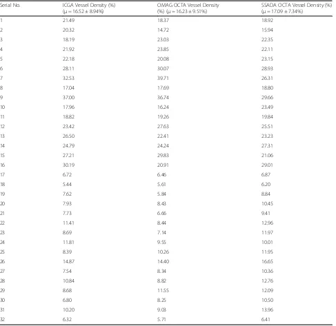
length to achieve precise focus on the B-scan area of interest [10]. Anterior-segment scans using OCTA and ICGA centred on the corneal vasculature were evaluated for vessel density computations from week 1 and 2 follow-up time-points. A total of 32 images segmented at the whole B-scan depth (two time-point scans from 16 rabbits) were evaluated from each OCTA and ICGA system for vessel density comparison.
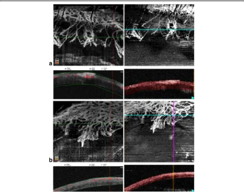
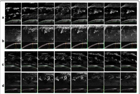
Representative OCTA images captured during Week 1 and Week 2 time-points at the same regions of interests are shown in Fig.1. The same representative images seg-mented at every 50μm of the cornea B-scan were ex-tracted from each OCTA volume in the two systems [8, 19] and compared as shown in Fig.2. The segmentation algorithm that was incorporated in the SSADA system was based on the macula B-scan layer segmentations, whereas the OMAG OCTA volume segmentation was based on the custom parallel layer segmentation devel-oped for research purposes.
Image processing
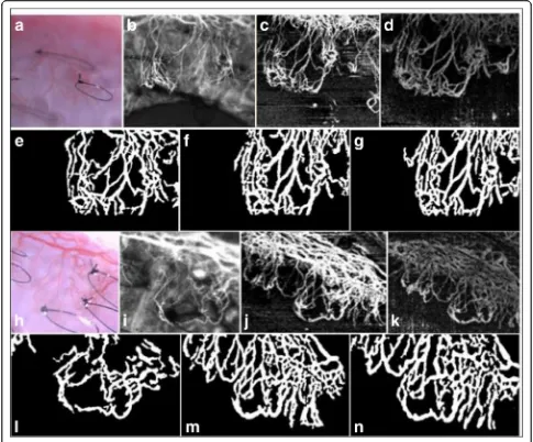
All image processing was performed using MATLAB R2017b (The MathWorks, Inc., Natick, Massachusetts, United States) similar to a previously described technique [5]. OCTA images from the SSADA system were extracted in the Portable Network Graphics and Bitmap image file for-mats from the OMAG system. ICGA images were extracted in the Joint Photographic Experts Group format. Briefly, the extracted images from the three systems were first automat-ically registered for matching overlap in the captured region of area. Thereafter, filters were applied to remove speckle and motion artefacts. Following which, binarization using Otsu’s method of intensity threshold based on automatic binarization-level decisions was performed, wherein white and black pixels represented the vasculature foreground and the background, respectively. Figure3 shows an illustrative example of binarized vessels performed in the processed images. Vessel density values were then computed from the binarized image as a ratio of the area of the white pixels (vessels) to the whole image pixel area.
Statistical analysis
represents what would be expected by chance. Negative values indicate potential disagreement between the ob-servers [20]. Comparison between the vessel densities of SSADA OCTA, OMAG OCTA and ICGA processed im-ages was computed using the paired t-test. Pearson correl-ation coefficient (r value) was used to determine the correlation between vessel density measurements of SSADA OCTA, OMAG OCTA and ICGA. Bland-Altman plots was evaluated to analyse the agreement between the three techniques; the difference of vessels’density measure-ments between the imaging modalities was plotted against the average vessels’density measurements of the methods. Further, vessel density values from the segmented en-face images using the two OCTA systems were also subjected to the Bland-Altman plot to show the different score measure-ments at the various depth segmentation ranges.
Results
scores were found to be comparable (p= 0.076) with good inter-observer agreement (κ= 0.704). Using Fig.3as a rep-resentative example, the higher vessel density observation in OCTA is demonstrated due to its ability to capture more vessels than slit lamp photography or ICGA. As SLP and ICGA have limited lateral resolution, this could potentially explain the reason for their reduced vascular acquisition. Table1lists the vessel density percentages computed from the 32 sets of matched images.
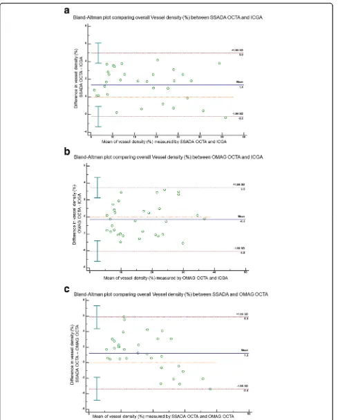
Figure 4 demonstrates the Bland-Altman plots agree-ments comparing the angiography techniques computed from the 32 sets of matched images. There was good agreement between all three imaging modalities in terms of vessel density measurements. ICGA vs. SSADA OCTA (r> 0.7) LOA lower limit−15.44μm (95% CI:− 20.072 to −10.825μm); upper limit 13.657μm (95% CI: 9.033 to 18.280μm); ICGA vs. OMAG OCTA (r > 0.9) LOA lower limit −9.713μm (95% CI: −12.547 to − 6.880μm); upper limit 8.125μm (95% CI: 5.292 to 10.959μm); SSADA OCTA vs. OMAG OCTA (r > 0.7)
LOA lower limit −12.585μm (95% CI: −16.550 to − 8.619μm); upper limit 12.381μm (95% CI: 8.415 to 16.347μm).
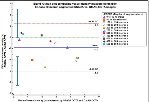
We also obtained a good correlation value (r= 0.993) when comparing vessel density measurements of the en-face segmentations at every 50μm between the OCTA systems. In superficial depth segmentations, the OMAG OCTA provided higher vessel density values than the SSADA OCTA system (mean vessel density 6.172 ± 3.6% vs. 4.377 ± 2.2%, respectively, p < 0.001). However, in segmentation layers greater than 400μm deep, the SSADA OCTA system mean vessel density measurements were higher (4.438 ± 2.127%) compared with the other system (4.041 ± 1.803%). The difference in the trend of vascular densities captured from the two devices is also shown in Fig. 2 as a representative example. In the last few segmentation depths of the SSADA OCTA system it is seen that it additionally captures the projection from the superficial segmenta-tions. The vessel density extracted from each of the
a
b
c
d
depth range segmentations from the two OCTA systems were plotted for their difference scores as a Bland-Altman graph as shown in Fig. 5. Good agree-ment between OMAG OCTA and SSADA OCTA was observed with a mean difference of 1.872 ± 1.942% (95% CI: 1.956 to 7.473%),P= 0.218.
Discussion
Using the SSADA and OMAG algorithm-based OCTAs, we have demonstrated that the visualization of both dense and fine vasculature across the entire cornea are compar-able to ICGA circulations without significant differences. It was observed that in the SSADA implemented images of SSADA OCTA, less axial bulk noise and smoother signal was present as compared to the OMAG algorithm-derived
OCTA images. This observation can be attributed to the SSADA OCTA’s volume averaged acquisition from two re-peated consecutive B-scans (each taking 3–4 s) with inbuilt motion correction software, as compared to the OMAG OCTA system that takes 5–6 s for one full scan, resulting in more motion artefacts in subjects with poor fixation [21]. Further, the improved signal-to-noise ratio could also be a result of the system being independent of phase infor-mation and is thereby insensitive to phase noise, giving rise to better signal strength. However, this advantage is also at the expense of the degradation of its axial resolution equalled to its transverse dimension, which can introduce undesirable projection artefacts [22].
projections of the SSADA system for the reconstruction of deeper layer vasculature segments [18]. This limitation of the SSADA system is a possible reason for the observa-tion of significantly higher vessel density values in the SSADA system than the OMAG system in the deeper layers of the cornea. On the other hand, the OMAG OCTA system overcomes these limitations of the SSADA system as it is not associated with projection artefact is-sues. The system processes images using both phase and amplitude information, using the algorithm of complex OCT signal difference (CODAA). This allows for the add-itional inclusion of flow-induced changes from the phase
of OCT signal, thus providing ultra-high sensitivity for the detection of micro-circulations [17, 23]. The phase vari-ance method, which is known to be the best method amongst all others to offer good contrast-to-noise ratio, also allows for effective suppression of the static tissue noise [21]. These strengths of the CODAA system support our findings that vessel density values of the ICGA images are more comparable to the OMAG OCTA system than the SSADA system. In overall comparison to ICGA, it was generally observed that the OMAG OCTA provided bet-ter quantitative agreement and the SSADA OCTA showed slightly better performance in quality.
Table 1Vessel density measurements computed from ICGA and OCTA in 16 rabbits at two consecutive follow-up time-points
Serial No. ICGA Vessel Density (%) (μ= 16.52 ± 8.94%)
OMAG OCTA Vessel Density (%) (μ= 16.23 ± 9.51%)
SSADA OCTA Vessel Density (%) (μ= 17.09 ± 7.34%)
1 21.49 18.37 18.92
2 20.32 14.72 15.94
3 18.19 23.03 22.35
4 21.92 23.85 22.11
5 22.18 20.08 23.15
6 28.11 30.07 28.93
7 32.53 39.71 26.31
8 17.04 17.69 18.80
9 37.00 36.74 29.66
10 17.96 16.24 23.49
11 18.82 19.26 19.84
12 23.42 27.63 25.51
13 26.50 22.41 23.23
14 24.79 24.24 27.31
15 27.21 29.83 21.06
16 30.19 20.91 29.01
17 6.72 6.46 6.87
18 5.44 5.61 6.20
19 7.62 5.84 8.84
20 7.93 8.43 10.45
21 7.73 6.66 9.41
22 11.41 8.44 12.96
23 8.69 7.14 11.97
24 11.81 9.55 10.01
25 8.39 10.26 11.95
26 14.87 14.40 16.65
27 7.54 8.34 10.36
28 10.84 8.82 12.76
29 8.68 11.55 12.09
30 6.80 8.25 10.50
31 10.20 9.03 13.96
In the second part of the analysis in the study, we reported for the first time the comparisons of en-face segmentations at every 50μm between two anterior seg-ment OCTA systems. The vessel density measureseg-ments at all depth segmentations correlated well in both systems, with no significant difference. It was observed that the OMAG OCTA system was found to have higher vessel density measurements than the SSADA OCTA system, in segmentation depth ranging from 0 to 350μm. This may be because of the higher contrast and working wavelength exhibited by the OCTA system. Conversely, in deeper seg-mentation layers (> 400μm), the SSADA system over-estimated the measurement, which could have been associated with the inaccuracy of vessel density
projections from the more superficial layers. Although the three-dimensional en-face scan tomography provided rea-sonable and reliable segmentation profiles for the cornea analysis, it should be noted that the extracted image out-comes may not be precise as they were not based on seg-mentation algorithms developed for anterior-segment B-scans and is less robust [13]. Non-parallel segmenta-tions and layer identification artefacts may contribute to errors in the en-face segmentations, especially in poor quality OCTA scans, where the segmentation lines were not oriented parallel to the corneal surface [15].
As a result, despite our study showing direct comparisons of the two OCTA systems for the same regions in the same subjects, factors associated with differences in segmentation
(See figure on previous page.)
Fig. 4Bland-Altman plots comparing vessels density measurements from OCTA with ICGA. The Bland-Altman plot between the differences (y-axis) of vessels density measurements from (a) SSADA OCTA and ICGA, (b) OMAG OCTA and ICGA and (c) SSADA OCTA and OMAG OCTA as the deviation from the mean vessels density values comparing the corresponding two methods (x-axis)—showing good agreement of vessels density between all imaging methods. Solid line = mean of the difference. Short dashed line = reference zero. Long dashed line = upper and lower 95% limits of agreement (mean + 1.96 SD, mean−1.96 SD). SD = standard deviation of the mean difference
and acquisition protocols in the two OCTA systems may not account for one-to-one comparison of their perfor-mances and analyses. Furthermore, device-dependent parameters such as the difference in speed, operating wavelengths, contrast-to-noise ratio, signal-to-noise ratio and sensitivity were not taken considered while comparing the vessel density outcomes processed from the two OCTA systems [13].
Therefore, we observed that there is generally a better agreement of the OMAG OCTA system to ICGA. How-ever, it cannot be conclusively decided that the OMAG OCTA system performs better than the SSADA system. Both the OCTA systems are found to be comparable to the ICGA imaging system to image the vasculature in the anterior-segment eye and are associated with their re-spective advantages and limitations based on their imple-mented algorithm. For example, the amplitude decorrelated images obtained from the SSADA system, provided a better signal-to-noise ratio, but was susceptible to bulk tissue motion noise and projection artefacts [18]. On the other hand, while the phase-variance method employed in the OMAG system provided higher sensitiv-ity to vascular details and independence of projection artefacts, it was still subject to greater background noise and motion artefacts. It is important to note that these findings are relevant in the case of optimal operation of the system comparable to the animal model setting where there is control of eye movements and limited motion artefacts present. In the clinical setting, the quality of the images and volume of artefacts may vary considerably.
Conclusion
In this experimental study, we compared and validated two OCTA systems with ICGA to delineate corneal vessels in an animal model. The overall vessel density measurements for both systems were comparable to the ICGA technique, where there was less difference between ICGA and OMAG OCTA than ICGA and SSADA OCTA system in the same region of corneal vascularisation. The en-face segmentation analysis of the two systems showed that the SSADA OCTA relative to the OMAG OCTA under-estimated vessel density in the superficially segmented angiography layers whereas, the OMAG OCTA under-estimated the same in deeper vasculature layers. Future studies are required to validate the differences between OCTA systems with hist-ology, compare for repeatability assessments and use seg-mentation algorithms implemented for the cornea. With OCTA technology advancing at a rapid rate than the com-munity’s experience with the technique, the need for the standardisation of anterior segment protocols and accurate segmentation software across competing OCTA technolo-gies for its image acquisition and interpretation is demand-ing. Evaluation of OCTA into a multimodal platform alongside other established imaging techniques will provide
us with a better understanding to correctly assess the vas-culature of the cornea and ocular surface diseases. This will enable the advancement of OCTA into clinical practice as a more precise and efficient diagnostic modality for the cornea.
Abbreviations
CI:Confidence interval; ICG: Indocyanine green; ICGA: Indocyanine green angiography; LOA: Limits of agreement; OCT: Optical coherence tomography; OCTA: Optical coherence tomography angiography; OMAG: Optical micro-angiography; SD: Spectral domain; SLP: Slit lamp photography; SSADA: Split-spectrum amplitude decorrelation angiography
Acknowledgements
Not applicable.
Funding
Not applicable.
Availability of data and materials
The images used in the analysis of this study are available by contacting the corresponding author.
Authors’contributions
KD designed and planned the experiments. KD, HSO and NCL conducted the experiments. KD and MA analysed the results. KD performed statistical analysis. MA, HSO, LS, JC and JM supervised the experiments and corrected the manuscript. MA coordinated the project and handled funding. KD drafted the main manuscript text. WDL and KD prepared all figures. All authors revised the manuscript for significant intellectual content and gave final approval of the version to be published.
Ethics approval
The work was carried out with prior approval from the Institutional Animal Care and Use Committee of SingHealth. All experimental procedures were carried out in accordance with the guidelines of the Association for Research in Vision and Ophthalmology for the use of animals in ophthalmic and vision research.
Consent for publication
Not applicable.
Competing interests
The authors declare that they have no competing interests.
Author details
1Singapore Eye Research Institute, Singapore, Singapore.2Singapore National
Eye Center, Singapore, Singapore.3Eye-ACP, Duke-NUS Graduate Medical School, Singapore, Singapore.4Department of Clinical Pharmacology, Medical
University of Vienna, Vienna, Austria.5Center for Medical Physics and
Biomedical Engineering, Medical University of Vienna, Vienna, Austria.
6
Nanyang Technological University, Singapore, Singapore.
Received: 14 September 2018 Accepted: 24 December 2018
References
1. Chang JH, Garg NK, Lunde E, Han KY, Jain S, Azar DT. Corneal neovascularization: an anti-VEGF therapy review. Surv Ophthalmol. 2012;57(5):415–29.
2. Spiteri N, Romano V, Brunner M, Steger B, Kaye SB. The management of corneal neovascularisation- update on new clinical data and recommendations of treatment. European Ophthalmic Review. 2016;10:86–93.
3. Tian S, Wang S, He Y, Liu X, Li Y, Su G, et al. Review of the progress in corneal neovascularization animal models. Am J Biochem Biotechnol. 2015;11(4):221–7. 4. Anijeet DR, Zheng Y, Tey A, Hodson M, Sueke H, Kaye SB. Imaging and
evaluation of corneal vascularization using fluorescein and indocyanine green angiography. Invest Ophthalmol Vis Sci. 2012;53(2):650–8. 5. Stanzel TP, Devarajan K, Lwin NC, Yam GH, Schmetterer L, Mehta JS, et al.
green angiography and slit lamp photography for corneal vascularization in an animal model. Sci Rep. 2018;8(1):11493.
6. Kirwan RP, Zheng Y, Tey A, Anijeet D, Sueke H, Kaye SB. Quantifying changes in corneal neovascularization using fluorescein and indocyanine green angiography. Am J Ophthalmol 2012;154(5):850–8.e2.
7. Ang M, Cai Y, Shahipasand S, Sim DA, Keane PA, Sng CC, et al. En face optical coherence tomography angiography for corneal neovascularisation. Br J Ophthalmol. 2016;100(5):616–21.
8. Ang M, Tan ACS, Cheung CMG, Keane PA, Dolz-Marco R, Sng CCA, et al. Optical coherence tomography angiography: a review of current and future clinical applications. Graefes Arch Clin Exp Ophthalmol. 2018;256(2):237–45. 9. Ang M, Devarajan K, Das S, Stanzel T, Tan A, Girard M, et al. Comparison of anterior segment optical coherence tomography angiography systems for corneal vascularisation. Br J Ophthalmol. 2018;102(7):873–7.
10. Cai Y, Alio Del Barrio JL, Wilkins MR, Ang M. Serial optical coherence tomography angiography for corneal vascularization. Graefes Arch Clin Exp Ophthalmol. 2017; 255:135–9.
11. Brunner M, Romano V, Steger B, Vinciguerra R, Lawman S, Williams B, et al. Imaging of corneal neovascularization: optical coherence tomography angiography and fluorescence angiography. Invest Ophthalmol Vis Sci. 2018;59(3):1263–9. 12. Gao SS, Jia Y, Zhang M, Su JP, Liu G, Hwang TS, et al. Optical coherence
tomography angiography. Invest Ophthalmol Vis Sci. 2016;57(9):OCT27–36. 13. Rodríguez FJ, Staurenghi G, Gale R. Vision Academy Steering Committee.
The role of OCT-A in retinal disease management. Graefes Arch Clin Exp Ophthalmol. 2018;256(11):2019–26.
14. Spaide RF, Fujimoto JG, Waheed NK. Image artifacts in optical coherence tomography angiography. Retina. 2015;35(11):2163–80.
15. Tahiri Joutei Hassani R, Liang H, El Sanharawi M, Brasnu E, Kallel S, Labbé A, et al. En-face optical coherence tomography as a novel tool for exploring the ocular surface: a pilot comparative study to conventional B-scans and in vivo confocal microscopy. Ocul Surf. 2014;12(4):285–306.
16. Ang M, Cai Y, Tan AC. Swept source optical coherence tomography angiography for contact lens-related corneal vascularization. J Ophthalmol. 2016;2016:9685297.
17. Zhang A, Zhang Q, Chen CL, Wang RK. Methods and algorithms for optical coherence tomography-based angiography: a review and comparison. J Biomed Opt. 2015;20(10):100901.
18. Jia Y, Tan O, Tokayer J, Potsaid B, Wang Y, Liu JJ, et al. Split-spectrum amplitude-decorrelation angiography with optical coherence tomography. Opt Express. 2012;20(4):4710–25.
19. Savastano MC, Rispoli M, Savastano A, Lumbroso B. En face optical coherence tomography for visualization of the choroid. Ophthalmic Surg Lasers Imaging Retina. 2015;46(5):561–5.
20. Hallgren KA. Computing inter-rater reliability for observational data: an overview and tutorial. Tutor Quant Methods Psychol. 2012;8(1):23–34. 21. Gorczynska I, Migacz JV, Zawadzki RJ, Capps AG, Werner JS. Comparison of
amplitude-decorrelation, speckle-variance and phase-variance OCT angiography methods for imaging the human retina and choroid. Biomed Opt Express. 2016;7(3):911–42.
22. Munk MR, Giannakaki-Zimmermann H, Berger L, Huf W, Ebneter A, Wolf S, et al. OCT-angiography: a qualitative and quantitative comparison of 4 OCT-A devices. PLoS One. 2017;12(5):e0177059.