R E S E A R C H
Open Access
Genome-wide identification of transcriptional
targets of RORA reveals direct regulation of
multiple genes associated with autism spectrum
disorder
Tewarit Sarachana
1,2and Valerie W Hu
1*Abstract
Background:We have recently identified the nuclear hormone receptorRORA(retinoic acid-related orphan receptor-alpha) as a novel candidate gene for autism spectrum disorder (ASD). Our independent cohort studies have consistently demonstrated the reduction ofRORAtranscript and/or protein levels in blood-derived
lymphoblasts as well as in the postmortem prefrontal cortex and cerebellum of individuals with ASD. Moreover, we have also shown thatRORAhas the potential to be under negative and positive regulation by androgen and estrogen, respectively, suggesting the possibility thatRORAmay contribute to the male bias of ASD. However, little is known about transcriptional targets of this nuclear receptor, particularly in humans.
Methods:Here we identify transcriptional targets ofRORAin human neuronal cells on a genome-wide level using chromatin immunoprecipitation (ChIP) with an anti-RORA antibody followed by whole-genome promoter array (chip) analysis. Selected potential targets ofRORAwere then validated by an independent ChIP followed by quantitative PCR analysis. To further demonstrate that reducedRORAexpression results in reduced transcription ofRORAtargets, we determined the expression levels of the selected transcriptional targets inRORA-deficient human neuronal cells, as well as in postmortem brain tissues from individuals with ASD who exhibit reducedRORAexpression.
Results:The ChIP-on-chip analysis reveals that RORA1, a major isoform of RORA protein in human brain, can be recruited to as many as 2,764 genomic locations corresponding to promoter regions of 2,544 genes across the human genome. Gene ontology analysis of this dataset of genes that are potentially directly regulated by RORA1 reveals statistically significant enrichment in biological functions negatively impacted in individuals with ASD, including neuronal differentiation, adhesion and survival, synaptogenesis, synaptic transmission and plasticity, and axonogenesis, as well as higher level functions such as development of the cortex and cerebellum, cognition, memory, and spatial learning. Independent ChIP-quantitative PCR analyses confirm binding of RORA1 to promoter regions of selected ASD-associated genes, includingA2BP1, CYP19A1, ITPR1, NLGN1,andNTRK2, whose expression levels (in addition to HSD17B10) are also decreased inRORA1-repressed human neuronal cells and in prefrontal cortex tissues from individuals with ASD.
(Continued on next page)
* Correspondence:valhu@gwu.edu
1Department of Biochemistry and Molecular Medicine, The George Washington University School of Medicine and Health Sciences, 2300 I Street NW, Washington, DC 20037, USA
Full list of author information is available at the end of the article
© 2013 Sarachana and Hu; licensee BioMed Central Ltd. This is an Open Access article distributed under the terms of the Creative Commons Attribution License (http://creativecommons.org/licenses/by/2.0), which permits unrestricted use, distribution, and reproduction in any medium, provided the original work is properly cited.
(Continued from previous page)
Conclusions:Findings from this study indicate that RORA transcriptionally regulatesA2BP1, CYP19A1, HSD17B10, ITPR1, NLGN1,andNTRK2, and strongly suggest that reduction of this sex hormone-sensitive nuclear receptor in the brain causes dysregulated expression of these ASD-relevant genes as well as their associated pathways and functions which, in turn, may contribute to the underlying pathobiology of ASD.
Keywords:RORA, Autism, Nuclear hormone receptor, Transcriptional targets, Chromatin immunoprecipitation, Promoter microarray
Background
Autism spectrum disorder (ASD) is a neurodevelopmental disorder that is characterized by deficits in social under-standing and interactions, aberrant communication, and repetitive, stereotyped behaviors, often with restricted interests [1-4]. With an overall prevalence of 1 in 88 indi-viduals in the United States [5], autism is inexplicably biased towards males by a ratio of at least 4:1, although some recent studies [6,7] have reported ratios closer to 2:1, depending on the population studied. The consistently observed male bias, however, has prompted theories that elevated fetal or neonatal testosterone levels may be a risk factor for ASD [8], and the recent association of various autism traits in individuals who presented prenatally with elevated testosterone levels in amniotic fluid has sup-ported this hypothesis [9,10]. However, the molecular and physiological mechanism(s) that lead to elevated testoster-one levels in individuals with ASD, both prenatally and postnatally, remain essentially unknown.
We have recently identified a novel autism candidate gene, retinoic acid-related (RAR) orphan receptor-alpha (RORA) [11] which is regulated by male and female hor-mones in a manner that may provide an explanation for the higher testosterone levels and, possibly, sex bias in ASD [12]. RORA is a ligand-dependent orphan nuclear hormone receptor that, in combination with co-regulator proteins, serves as a transcriptional regulator. Although RORA has never before been associated with ASD, our recent studies have demonstrated: reduced expression of RORA in lymphoblastoid cell lines (LCL) derived from individuals with autism [13]; increased methylation lead-ing to reduced expression ofRORAin the LCL from cases vs. sibling controls [11]; and decreased expression of RORA protein in the prefrontal cortex and the cerebellum of individuals with autism [11]. Together, these results link these molecular changes in RORA in blood-derived peripheral cells to molecular pathology in the brain tissues of individuals with autism.
These findings are notable because studies on the Rora-deficientstaggerermouse model indicate that Rora is involved in several processes potentially relevant to autism, including Purkinje cell differentiation [14,15], cerebellar development [16,17], protection of neurons
against oxidative stress [18], suppression of inflamma-tion [19], and regulainflamma-tion of circadian rhythm [20]. In-deed, the involvement of Purkinje cells and cerebellar abnormalities as well as neuroinflammation and oxida-tive stress in the autistic brain has been comprehensively discussed in a consensus report on the pathological role of the cerebellum in autism [21]. Recently, the proposed circadian dysfunction in ASD [22,23] has also been sup-ported by both genetic studies that have identified poly-morphisms in“clock” (circadian regulator) genes [24] as well as gene expression analyses that identifiedRORAas one of the 15 differentially expressed circadian genes in a phenotypic subgroup of individuals with ASD with severe language impairment [13]. The known functions of Rorain the mouse model thus appear to be relevant to the observed pathological findings in humans with ASD. Moreover, behavioral studies on the staggerer mouse, primarily used as a model to study ataxia and dystonia [16], show that RORA is also associated with restricted behaviors reminiscent of autism, such as per-severative tendencies [25], limited maze patrolling [26], anomalous object exploration [27], and deficits in spatial learning [28]. Although there are currently no reported studies on the social behaviors of staggerer mice, it is clear that RORA is associated with at least some of the symptomatology and pathology of autism.
exhibit altered expression of several genes involved in triglyceride synthesis and storage (for example, Cidec, Cidea, Mogat1) [31], thus demonstrating pleiotropic effects ofRoradepending on tissue type. However, little is known about transcriptional targets of RORA in humans, particularly in the central nervous system. We therefore sought to identify, at the genome-wide level, putative tran-scriptional targets of RORA in human neuronal cells, and to validate a functionally relevant subset of targets that may play a role in ASD. Since we have previously demon-strated decreased RORAexpression in the frontal cortex of individuals with autism relative to that of unaffected controls [11,12], we also investigated mRNA expression of the confirmed RORA targets in postmortem brain tissues of individuals diagnosed with autism in comparison with the expression of those genes in the brain of sex-matched and age-matched unaffected individuals.
Methods
Cell culture
The human neuroblastoma cells SH-SY5Y (ATCC, Manassas,VA, USA) were cultured in 1:1 Modified Eagle’s Medium (MEM) and Ham’s F12 media (MediaTech, Manassas, VA, USA) supplemented with 15% (v/v) fetal bovine serum (Atlanta Biologicals, Lawrenceville, GA, USA) and 1% penicillin/streptomycin (MediaTech). Cells were maintained at 37°C with 5% CO2, and split 1:2 every 3 or 4 days when the cells reached ~80% confluency. For harvesting, the cells were treated with trypsin-ethylenediamine tetraacetic acid (MediaTech) for 2 to 3 minutes to release them from the surface of the culture flask. Complete growth medium was then added to the flask containing suspension cells to inactivate trypsin. Cells were transferred to a sterile centrifuge tube and pelleted by spinning at 800 rpm for 5 minutes at 4°C and gently washed twice with ice-cold PBS.
Frozen human brain tissues
Frozen postmortem prefrontal cortex (BA9/10) tissues from male individuals with autism (n= 3) and from age-matched typically developing males (n= 3) were obtained through the Autism Tissue Program (San Diego, CA, USA). All frozen brain tissues were preserved by the Harvard Brain Tissue Resource Center, Harvard Medical School. The list of human brain tissues used in this study is shown in Additional file 1, along with information on the age of the donor, the postmortem interval (PMI) be-tween death and tissue collection, and the cause of death when known.
Chromatin immunoprecipitation (ChIP)-on-chip analysis Chromatin immunoprecipitation (ChIP) for promoter array analysis was performed using the Millipore EZ-ChIP Chromatin Immunoprecipitation Kit (Millipore, Billerica,
MA, USA) according to the manufacturer's protocol. Briefly, the human neuronal cell line SH-SY5Y was cultured in complete growth medium in a T-175 flask until ~80% confluency (approximately 1.5 × 10⁷ cells). The medium was then carefully removed without distur-bing the cells. The cells were fixed with 37% formaldehyde for exactly 10 minutes to crosslink chromatin. The crosslinking reaction was terminated by addition of 10% glycine, after which ice-cold PBS supplemented with protease inhibitor cocktail was added to the culture flask to wash and chill the cells. The crosslinked cells were dislodged from the flask by scraping gently with a cell scraper and then transferred to a pre-chilled centrifuge tube. Crosslinked cells were pelleted and nuclear extrac-tion was performed using the Active Motif Nuclear Extract Kit (Active Motif, Carlsbad, CA, USA) according to the manufacturer's protocol. Nuclear pellets were resuspended in SDS Lysis Buffer and sonicated on wet ice to shear crosslinked chromatin to 200–1,000 bp using a Heat Systems-Ultrasonics W-380 sonicator (Heat Systems-Ultrasonics/Misonix, Farmingdale, NY, USA) set to 30% of maximum output power for 20 × 10 seconds with 1-minute intervals. Sonicated chromatin was divided into several aliquots for immunoprecipitation reactions and stored at−80°C until use. Each immunoprecipitation reaction was conducted using 1 μg of goat anti-RORA1 or normal goat IgG antibody (Santa Cruz Biotechnology, Santa Cruz, CA, USA). Immunoprecipitated chromatin was then reverse-crosslinked by adding 5 M NaCl to the final concentration of 0.2 M and incubated at 65°C over-night. DNA from the immunoprecipitated chromatin was isolated and purified using Millipore DNA purification columns (Millipore) and then submitted to the Genomics Core Facility at The George Washington University for analysis on Affymetrix human promoter tiling arrays (Affymetrix, Santa Clara, CA, USA).
Analysis of RORA binding sites on Affymetrix promoter tiling arrays
Purified DNA from the chromatin
immunopre-cipitated with anti-RORA1 or control IgG antibodies was amplified using the Whole Genome Amplification 2 Kit (Sigma Aldrich, St. Louis, MO, USA) according to the manufacturer’s protocol. The amplification prod-uct of each immunoprecipitation reaction was purified and hybridized on Affymetrix GeneChip Human Pro-moter 1.0R microarrays. Probes significantly enriched in RORA1-immunoprecipitated DNA relative to IgG-immunoprecipitated DNA were identified using the Partek Genomics Suite software (Partek, St. Louis, MO, USA), as described in detail below.
The Affymetrix Human Promoter 1.0R array contains over 4.6 × 10⁶probes tiled over 25,500 promoter regions of annotated genes. Each gene promoter is thus interrogated
Sarachana and HuMolecular Autism2013,4:14 Page 3 of 19
on average by over 150 probes or “tiles”. For this study, DNA immunoprecipitated from SH-SY5Y cells with either a RORA1-specific antibody or a non-specific IgG as a con-trol was hybridized on separate chips in triplicate, and the amount of probe hybridized to each element on the chips was determined. Partek Genomics Suite Software was used to analyze the intensities of the probe elements across the promoter array using the workflow recommended by Affymetrix for tiling arrays. In brief, the data normaliza-tion procedures include adjusting for probe sequence, RMA background correction, quantile normalization, and log (base 2) transformation of intensity data. Two-way analysis of variance was then used to determine differences between the RORA1-co-immunoprecipitated DNA and IgG-co-immunoprecipitated control DNA in hybridizing to specific probes. The MAT (model-based analysis of tiling arrays) peak-seeking algorithm [32] was subsequently used to detect enriched regions in the RORA1-co -immunoprecipated DNA versus IgG control samples. NominalP value cutoff ≤0.05 (corresponding to false dis-covery rate (FDR) <7%), MAT score >0, minimum number of probes for each region >10, and average length for the ChIP fragment >600 bp were used as the peak detection pa-rameters in this discovery phase of high-throughput screen-ing for potential RORA targets. The microarray data from this study have been deposited into the Gene Expression Omnibus repository [GEO: GSE45756]. Positive MAT scores for a specific region indicate that RORA binding is enriched in that region relative to IgG controls. The genes mapping to the RORA-enriched promoter regions were identified using the gene annotation database (hg18) pro-vided by Affymetrix. To further aid in the selection of gene promoters for confirmation analyses, an additional level of analysis was performed to determine the average intensity of probes across the regions hybridizing to the RORA-immunoprecipitated DNA relative to that hybridizing to the control DNA. These intensities were used to calculate the fold-change or enrichment across each gene promoter region for binding of the RORA-immunoprecipitated DNA over that of the IgG-precipitated DNA. Among the RORA-enriched promoter sites, the site corresponding to ITPR1 was used as a positive control since Itpr1 had been identified as a transcriptional target of Rora in mice [29]. Because theITPR1promoter region exhibited a fold-change of 1.29, we used this value as a minimum average fold-change in selecting target genes for further confirmation.
Rationale for selection of putative target genes for further confirmation
Of the 2,544 genes identified as putative targets of RORA by the ChIP-on-chip analysis (see Results), we selected ITPR1, CYP19A1, HSD17B10, A2BP1 (RBFOX1), NLGN1, andNTRK2for further confirmation by ChIP-quanititative
PCR analyses and by functional knockdown of RORA. Among these putative target genes,A2BP1, ITPR1, NLGN1, and NTRK2 have been previously identified as candidate genes for ASD [33-35]. The protein product of CYP19A1, also known as aromatase, is involved in the enzymatic con-version of androgen to estrogen via testosterone intermedi-ates. Although CYP19A1 had previously been reported to be a target of RORA in human breast cancer cell lines [36], we recently demonstrated that its promoter region was a site of RORA binding in a human neuronal cell line [12]. Here, we wished to further confirm CYP19A1 as a tran-scriptional target of RORA by functional knockdown stud-ies. Like CYP19A1, HSD17B10 also codes for an enzyme involved in the conversion of androgen to estrogen, but via 19-hydroxyandrostenedione, 19-oxyandrostenedione and estrone intermediates. Because of our earlier study demon-strating significant and correlated reductions in RORA and CYP19A1 protein levels in the autistic brain [12], we were interested in determining whether a deficiency in RORA could also impact this alternate biochemical pathway through down-regulation of HSD17B10, thus potentially reinforcing the buildup of androgen and reduction of estro-gen in neuronal cells and tissues. The relevance of these genes to ASD is further elaborated in the Discussion.
Prediction of RORA binding elements
Putative binding elements of RORA in the promoter regions of A2BP1, CYP19A1, HSD17B10, ITPR1, NLGN1, and NTRK2 were predicted using PROMO 3.0 [37,38], JASPAR [39], and the EpiTect ChIP Search Portal of SABiosciences (Valencia, CA, USA). For each putative target gene, a total of three or four predicted transcription factor binding sites within 10 kb upstream of the tran-scription start site were selected for ChIP-quantitative PCR analyses.
ChIP-quantitative PCR analysis
centrifugal force) at 4°C, and then resuspended in 1 ml ice-cold Active Motif Lysis Buffer (Active Motif ). The crosslinked cells were transferred to an ice-cold dounce homogenizer and the nuclei were released from the cells by douncing with a tight pestle. Optimal cell lysis was assessed under a phase contrast microscope using a hemacytometer. The nuclei were then transferred to an ice-cold 1.7 ml microcentrifuge tube and pelleted by cen-trifugation for 10 minutes at 5,000 rpm (2,400 relative centrifugal force) at 4°C. Chromatin was then isolated from the nuclear pellets and sheared into 150 to 1,000 bp fragments by incubating with 10 U/ml (final concentra-tion) Enzymatic Shearing Cocktail (Active Motif ) at 37°C for exactly 10 minutes. The enzymatic shearing reaction was stopped by adding ethylenediamine tetraacetic acid to a final concentration of 10 mM and chilling the reaction tube on ice for 10 minutes. Optimal shearing was assessed by agarose gel electrophoresis. For each ChIP reaction, enzymatically-sheared chromatin containing ~7 to 25 μg chromatin DNA was immunoprecipitated using 1μg anti-body and 25μl Protein G Magnetic Beads (Active Motif ). The list of antibodies for ChIP analyses is shown in Additional file 2. Immunoprecipitated chromatin was reverse-crosslinked according to the ChIP-IT Express Enzymatic Kit protocol and DNA was isolated and puri-fied from the chromatin using the ChIP DNA Purifica-tion Kit (Active Motif ).
Real-time quantitative PCR analysis was conducted using the Applied Biosystems 7300 Real-Time PCR System (Applied Biosystems, Foster City, CA, USA) to determine the enrichment of each RORA binding element in immunoprecipitated DNA. Primers for quantitative PCR analysis were designed using Primer3 software [40] and were synthesized by Integrated DNA Technologies (Coralville, IA, USA). Input DNA was diluted into five 10-fold serial dilutions and included in quantitative PCR analyses. Relative enrichment values of RORA binding ele-ments in each immunoprecipitated chromatin were calcu-lated using standard curves obtained from the enrichment of RORA binding elements in the 10-fold serial dilutions of respective input DNA. The list of primers is shown in Additional file 3.
Short hairpin RNA (shRNA) transfection
SH-SY5Y cells were cultured in a six-well culture plate containing complete growth medium without antibi-otics (approximately 2.5 × 10⁵ cells per well) until cells were ~70% confluent. For each well, the cells were transfected with 2.50μgRORA1shRNA (Santa Cruz Bio-technology) or 2.50 μg negative control shRNA (Santa Cruz Biotechnology) using Lipofectamine LTX and PLUS reagent (Invitrogen, Carlsbad, CA, USA) according to the manufacturer's protocol. Briefly, shRNA (2.50 μg) was diluted in 500 μl Opti-MEM I Reduced Serum Medium
(Invitrogen) and 1.25 μl PLUS reagent was added to the diluted shRNA solution. Lipofectamine LTX (25 μl) was then added to the shRNA-PLUS solution, incubated for 25 minutes at room temperature to form shRNA-Lipofectamine-LTX-PLUS complexes, and added to the cells. The cells were then incubated with the shRNA-Lipofectamine-LTX-PLUS complexes at 37°C and 5% CO2 for 24 hours before harvesting. The list of shRNAs is shown in Additional file 2.
RNA isolation and quantitative RT-PCR analysis
Quantitative RT-PCR analyses were performed as pre-viously described [41]. Total RNA from the shRNA-transfected cells was isolated using TRIzol (Invitrogen) and purified using the RNeasy Mini Kit (Qiagen, Gaithersburg, MD, USA) following the manufacturers' instructions. Hu-man brain tissues were homogenized in the Bullet Blender Homogenizer (Next Advance, Averill Park, NY, USA) using nuclease-free glass beads, after which total RNA from ho-mogenized brain tissues was isolated using the RNeasy Mini Kit (Qiagen). A total of 1μg purified total RNA was used for cDNA synthesis using the iScript cDNA Synthesis Kit (BioRad, Hercules, CA, USA) according to the manu-facturer’s protocols. The reaction (20μl) was incubated at 25°C for 5 minutes, followed by 42°C for 30 minutes, and ending with 85°C for 5 minutes. After reverse transcription, the cDNA reaction mixture was diluted to a volume of 50 μl with nuclease-free water and used as a template for quantitative PCR analyses. Real-time PCR analyses were conducted using the Applied Biosystems 7300 Real-Time PCR System (Applied Biosystems). Primers for quantitative RT-PCR analyses designed by Primer3 software are listed in Additional file 3. The relative quantity of transcripts in each sample was calculated using standard curves based on the relative quantity of 18S RNA transcript in 10-fold serial dilutions of the respective sample.
Statistical analysis
A paired, two-sided, Student’s t test was performed to determine significance of the differences in numerical data obtained by quantitative PCR analyses.P <0.05 was considered statistically significant.
Hypergeometric distribution analyses [42] were used to determine the statistical significance of enrichment in autism candidate genes among the transcriptional targets identified by ChIP-on-chip analysis, relative to the genes present in AutDB [34], AutismKB [35], or a combination of the two databases of autism-associated genes.
Pathway and gene ontology analyses
Network prediction, functional, and gene ontology ana-lyses were accomplished using licensed Ingenuity Pathway Analysis (IPA) and Pathway Studio7.0 software, as well as the open-access DAVID Bioinformatics Resources 6.7
Sarachana and HuMolecular Autism2013,4:14 Page 5 of 19
[43,44]. For functional and network analyses, Fisher exact P values were calculated using the entire set of genes in the Ingenuity Knowledge Base as the reference set.
Results
Identification of RORA transcriptional targets using whole-genome promoter array analysis and gene ontology analyses
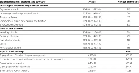
ChIP-on-chip analysis revealed that a total of 2,764 probes (corresponding to 2,544 unique genes) were significantly enriched in RORA-immunoprecipitated chromatin rela-tive to IgG-immunoprecipitated chromatin(P<0.05; FDR <7%). The complete list of gene-associated regions from the ChIP-on-chip analyses is shown in Additional file 4. Gene ontology analysis of this complete list of genes using DAVID Bioinformatics Resources 6.7 shows that neuron differentiation and development as well as axonogenesis are significantly over-represented among biological pro-cesses shown in Annotation cluster 1 which has a highly significant Enrichment Score (ES) of 5.107 (Table 1). The dataset is also significantly enriched for genes involved in synaptic transmission and plasticity (Annotation cluster 4 with enrichment score 3.456) and post-synaptic density (Annotation cluster 5 with enrichment score 3.176). Fur-thermore, it is remarkable thatallof genes in annotation clusters 1 to 5 are contained in AutDB and/or AutismKB databases. (See Additional file 5 for a full list of genes and functions associated with annotation clusters 2 to 5.) This is noteworthy because, although this nuclear hormone re-ceptor is known to have pleiotropic functions in different tissues, neurological functions are clearly enriched among the putative transcriptional targets of RORA within the context of a human neuronal cell line.
To further mine the dataset of potential transcriptional targets for higher level biological functions, disorders, and canonical pathways, we conducted network predic-tion and funcpredic-tional analysis on the RORA-bound genes using Ingenuity Pathway Analysis (IPA) software. Table 2 summarizes the functional and pathway analysis which reveals that nervous system development and function, neurological disease, and axonal guidance signaling are among the top five most significantly over-represented biological functions, disorders, and canonical pathways, respectively, associated with the RORA-enriched gene dataset. More detailed investigation into the specific neurological functions associated with the genes showed significant enrichment of genes involved in development of the brain and nervous system, axonogenesis, cell-cell adhesion of neurons, long-term potentiation of granule cells, neuritogenesis, and development of the cerebellum (Table 3). The genes associated with neurological disor-ders/diseases were enriched for schizophrenia, Huntington’s disease, movement disorders, dyskinesia, and seizure dis-order, the latter three of which often present with the most
severe subtype of ASD, which we have found to be associ-ated with RORA deficiency [13]. Interestingly, a number of behaviors that are often disrupted or impaired in ASD, such as cognition, learning, circling behavior, emotional behavior, memory and spatial learning, are also significantly over-represented in the dataset of putative transcriptional targets of RORA. The complete list of genes associated with each of the biological functions, diseases/disorders, and behav-iors described in Table 3 are provided in Additional file 6.
To determine whether the ChIP-on-chip-identified putative targets of RORA were enriched in autism candi-date genes, hypergeometric distribution analyses were performed to calculate P values for over-representation of genes from two autism gene databases (AutDB and AutismKB) within the target gene list. The results of the hypergeometric distribution analyses (Table 4) indicate that autism candidate genes, identified through various other studies, are indeed enriched among our transcrip-tional targets.
Network prediction of selected transcriptional targets to assess relevance to autism spectrum disorder (ASD) We selected six potential gene targets, ITPR1, CYP19A1, A2BP1, HSD17B10, NLGN1, and NTRK2, for confirm-ation by independent ChIP-quantitative PCR and func-tional analyses based on the reasons given in Methods. The probe enrichment data and genomic locations for these genes are shown in Table 5. Functional analysis of these six genes using the Pathway Studio 7.0 network pre-diction program revealed an association of these genes with neurological disorders, including autism and ataxia, as well as autism-related neurological functions, including synaptogenesis, synaptic transmission, long-term potenti-ation, learning, and memory (Figure 1). Interestingly, other autism candidate genes, includingNLGN3,NRXN1,RELN, and GABAA, were also included in the predicted gene network, thus supporting the investigation of our selected genes as ASD-relevant transcriptional targets of RORA.
Confirmation of RORA binding to selected transcriptional targets in human neuronal cells
Table 1 Top annotation cluster from gene ontology analysis of 2,544 potential transcriptional targets of RORA
Annotation cluster 1: enrichment score 5.107
GO term Count PValue Benjamini* Genes
GO:0030182-neuron differentiation
35 1.52E-08 3.56E-05 RAB3A, CDK5R1, ADORA2A, PAX3, RORA, RTN1, EPHB2, ARX, ATP2B2, BDNF, SLC1A3, LAMB2, CD44, CXCR4, ANK3, DMD, ROBO3, LHX8, LMX1B, MDGA2, PTPRR, NTNG1, RPGRIP1, NUMBL, SOD2, PTPN11, CTNNA2, SLITRK1, NTRK1, NTRK2, FOXG1, CNTN4, CACNA1F, CUX1, NTM
GO:0048666-neuron development
28 2.96E-07 3.48E-04 RAB3A, CDK5R1, ADORA2A, EPHB2, ARX, ATP2B2, BDNF, SLC1A3, LAMB2, CD44, ANK3, CXCR4, DMD, ROBO3, LHX8, LMX1B, NTNG1, RPGRIP1, CTNNA2, PTPN11, SOD2, NUMBL, SLITRK1, FOXG1, NTRK2, CNTN4, CACNA1F, NTM
GO:0000904-cell morphogenesis involved in differentiation
22 1.86E-06 1.45E-03 RAB3A, CDK5R1, NTNG1, PTPN11, CTNNA2, EPHB2, NUMBL, ARX, SLITRK1, ATP2B2, DAB2, BDNF, SLC1A3, LAMB2, CXCR4, ANK3, LAMA5, FOXG1, CNTN4, ROBO3, CACNA1F, FN1
GO:0000902-cell morphogenesis 27 2.53E-06 1.48E-03 RAB3A, CDK5R1, ADORA2A, EPHB2, ARX, ATP2B2, BDNF, DAB2, SLC1A3, LAMB2, ANK3, CXCR4, DMD, MKKS, ROBO3, FN1, NTNG1, MARK2, CTNNA2, PTPN11, NUMBL, SLITRK1, LAMA5, FOXG1, CNTN4, CACNA1F, CDC42BPB
GO:0032989-cellular component morphogenesis
28 6.15E-06 2.88E-03 RAB3A, CDK5R1, ADORA2A, EPHB2, ARX, ATP2B2, DAB2, BDNF, SLC1A3, LAMB2, ANK3, CXCR4, DMD, OBSL1, MKKS, ROBO3, FN1, NTNG1, MARK2, CTNNA2, PTPN11, NUMBL, SLITRK1, LAMA5, FOXG1, CNTN4, CACNA1F, CDC42BPB
GO:0048858-cell projection morphogenesis
21 7.30E-06 2.14E-03 RAB3A, CDK5R1, ADORA2A, NTNG1, PTPN11, CTNNA2, EPHB2, NUMBL, ARX, SLITRK1, BDNF, LAMB2, CXCR4, ANK3, LAMA5, DMD, FOXG1, MKKS, CNTN4, ROBO3, CACNA1F
GO:0048667-cell morphogenesis involved in neuron differentiation
19 1.01E-05 2.63E-03 RAB3A, CDK5R1, NTNG1, PTPN11, CTNNA2, EPHB2, NUMBL, ARX, SLITRK1, ATP2B2, BDNF, SLC1A3, LAMB2, CXCR4, ANK3, FOXG1, CNTN4, ROBO3, CACNA1F
GO:0048812-neuron projection morphogenesis
19 1.31E-05 3.07E-03 RAB3A, CDK5R1, ADORA2A, NTNG1, PTPN11, CTNNA2, EPHB2, NUMBL, ARX, SLITRK1, BDNF, LAMB2, CXCR4, ANK3, DMD, FOXG1, CNTN4, ROBO3, CACNA1F
GO:0030030-cell projection organization
26 1.39E-05 2.96E-03 MTSS1, RAB3A, CDK5R1, DNAH9, ADORA2A, EPHB2, ARX, ATP2B2, BDNF, LAMB2, CD44, ANK3, CXCR4, DMD, MKKS, ROBO3, FGD3, NTNG1, CTNNA2, PTPN11, NUMBL, SLITRK1, LAMA5, FOXG1, CNTN4, CACNA1F GO:0032990-cell part
morphogenesis
21 1.40E-05 2.73E-03 RAB3A, CDK5R1, ADORA2A, NTNG1, PTPN11, CTNNA2, EPHB2, NUMBL, ARX, SLITRK1, BDNF, LAMB2, CXCR4, ANK3, LAMA5, DMD, FOXG1, MKKS, CNTN4, ROBO3, CACNA1F
GO:0031175-neuron projection development
20 4.65E-05 6.80E-03 RAB3A, CDK5R1, ADORA2A, NTNG1, PTPN11, CTNNA2, EPHB2, NUMBL, ARX, SLITRK1, BDNF, LAMB2, CD44, CXCR4, ANK3, DMD, FOXG1, CNTN4, ROBO3, CACNA1F
GO:0007409-axonogenesis 17 4.98E-05 6.86E-03 RAB3A, CDK5R1, NTNG1, PTPN11, CTNNA2, EPHB2, NUMBL, ARX, SLITRK1, BDNF, LAMB2, CXCR4, ANK3, FOXG1, CNTN4, ROBO3, CACNA1F
GO:0007411-axon guidance 9 7.12E-03 2.67E-01 ARX, CDK5R1, BDNF, CXCR4, ANK3, FOXG1, CNTN4, ROBO3, EPHB2
Results were obtained using DAVID Bioinformatics Resources 6.7. The enrichment score is the negative logarithm of the average ofPvalues for items within each cluster. *Pvalues corrected for multiple testing.
Sarachana
and
Hu
Molecular
Autism
2013,
4
:14
Page
7
o
f
1
9
http://ww
w.moleculara
utism.com/c
ontent/4/1
RORAsuppression reduces expression of selected transcriptional targets in human neuronal cells
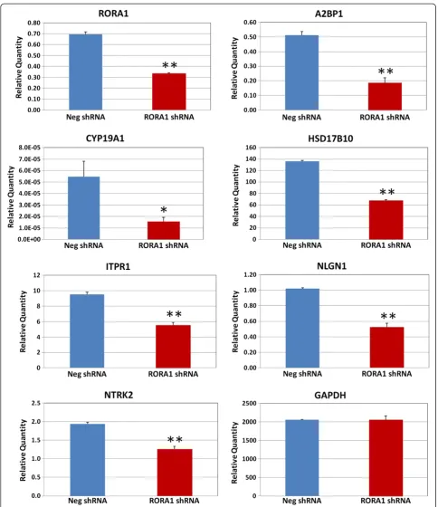
To further validate that RORA regulates expression of A2BP1, CYP19A1, HSD17B10, ITPR1, NLGN1, and NTRK2, and to examine whether reduction of RORA expression leads to reduced expression of these selected RORA transcriptional targets, shRNA-mediated knock-down of RORA was performed in SH-SY5Y cells using RORA1 shRNA, and quantitative RT-PCR analyses were conducted to measure expression of RORA1 and the potential targets. Expression ofRORA1in SH-SY5Y cells was significantly down-regulated by approximately 50% in comparison with cells transfected with negative control shRNA, indicating that shRNA-mediated knockdown of RORA1 was successful. Notably, expression of all selected RORA transcriptional targets was significantly reduced (Figure 3), indicating that expression of these genes is regulated by RORA and that these genes indeed are RORA transcriptional targets in human neuronal cells. As a negative control, the expression level ofGAPDH, which is not a transcriptional target of RORA, is unaffected by transfection of the SH-SY5Y cells with RORA1 shRNA.
Expression of transcriptional targets of RORA is relatively reduced in the frontal cortex of individuals with autism in comparison with age-matched controls
We have previously reported reduction of RORA tran-script and/or protein in four independent cohorts using
LCL as well as tissues from the prefrontal cortex and the cerebellum of individuals diagnosed with autism [11-13]. To examine whether RORA reduction in the brain of in-dividuals with autism may be associated with aberrant expression of the transcriptional targets identified in this study, a pilot study involving quantitative RT-PCR ana-lysis of frozen postmortem prefrontal cortex tissues from individuals with autism (n = 3) and age-matched con-trols (n = 3) was performed. As shown in Table 6, the average expression of each of the six gene targets in the combined cases is reduced relative to the average ex-pression levels of the respective genes in the combined controls, although thePvalues (allP>0.05) indicate that the differences are not statistically significant.
Discussion
Based on reduced expression of RORA in LCL and in postmortem brain tissues of individuals with ASD versus unaffected controls, coupled with its known functions in cerebellar development and neuroprotection against in-flammation and oxidative stress in mice, we postulated that this nuclear hormone receptor may be responsible for at least some of the pathobiology associated with ASD. In particular, our recent finding that RORA speci-fically binds the promoter region of aromatase, whose protein expression in the human brain is highly corre-lated with that of RORA, suggests a molecular explan-ation for the increased levels of testosterone observed in
Table 2 Top five biological functions, disorders, and canonical pathways associated with 2,544 potential transcriptional targets of RORA
Biological functions, disorders, and pathways Pvalue Number of molecules Physiological system development and function
Organismal survival 3.56E-08 to 6.82E-04 305
Nervous system development and function 5.83E-08 to 4.28E-03 344
Tissue morphology 5.83E-08 to 4.72E-03 419
Cardiovascular system development and function 8.89E-08 to 4.72E-03 268
Embryonic development 9.01E-08 to 4.72E-03 370
Diseases and disorders
Hereditary disorder 8.09E-06 to 1.56E-03 204
Neurological disease 8.09E-06 to 4.72E-03 292
Psychological disorders 8.09E-06 to 8.09E-06 107
Cancer 1.65E-05 to 4.77E-03 731
Hematological disease 1.65E-05 to 4.67E-03 166
Top canonical pathways Ratio
Superpathway of inositol phosphate compounds 4.97E-04 37/227
Production of nitric oxide and reactive oxygen species in macrophages 1.29E-03 35/210
Axonal guidance signaling 2.47E-03 69/468
3-phosphoinositide biosynthesis 2.52E-03 29/175
Small cell lung cancer signaling 2.60E-03 17/89
some individuals with ASD [12]. Specifically, we anticipate that downregulation of aromatase would lead to a buildup of its substrate, testosterone, with a corresponding reduc-tion in estrogen synthesis. Moreover, the downregulareduc-tion of RORA expression in response to androgen and upre-gulation in response to estrogen suggest a mechanism for the introduction of sex bias in ASD.
As a nuclear hormone receptor, RORA, in combination with various co-regulator proteins, can potentially regulate the transcription of a large number of gene targets. This study was therefore conducted in order to identify add-itional transcriptional targets of RORA, specifically within the context of a human neuronal cell model. To our know-ledge, this is the first ChIP-on-chip study directed towards the comprehensive identification of transcriptional targets of RORA at the genome-wide level in any species. Conse-quently, in this pilot study aimed at discovery of genes that may be regulated by RORA, we chose to use less stringent P values (P ≤0.05; FDR <7%) for identification of RORA-enriched regions in order to capture as many potential gene targets of RORA as possible. Not surprisingly, our ChIP-on-chip analysis identified 2,764 promoter regions
Table 3 Top neurological diseases, disorders, and behaviors associated with the 2,544 potential transcriptional targets of RORA
Function annotation (number of molecules) Pvalue Nervous system development and function
Quantity of neurons (67) 5.83E-08
Development of brain (102) 9.01E-08
Morphology of nervous system (161) 3.96E-07
Morphology of nervous tissue (118) 5.44E-07
Development of central nervous system (122) 1.50E-06
Axonogenesis (43) 1.68E-05
Morphology of nerves (30) 7.32E-05
Cell-cell adhesion of neurons (6) 1.48E-04
Morphology of central nervous system (98) 1.90E-04
Morphology of rhombencephalon (35) 2.41E-04
Quantity of sensory neurons (19) 2.81E-04
Development of nerves (19) 2.81E-04
Abnormal morphology of neurons (51) 2.85E-04
Morphology of neurons (55) 3.37E-04
Antinociception (13) 3.84E-04
Development of neurons (37) 3.98E-04
Morphology of brain (88) 4.40E-04
Development of interneurons (7) 4.56E-04
Migration of neurons (46) 4.79E-04
Abnormal morphology of cerebellum (21) 5.58E-04
Development of metencephalon (24) 5.88E-04
Loss of neurons (29) 8.01E-04
Long-term potentiation of granule cells (4) 8.18E-04
Abnormal morphology of cranial nerve (19) 8.60E-04
Neuritogenesis (83) 1.11E-03
Development of cerebellum (23) 1.22E-03
Development of forebrain (45) 1.26E-03
Fasciculation of nervous tissue (9) 1.34E-03
Abnormal morphology of granule cells (12) 1.42E-03
Development of globus pallidus (3) 1.56E-03
Activation of hippocampus (3) 1.56E-03
Abnormal morphology of hair cells (9) 1.84E-03
Morphogenesis of neurites (58) 1.85E-03
Adhesion of neuronal cells (14) 1.88E-03
Abnormal morphology of outer hair cells (6) 2.01E-03
Sensory system development (23) 2.09E-03
Development of rhombencephalon (28) 2.15E-03
Chemotaxis of granule cells (4) 2.23E-03
Guidance of axons (31) 2.24E-03
Morphology of mechanosensory neurons (10) 2.49E-03
Cell viability of neurons (39) 2.51E-03
Analgesia (18) 2.62E-03
Synaptic transmission of cerebral cortex cells (11) 3.03E-03
Memory (39) 3.52E-03
Table 3 Top neurological diseases, disorders, and behaviors associated with the 2,544 potential transcriptional targets of RORA(Continued)
Neurotransmission (71) 3.75E-03
Development of cranial nerve (11) 3.79E-03
Cell viability of granule cells (8) 4.28E-03
Neurological disease
Schizophrenia (107) 8.09E-06
Chorea (117) 3.23E-04
Huntington's Disease (116) 4.09E-04
Movement Disorders (183) 9.11E-04
Dyskinesia (118) 1.27E-03
Neurological signs (120) 1.45E-03
Familial transthyretin amyloidosis (3) 1.56E-03
Seizure disorder (60) 3.92E-03
Disorder of basal ganglia (135) 4.31E-03
Polymicrogyria (4) 4.72E-03
Behavior
Behavior (180) 2.74E-07
Cognition (79) 1.70E-05
Learning (74) 3.41E-05
Circling behavior (12) 1.11E-03
Emotional behavior (42) 3.04E-03
Memory (39) 3.52E-03
Spatial learning (33) 4.23E-03
These data were obtained using Ingenuity Pathway Analysis software.Pvalues were calculated using Fisher's exact test, which was performed using the entire set of genes within the Ingenuity Knowledge Base as the reference set.
Sarachana and HuMolecular Autism2013,4:14 Page 9 of 19
enriched for RORA binding sites, which corresponded to 2,544 unique genes. Interestingly, gene ontology analysis of this complete set of putative target genes revealed a strik-ingly high enrichment in genes associated with neuron differentiation and development, neuron projection mor-phogenesis, axonogenesis, and axon guidance in the top functional annotation cluster (enrichment score 5.107). In-triguingly,allof the genes associated with these processes are listed in the AutDB (SFARI Gene) and/or AutismKB databases of autism-associated genes, indicating relevance of our target gene set to ASD. Two additional highly significant functional annotation categories of genes are re-lated to synaptic transmission and plasticity (enrichment score 3.456) and postsynaptic density (enrichment score 3.176). Again, all of the genes in both annotation categories are contained within one of the two aforementioned data-bases. The statistically significant enrichment of autism candidate genes within our dataset of genes identified by ChIP-on-chip analysis was also confirmed by hypergeo-metric distribution analyses which used the number of genes in either or both of the autism databases as the total number of interesting markers and the overlap between the genes in our dataset (that is, the selected markers) and the genes in either database as the number of selected
interesting markers. The total number of annotated genes on the microarray represents the total number of general markers (that is, the population).
Biological network and pathway analysis using Ingenu-ity Pathway Analysis software further revealed that these potential transcriptional targets of RORA are signifi-cantly associated with nervous system development and function including development of cortex and cerebel-lum, axonogenesis, adhesion of neuronal cells, neuronal migration, neuritogenesis, neurotransmission, and syn-aptic density, all of which have been associated with autism [45-50]. It is also noteworthy that neurological disorders known to be co-morbid with autism, including schizophrenia [51,52], movement disorder [53-57], and seizure disorder [58-61], were also over-represented among the genes in our dataset. Of particular relevance to behavioral deficits in ASD, genes related to cognition, learning, repetitive behaviors (for example, circling), memory, and spatial learning are also associated with these potential targets of RORA.
Among the selected targets of RORA investigated in this study, we confirmed ITPR1 and CYP19A1 as tran-scriptional targets in human neuronal cells by both ChIP-quantitative PCR and functional knockdown of
Table 4 Hypergeometric distribution analysis results to determine enrichment of autism candidate genes from AutDB and AutismKB
Autism gene database Total number of genes in database
Total promoter regions on array
Total IP RORA-binding promoter regions
RORA-binding autism genes (overlap) P
value
AutDB (SFARI gene) 328 25,500 2,544 49 0.0028
AutismKB (syndromic + non-syndromic) 3,050 25,500 2,544 426 <0.001
AutDB + AutismKB 3,158 25,500 2,544 438 <0.001
For the hypergeometric distribution analyses, the total number of general markers (that is, the population) is the number of genes (25,500) whose promoter regions are represented on the array, and the total number of selected markers is the number of unique genes (2,544) identified as targets of RORA by the ChIP-on-chip analysis. The total interesting markers are the number of autism candidate genes in the respective or combined databases, and the number of selected interesting markers are the genes within our dataset that overlap with those in either or the combined databases.
Table 5 Enrichment data and genomic location of the RORA-binding enriched regions closest to the selected genes
Nearest gene Entrez gene name MAT score on T (R vs. G)
Pvalue (region)
FDR (%)
Average intensity fold-change
(R vs. G)
Chromosome (strand)
Region start
Length (bps)
Number of probes in
region
ITPR1 Inositol 1,4,5-triphosphate receptor, type 1
17.19 0.005 <5 1.29 chr3 (+) 4,724,792 2,804 71
HSD17B10 Hydroxysteroid (17-β) dehydrogenase 10
14.11 0.007 <5 11.86 chrX (−) 53,483,92 1,723 42
A2BP1 (RBFOX1) RNA binding protein, fox-1 homolog
12.86 0.009 <5 1.29 chr16 (+) 7,315,418 2,636 68
NLGN1 Neuroligin 1 7.30 0.036 <5 3.27 chr3 (+) 174,777,672 639 18
CYP19A1 (aromatase)
Cytochrome P450, family 19, subfamily A, polypeptide 1
7.02 0.040 <5 1.61 chr15 (−) 49,415,636 2,111 54
NTRK2 Neurotrophic tyrosine kinase, receptor, type 2
6.72 0.044 <7 2.17 chr9 (+) 86,412,498 966 21
RORA with shRNA. As mentioned earlier, both genes had been implicated as targets of RORA by earlier studies with Rora-deficient mice [29] as well as in hu-man breast cancer [36]. Our current study, however, also reveals four novel transcriptional targets of RORA, thus expanding the repertoire of genes and pathways that may be impacted by RORA deficiency in humans. These novel target genes are A2BP1 (RBFOX1), HSD17B10, NLGN1, and NTRK2. Furthermore, our pilot study exa-mining the expression of these six transcriptional targets of RORA in postmortem brain tissues from individuals with ASD and that of age-matched controls shows an average reduction of all targets in the autism samples, al-though the differences between combined cases and combined controls were not statistically significant, all exhibiting P >0.05. Surprisingly, the least significant differences in average expression levels were observed forRORAandCYP19A1. Inasmuch as we had previously detected significant differences (P <0.05) between the protein expression levels of both RORA and CYP19A1 in the frontal cortex of 22 cases versus 12 controls by confocal immunofluorescence analyses [12], we suggest that the lack of statistical significance for reduced expression of these two transcripts as well as the other targets in this study is probably due to the limitation in sample size (n= 3 for each group) coupled with the nat-ural variability of gene expression among the individuals.
Nevertheless, the data from this limited sampling of cases and controls suggest a trend towards reduced ex-pression of transcriptional targets in autism brain tissues exhibiting reduced RORA. As discussed below, an inde-pendent review of the literature revealed that these six RORA targets were reliably associated with autism.
Relevance ofA2BP1, CYP19A1, HSD17B10, ITPR1, NLGN1, andNTRK2to the pathobiology of autism
A2BP1 (ataxin 2-binding protein 1), also known as RBFOX1, is an RNA-binding protein that regulates neuron-specific alternative splicing [62]. Several studies, including genetic and gene expression analyses, have established the link between this gene and autism. Using fluorescent in situ hybridization and quantitative PCR analyses, Martin and colleagues found a cryptic deletion of theA2BP1gene in a female with autism, epilepsy, and global development delay [33]. Reduction of the A2BP1 transcript level was also observed in the lymphocytes of this individual, suggesting that the deletion causes aber-rant expression of this gene in this autism case. By geno-typing 27 SNPs across this gene in 206 parent–child trios, they identified two regions exhibiting a nominal association with autism. Moreover, a recent noise-reduction genome-wide association study (GWAS) of two autism GWAS datasets (with 597 and 696 families) from the Autism Genetic Resource Exchange revealed Figure 1Pathway analysis of selected potential transcriptional targets of RORA.A biological network was created using the Pathway Studio 7.0 program to identify biological functions and disorders associated with potential RORA transcriptional targets selected for confirmation (that is,ITPR1,CYP19A1,A2BP1,HSD17B10,NLGN1, andNTRK2which are highlighted with a blue halo).
Sarachana and HuMolecular Autism2013,4:14 Page 11 of 19
1,535 significant linkage disequilibrium blocks overlapping 431 genes [63]. Interestingly, regions in the A2BP1 gene were identified among the most significant linkage dis-equilibrium blocks (P= 3.6×10⁻⁵). Furthermore, Voineagu and colleagues also conducted transcriptomic analysis of postmortem frontal and temporal cortex tissues from 19 individuals with autism and 17 controls using microarrays [64]. They found thatA2BP1expression was significantly reduced in both frontal and temporal cortex tissues from individuals with autism relative to controls. Using high-throughput RNA sequencing and semi-quantitative RT-PCR analyses, they also demonstrated that splicing of A2BP1-dependent alternative exons in the brain of indi-viduals with autism was dysregulated [64], suggesting that aberrant expression of A2BP1 results in dysregulation of alternative splicing in autism. Findings from several copy number variation (CNV) studies [65-70] and linkage
studies [71-75] have also reported that A2BP1 is associ-ated with autism. Aside from its association with ASD, re-cent studies on the physiological function of A2BP1/ RBFOX1 demonstrate its involvement in synaptic trans-mission and neuronal excitation [76] as well as its role in the regulation of transcriptional networks involved in neuronal development [77].
CYP19A1 encodes aromatase, a key enzyme that
converts androstenedione to estrone and testosterone to estradiol. Although its neuronal function is most often associated with the regulation of reproductive behaviors through the sex hormones, more recent studies have revealed unexpected functions of aromatase in the brain, including neurogenesis, neuronal differentiation, synap-tic activity and plassynap-ticity, and protection against oxida-tive stress [50,78-82]. Another interesting observation is the colocalization of aromatase with oxytocin in several Figure 2Chromatin immunoprecipitation-quantitative PCR of RORA transcriptional targets.Chromatin immunoprecipitation followed by quantitative PCR analysis was conducted to determine whether RORA protein binds toA2BP1,CYP19A1,HSD17B10,ITPR1,NLGN1, andNTRK2
Figure 3Quantitative RT-PCR analysis of RORA short hairpin RNA-transfected SH-SY5Y cells.SH-SY5Y cells were transfected with RORA1 short hairpin RNA (shRNA) or negative control shRNA for 24 hours and quantitative RT-PCR analysis (n= 3) was performed to determine expression ofRORA1and potential transcriptional targets in the transfected cells.GAPDHwas included as a negative control. Error bars indicate standard error of the mean. *P<0.05, **P <0.01.
Sarachana and HuMolecular Autism2013,4:14 Page 13 of 19
regions of the rat brain, including the periventricular nu-cleus of the hypothalamus and the zona incerta, which suggests a role for this gene in oxytocinergic neurons in the limbic system [83], which may have implications for social cognition.CYP19A1has also been associated with autism by genetic studies. Allen-Brady and colleagues conducted genome-wide screening of 70 families (192 individuals with autism and 461 unaffected relatives) and identified three regions shown to be highly signifi-cant in the linkage analysis [84]. Interestingly, one of the three regions is 15q21.1-15q22.2 (heterogeneity loga-rithm (base 10) of the odds = 5.31) where theCYP19A1 gene is located. Moreover, Chakrabarti and colleagues studied SNPs in 68 candidate genes for autism to identify common genetic variations associated with autistic traits and Asperger syndrome, a high-functioning subgroup of autism. A case–control association analysis of individuals with Asperger syndrome (n= 174) revealed regions in 14 genes, includingCYP19A1, that showed a nominally sig-nificant association with Asperger syndrome [85].
HSD17B10 encodes 3-hydroxyacyl-CoA dehydrogenase type-2, a mitochondrial enzyme involved in mitochondrial integrity and oxidation of fatty acids and steroids. As men-tioned earlier, like CYP19A1, HSD17B10 is involved in the conversion of androgens to estradiol via androstenedione and estrone intermediates. A reduction in HSD17B10 would thus be expected to lead to increased androgen and reduced estrogen, either of which may have serious conse-quences for brain development [86]. Indeed, reduced expression of HSD17B10 causes X-linked mental retard-ation, choreoathetosis, language impairment, and abnormal
behavior [87-89]. These associated disorders are particu-larly interesting since we have identified RORA deficiency (specifically, reduced expression) in the phenotypic sub-type of ASD associated with severe language impairment [13]. Using chromosome microarray analysis, Edens and colleagues reported duplications involving Xp11.22-p11.23, a region whereHSD17B10gene is located, in two females with autism and epilepsy [90].
ITPR1 encodes inositol 1,4,5-triphosphate receptor type 1, a ligand-gated ion channel that is activated by cytosolic calcium and inositol triphosphate, and that is involved in synaptogenesis and formation of dendritic contacts [91]. Computational modeling also suggests that the biochemical and biophysical properties of ITPR1 coupled with its high density and lower sensitivity to IP3 in cerebellar Purkinje cells are critical to its postulated role in long-term depression [92]. With respect to ASD, transcriptomic analysis of frontal and temporal cortex tissues from 16 individuals with autism and 16 controls revealed down-regulation ofITPR1[64]. Bremer and col-leagues used high-resolution whole genome array-based comparative genomic hybridization to screen 223 indi-viduals with autism to identify gene dose alterations as-sociated with autism susceptibility. They found regions in chromosome 3p25.3, where ITPR1is located, signifi-cantly enriched in individuals with autism [93]. Another CNV study of 1,124 families, each of which included a single proband, his/her unaffected parents and, in most cases, an unaffected sibling, revealed as many as 234 CNV regions in several genes, includingITPR1, to be as-sociated with autism [69]. Moreover, Levy and colleagues
Table 6 Quantitative RT-PCR analysis of post-mortem brain tissues from individuals with autism
Sample comparison (A vs. C)
Age (A, C) RORA CYP19A1 ITPR1 A2BP1
Control Autistic A/C Control Autistic A/C Control Autistic A/C Control Autistic A/C
5144A vs. 5251C 20, 19 6.4 2.0 0.316 1.7E-04 5.2E-05 0.307 127.7 70.8 0.554 553.1 105.3 0.190
5173A vs. 5873C 30, 28 38.4 25.4 0.661 1.2E-02 5.9E-03 0.487 246.4 27.3 0.111 830.1 74.2 0.089
6337A vs. 5718C 22, 22 7.9 0.4 0.046 7.7E-06 1.0E-07 0.013 217.4 176.3 0.811 246.0 64.6 0.263
Average ratio (A/C) 17.6 9.3 0.527 4.1E-03 2.0E-03 0.484 197.2 91.5 0.464 543.1 81.4 0.150
Pvalue (unpairedttest) 0.562 0.660 0.137 0.052
HSD17B10 NLGN1 NTRK2
Control Autistic A/C Control Autistic A/C Control Autistic A/C
5144A vs. 5251C 20, 19 128.7 15.7 0.122 178.3 23.8 0.133 3218.9 544.6 0.169
5173A vs. 5873C 30, 28 126.3 41.1 0.325 255.3 44.9 0.176 2600.1 1077.2 0.414
6337A vs. 5718C 22, 22 47.6 39.2 0.824 84.8 61.5 0.725 929.8 1265.1 1.361
Average ratio (A/C) 100.9 32.0 0.317 172.8 43.4 0.251 2249.6 962.3 0.428
Pvalue (unpairedttest) 0.069 0.062 0.147
Gene expression in brain tissues from individuals with autism (indicated by’A’) was compared with that in controls (indicated by’C’). The numbers preceding‘A’
studied genomic copy-number variation in a large co-hort of families with a single affected child and at least one unaffected sibling. Among significant regions identi-fied to be de novo CNVs were regions containing the ITPR1gene [94].
NLGN1 encodes neuroligin-1, a neuronal cell surface adhesion molecule that interacts with neurexin mole-cules. NLGN1 is involved in formation and remodeling of synapses in the central nervous system [95]. In addition to synaptogenesis, a recent study has also dem-onstrated a role for NLGN1, acting together with NRXN1β, in neuritogenesis and the formation of neu-ronal circuits upon activation of the fibroblast growth factor receptor-1 [96]. In the context of ASD, Glessner and colleagues conducted a whole-genome CNV analysis of 859 individuals with autism and 1,409 typically deve-loping children of European ancestry who were geno-typed with ~550,000 SNP markers to comprehensively identify CNVs conferring susceptibility to autism [97]. Positive findings were evaluated in an independent co-hort of 1,336 individuals with autism and 1,110 controls of European ancestry. Interestingly, a CNV region in the NLGN1 gene was significantly enriched in individuals with autism. Moreover, a family-based association ana-lysis of 100 families with autism found a modest signi-ficant association at a region in NLGN1 gene [98]. A
high-density SNP, genome-wide linkage study also showed that the chromosomal region 3q26.31-q27.3 was signifi-cantly associated with autism [99]. Deletion of this gene was also reported to cause impaired spatial memory and increased repetitive behavior [100].
NTRK2encodes neurotrophic tyrosine kinase receptor type 2 (also known as TRKB) that is a catalytic receptor for several neurotrophins, including BDNF, NT4, and NT3. NTRK2 is involved in axon guidance signaling as well as synapse formation and plasticity [101-104]. Its role in ASD is implicated by genetic association analysis of 146 children with autism and 50 typically developing controls which identified six SNPs and multiple haplo-types in theNTRK2 gene [105]. This study also showed that BDNF levels were significantly increased in children with autism relative to that in controls, supporting a role of NTRK2/BDNF signaling as a susceptibility factor for the disorder.
Study limitations and future directions
Because little is known with regard to transcriptional targets of RORA, particularly in human neuronal cells, this ChIP-on-chip analysis was undertaken as a discov-ery tool to screen for potential transcriptional targets at the genome-wide level. As a result, we used a less strin-gent P-value of 0.05 as a filter to select probes enriched
Figure 4Possible downstream consequences of deregulation of the six confirmed transcriptional targets of RORA.This figure summarizes findings from this study. ChIP, chromatin immunoprecipitation; qPCR, quantitative PCR.
Sarachana and HuMolecular Autism2013,4:14 Page 15 of 19
for RORA binding without applying correction for mul-tiple testing (which is not part of the standard Partek analytical workflow for tiling arrays). However, all of the regions identified using P ≤0.05 are retained in the dataset even after imposing FDR ≤7%. Thus, although the list of 2,544 genes that are identified as putative targets of RORA should be interpreted with caution pending independent validation, we suggest that this list may be used as a basis for hypothesis generation with re-gard to the genes, pathways and functions that may be impacted by RORA deficiency. Another caveat is that the list of RORA targets does not replicate all of the sev-eral targets identified in the mouse cerebellum by ChIP-quantitative PCR analyses [29] other thanITPR1. We do not know the precise reason for this difference, but it is possible that the target selection may be a function of the specific tissues and cell types as well as species, since the mouse targets were identified in primary cerebellar cells, and the human targets were identified in the SH-SY5Y human neuroblastoma cell line. Indeed, co-regulator availability is also tissue-specific, and transcrip-tional regulation by RORA requires association with gene-specific and tissue-specific co-regulator proteins, thus adding another layer of complexity to target selection.
Another potential complication is the possibility that target selection may be directed by circadian rhythm, as RORA is a regulator of the circadian cycle. Thus, it would be interesting to conduct ChIP-on-chip analysis on cells synchronized according to stage in the circadian cycle. Finally, as noted earlier, although the average expression levels of the RORA targets were reduced in the frontal cortex of individuals with ASD relative to that of controls as expected, the differences were not statistically signifi-cant. Notably, the protein expression levels of the genes exhibiting the least significant expression differences (that is, highest P values) in this study, RORA and CYP19A1, were found to be significantly different in our previous study in which 12 autism tissue samples were compared with 22 controls [12], thus emphasizing the need for an expanded investigation of RORA targets involving add-itional brain tissues from individuals with ASD and con-trols, including samples from different brain regions.
In summary, using a high-throughput genome-wide method for identifying potential transcriptional targets of RORA followed by independent validation of six targets with functional relevance to ASD, we have demonstrated that RORA deficiency can lead to a cascade of transcrip-tional deregulation that impacts a number of genes known to be associated with ASD. Figure 4 summarizes the prin-cipal findings from this study.
Conclusions
Using ChIP-on-chip analyses, we identify at the genome-wide level over 2,500 potential transcriptional targets of
RORA, a nuclear hormone receptor, and confirm six of these targets (four of them novel with respect to regula-tion by RORA) in human neuronal cells by ChIP-quantitative PCR and functional analyses. Moreover, we demonstrate that the transcript levels of these target genes are reduced in the postmortem frontal cortex of autistic males who exhibit reduced RORA expression relative to that of control males, further implicating RORA as an upstream regulator of these genes in hu-man brain. The significance of these findings is the like-lihood that any mechanism that contributes to reduced RORA expression, including elevated levels of male hor-mones [12] and epigenetic modifications that may be the result of as yet unknown environmental factors [11], may lead to increased risk for autism based on the aber-rant transcription of downstream targets of RORA, which we show are significantly enriched for autism can-didate genes based on hypergeometric distribution ana-lyses of our dataset in comparison with established databases of autism genes.
Additional files
Additional file 1:A table presenting a list of frozen human brain tissue samples from the Autism Tissue Program.
Additional file 2:A table presenting a list of antibodies and shRNAs.
Additional file 3:A table presenting a list of primers for ChIP-quantitative PCR, and ChIP-quantitative RT- PCR.
Additional file 4:A table presenting a list of potential
transcriptional targets of RORA identified by ChIP-on-chip analysis.
Additional file 5:A table presenting annotation clusters 2 to 5 resulting from gene ontology analysis of the ChIP-on-chip gene dataset using DAVID Bioinformatics Resources 6.7.
Additional file 6:A table presenting a complete list of genes associated with neurological functions, disorders, and behaviors identified by Ingenuity Pathway Analysis as significantly over-represented in ChIP-on-chip dataset of RORA-binding target promoters.
Abbreviations
A2BP1:Ataxin 2 binding protein 1; Acsl4: Acyl-CoA synthetase long-chain family member 4; Akt2: V-akt murine thymoma viral oncogene homolog 2; ASD: Autism spectrum disorder; BA: Brodmann area; BDNF: brain-derived neurotrophic factor; bp: base pairs; Calb1: Calbindin 1, 28kDa; Cals: Callose synthase; Cd36: Trombospondin receptor; ChIP: chromatin
immunoprecipitation; ChIP-on-chip: chromatin immunoprecipitation followed by microarray analysis; Cidea: Cell death-inducing DFFA-like effector a; Cidec: Cell death-inducing DFFA-like effector c; CNV: Copy number variant (s); CYP19A1: Cytochrome P450, family 19, subfamily A, polypeptide 1; FDR: False discovery rate; GABA: Gamma aminobutyric acid; Grm1: Glutamate receptor, metabotropic 1; Hif1a: Hypoxia inducible factor 1, alpha subunit; HSD17B10: Hydroxysteroid (17-β) dehydrogenase 10; IPA: Ingenuity Pathway Analysis; ITPR1: Inositol 1,4,5-trisphosphate receptor, type 1; LCL: Lymphoblastoid cell line; LXRa: Liver X receptor alpha; MEM: modified Eagle’s medium; Mogat1: Monoacylglycerol O-acyltransferase 1; NLGN1: Neuroligin 1; NLGN3: Neuroligin 3; NRXN1: Neurexin 1;
homolog; RELN: Reelin; RORA: Retinoic acid receptor-related orphan receptor alpha; RORA1: Retinoic acid receptor-related orphan receptor alpha, isoform 1; RT: Reverse transcription; Shh: Sonic hedgehog; shRNA: Short hairpin RNA; SH-SY5Y: Human neuroblastoma cell line; Slc1a6: Solute carrier protein 1, member 6; SNP: Single-nucleotide polymorphism; Srebp-1c: Sterol regulatory element-binding protein 1c.
Competing interests
The authors declare that they have no competing interests.
Authors' contributions
TS and VWH conceived of the study, contributed to the study design, performed the data analyses, and prepared the manuscript. TS conducted the experiments. All authors read and approved the final manuscript.
Acknowledgements
The authors wish to thank the families of the brain donors for their invaluable gift to autism research and the Autism Tissue Program (San Diego, CA, USA) for making frozen human brain tissues available for this work through the Harvard Brain Tissue Resource Center (Harvard University, Boston, MA, USA). They also thank Ms. Yi Hong for her assistance with the Affymetrix promoter array hybridization and analysis, and Ms. Jing Jing for help with RNA isolation. The authors are also grateful to Dr. Yinglei Lai (Department of Statistics at The George Washington University) for conducting the hypergeometric distribution analyses.
This study was supported in part by a generous gift from the LIFE Foundation (Aspen, CO, USA) and by an intramural grant from The George Washington University (Medical Faculty Associates award). Neither of the funding sources played any role in the study design, collection, analysis, and interpretation of data, writing of the manuscript, or decision to submit this study for publication. TS was a predoctoral student in the Institute for Biomedical Sciences at the George Washington University, who was supported by the Higher Educational Strategic Scholarship for Frontier Research Network (SFR scholarship) from the Office of the Commission on Higher Education of the Royal Thai Government, Thailand, through the Faculty of Allied Health Sciences, Chulalongkorn University.
Author details
1Department of Biochemistry and Molecular Medicine, The George Washington University School of Medicine and Health Sciences, 2300 I Street NW, Washington, DC 20037, USA.2Current address: Center for Biologics Evaluation and Research, Food and Drug Administration, 8800 Rockville Pike, Bethesda, MD 20892, USA.
Received: 12 February 2013 Accepted: 24 April 2013 Published: 22 May 2013
References
1. American Psychiatric Association:Task Force on DSM-IV: Diagnostic and statistical manual of mental disorders: DSM-IV-TR.4th edition. Washington, DC: American Psychiatric Association; 2000.
2. Moldin SO, Rubenstein JLR:Understanding autism: from basic neuroscience to treatment.CRC/Taylor & Francis: Boca Raton; 2006.
3. Volkmar F, Chawarska K, Klin A:Autism in infancy and early childhood.
Annu Rev Psychol2005,56:315–336.
4. Volkmar FR, Klin A, Siegel B, Szatmari P, Lord C, Campbell M, Freeman BJ, Cicchetti DV, Rutter M, Kline W:Field trial for autistic disorder in DSM-IV.
Am J Psychiatry1994,151:1361–1367.
5. Baio J:Prevalence of Autism Spectrum Disorders - Autism and Developmental Disabilities Monitoring Network, 14 Sites, United States, 2008.Centers for Disease Control and Prevention Morbidity and Mortality Weekly Report2012,61:3.
6. Kim YS, Leventhal BL, Koh Y, Fombonne E, Laska E, Lim E, Cheon K, Kim S, Kim Y, Lee H, Song D, Grinker RR:Prevalence of autism spectrum disorders in a total population sample.Am J Psychiatry2011,168:904–912. 7. Mattila M, Kielinen M, Linna S, Jussila K, Ebeling H, Bloigu R, Joseph RM,
Moilanen I:Autism spectrum disorders according to DSM-IV-TR and comparison with DSM-5 draft criteria: an epidemiological study.J Am Acad Child Adolesc Psychiatry2011,50:583–592.
8. Baron-Cohen S, Knickmeyer RC, Belmonte MK:Sex differences in the brain: implications for explaining autism.Science2005,310:819–823.
9. Auyeung B, Baron-Cohen S, Ashwin E, Knickmeyer R, Taylor K, Hackett G: Fetal testosterone and autistic traits.Br J Psychol2009,100(Pt 1):1–22. 10. Auyeung B, Taylor K, Hackett G, Baron-Cohen S:Foetal testosterone and
autistic traits in 18 to 24-month-old children.Mol Autism2010,1:11. 11. Nguyen A, Rauch TA, Pfeifer GP, Hu VW:Global methylation profiling of
lymphoblastoid cell lines reveals epigenetic contributions to autism spectrum disorders and a novel autism candidate gene, RORA, whose protein product is reduced in autistic brain.FASEB J2010,24:3036–3051. 12. Sarachana T, Xu M, Wu RC, Hu VW:Sex hormones in autism: androgens
and estrogens differentially and reciprocally regulate RORA, a novel candidate gene for autism.PLoS One2011,6:e17116.
13. Hu VW, Sarachana T, Kim KS, Nguyen A, Kulkarni S, Steinberg ME, Luu T, Lai Y, Lee NH:Gene expression profiling differentiates autism case-controls and phenotypic variants of autism spectrum disorders: evidence for circadian rhythm dysfunction in severe autism.Autism Res2009,2:78–97. 14. Boukhtouche F, Janmaat S, Vodjdani G, Gautheron V, Mallet J, Dusart I,
Mariani J:Retinoid-related orphan receptor alpha controls the early steps of Purkinje cell dendritic differentiation.J Neurosci2006,26:1531–1538. 15. Hadj-Sahraoui N, Frederic F, Zanjani H, Delhaye-Bouchaud N, Herrup K,
Mariani J:Progressive atrophy of cerebellar Purkinje cell dendrites during aging of the heterozygous staggerer mouse (Rora(+/sg)).Brain Res Dev Brain Res2001,126:201–209.
16. Gold DA, Gent PM, Hamilton BA:ROR alpha in genetic control of cerebellum development: 50 staggering years.Brain Res2007, 1140:19–25.
17. Harding HP, Atkins GB, Jaffe AB, Seo WJ, Lazar MA:Transcriptional activation and repression by RORalpha, an orphan nuclear receptor required for cerebellar development.Mol Endocrinol1997,11:1737–1746. 18. Boukhtouche F, Vodjdani G, Jarvis CI, Bakouche J, Staels B, Mallet J, Mariani
J, Lemaigre-Dubreuil Y, Brugg B:Human retinoic acid receptor-related orphan receptor alpha1 overexpression protects neurones against oxidative stress-induced apoptosis.J Neurochem2006,96:1778–1789. 19. Delerive P, Monte D, Dubois G, Trottein F, Fruchart-Najib J, Mariani J, Fruchart
JC, Staels B:The orphan nuclear receptor ROR alpha is a negative regulator of the inflammatory response.EMBO Rep2001,2:42–48.
20. Sato TK, Panda S, Miraglia LJ, Reyes TM, Rudic RD, McNamara P, Naik KA, FitzGerald GA, Kay SA, Hogenesch JB:A functional genomics strategy reveals Rora as a component of the mammalian circadian clock.Neuron
2004,43:527–537.
21. Fatemi SH, Aldinger KA, Ashwood P, Bauman ML, Blaha CD, Blatt GJ, Chauhan A, Chauhan V, Dager SR, Dickson PE, Estes AM, Goldowitz D, Heck DH, Kemper TL, King BH, Martin LA, Millen KJ, Mittleman G, Mosconi MW, Persico AM, Sweeney JA, Webb SJ, Welsh JP:Consensus paper: pathological role of the cerebellum in autism.Cerebellum2012, 11:777–807.
22. Bourgeron T:The possible interplay of synaptic and clock genes in autism spectrum disorders.Cold Spring Harb Symp Quant Biol2007, 72:645–654.
23. Wimpory D, Nicholas B, Nash S:Social timing, clock genes and autism: a new hypothesis.J Intellect Disabil Res2002,46(Pt 4):352–358. 24. Melke J, Goubran Botros H, Chaste P, Betancur C, Nygren G, Anckarsater H,
Rastam M, Stahlberg O, Gillberg IC, Delorme R, Chabane N, Mouren-Simeoni MC, Fauchereau F, Durand CM, Chevalier F, Drouot X, Collet C, Launay JM, Leboyer M, Gillberg C, Bourgeron T:Abnormal melatonin synthesis in autism spectrum disorders.Mol Psychiatry2008,13:90–98.
25. Lalonde R, Strazielle C:Discrimination learning in Rora(sg) and Grid2(ho) mutant mice.Neurobiol Learn Mem2008,90(2):472–474.
26. Goodall G, Gheusi G:Abnormal patterns of maze patrolling in the mutant mouse staggerer.Behav Neural Biol1987,47:307–320.
27. Lalonde R, Botez MI, Boivin D:Object exploration in staggerer mutant mice.Physiol Behav1987,41:115–117.
28. Lalonde R:Exploration and spatial learning in staggerer mutant mice.
J Neurogenet1987,4:285–291.
29. Gold DA, Baek SH, Schork NJ, Rose DW, Larsen DD, Sachs BD, Rosenfeld MG, Hamilton BA:RORalpha coordinates reciprocal signaling in cerebellar development through sonic hedgehog and calcium-dependent pathways.Neuron2003,40:1119–1131.
30. Raichur S, Fitzsimmons RL, Myers SA, Pearen MA, Lau P, Eriksson N, Wang SM, Muscat GE:Identification and validation of the pathways and functions regulated by the orphan nuclear receptor, ROR alpha1, in skeletal muscle.Nucleic Acids Res2010,38:4296–4312.
Sarachana and HuMolecular Autism2013,4:14 Page 17 of 19