R E S E A R C H
Open Access
Reduced expression of SOX7 in ovarian
cancer: a novel tumor suppressor through the
Wnt/
β
-catenin signaling pathway
Huidi Liu
1,2,4, Zi-Qiao Yan
1, Bailiang Li
1, Si-Yuan Yin
1, Qiang Sun
1, Jun-Jie Kou
1, Dan Ye
1, Kelsey Ferns
1,5,
Hong-Yu Liu
3and Shu-Lin Liu
1,2,4*Abstract
Background:Products of the SOX gene family play important roles in the life process. One of the members, SOX7, is associated with the development of a variety of cancers as a tumor suppression factor, but its relevance with ovarian cancer was unclear. In this study, we investigated the involvement of SOX7 in the progression and prognosis of epithelial ovarian cancer (EOC) and the involved mechanisms.
Methods:Expression profiles in two independent microarray data sets were analyzed for SOX7 between malignant
and normal tissues. The expression levels of SOX7 in EOC, borderline ovarian tumors and normal ovarian tissues were measured by immunohistochemistry. We also measured levels of COX2 and cyclin-D1 to examine their possible involvement in the same signal transduction pathway as SOX7.
Results:The expression of SOX7 was significantly reduced in ovarian cancer tissues compared with normal controls, strongly indicating that SOX7 might be a negative regulator in the Wnt/β-catenin pathway in ovarian cancer. By immunohistochemistry staining, the protein expression of SOX7 showed a consistent trend with that of the gene expression microarray analysis. By contrast, the protein expression level of COX2 and cyclin-D1 increased as the tumor malignancy progressed, suggesting that SOX7 may function through the Wnt/β-catenin signaling pathway as a tumor suppressor. In comparison between the protein expression levels of SOX7 with pathological features of the cancer, we found that SOX7 was down-regulated mainly in serous cystadenocarcinoma and advanced stages of the cancers.
Conclusions:The expression of SOX7 correlates with tumor progression as a tumor suppressor, possibly through the Wnt/β-catenin signaling pathway in ovarian cancers, suggesting that SOX7 may be a promising prognostic marker.
Keywords:SOX7, Tumor suppressor, Ovarian cancer, Wnt/β-catenin signaling pathway
Background
Ovarian cancer is one of the three most common can-cers in female with high morbidity and mortality rates [1]. The disease is also among the most lethal cancers in females, primarily because of its aggressive metastasis within the peritoneal cavity. It is hardly detectable at the early stage, largely due to the lack of specific symptoms and reliable screening [2,3]. Among all types, epithelial
ovarian cancer (EOC) is the most common, accounting for 85% to 90% of ovarian cancers [4], and this type is highly lethal. This situation calls for the investigation of pathogenesis of EOC and the identification of molecular markers for early diagnosis or for use as targets in treatment.
SOX7, a member of subfamily SOXF along with SOX17 and SOX18, has been identified as a developmental regu-lator in hematopoiesis and cardiogenesis [5-9]. The hu-man SOX7 gene is located at the chromosomal region of 8p23.1 and is approximately 7.7 kbps in length [10]; a fre-quently methylated CpG island on the promoter region of * Correspondence:slliu@ucalgary.ca
1
Genomics Research Centre, Harbin Medical University, Harbin 150081, China
2HMU-UCFM Centre for Infection and Genomics, Harbin, China
Full list of author information is available at the end of the article
the SOX7 gene plays a role in regulating SOX7 gene expression. SOX7 is frequently down-regulated in many human cancers, such as prostate, colon, lung, and breast cancers, and its reduced expression often correlates with poor prognoses [11-13]. It is reported that SOX7 can directly bind β-catenin and negatively regulate its ac-tivity [14,15].
The Wnt/β-catenin signal transduction pathway is widely acknowledged as playing a dominating role in human diseases, especially in the occurrence and devel-opment of cancer [16]. Although the molecular mechan-ism by which SOX7 produces its tumor suppressive effects has yet to be fully determined, SOX7 has been shown to interact with β-catenin and inhibit cell pro-liferation mediated by the Wnt signaling pathways. Evidence is accumulating to show that SOX7 may dis-rupt the transcriptional function of the β-catenin-TCF/ LEF complex and inhibit the activity of Wnt target genes including cyclin D1, c-Myc and COX-2 [15,17]. Add-itionally, recent studies indicate that SOX7 may act as a tumor suppressor [11,13,14]. Ectopic SOX7 expression is reported to have a regulating function against cell growth and promote apoptosis; meanwhile, SOX7 gene knock-down may lead to neoplastic transformation [14,18]. As SOX7 negatively regulates the Wnt/β-catenin signaling pathway by impeding the transcriptional machinery of β-catenin/TCF/LEF-1, inactivation of SOX7 may be associated with the pathogenesis of cancers, e.g., endo-metrial cancer and prostate cancer [14,15]. Additionally, SOX7 may be involved in aspirmediated growth in-hibition of COX2 colorectal cancer cells, in which as-pirin may up-regulate the expression of SOX7 by activating the MAPK [17]. There are also reports indi-cating that the decreased expression of SOX7 is an im-portant feature of lung adenocarcinoma [12]. Based on whole genome analysis to measure the expression of SOX7 on a series of non-small cell lung cancers (NSCLC), investigators have shown that SOX7 is a novel tumor suppressor gene silenced in the majority of NSCLC [19]. Numerous studies also suggest that promoter methylation might be an important mech-anism of SOX7 down-regulation in breast cancer and MDS [11,13]. On the other hand, reports are also available to show that SOX7 mRNA is significantly up-regulated in cancer, such as in pancreatic, gastric, and esophageal cancer cell lines, as well as in primary gastric cancer [20]. To date, nevertheless, the contri-butions and molecular mechanisms of SOX7 in ovarian cancer are largely unknown.
In this study, we investigated the changes and possible roles of SOX7 in ovarian cancer using the microarray gene expression techniques and validated the results with clinical tissues. Both kinds of data showed that the expression of SOX7 was significantly reduced in ovarian
cancer and the levels of expression were correlated with tumour progression. This effect might be mediated by the down-regulation of Wnt target genes cyclinD1 and COX2.
Methods
Chemicals and antibodies
Peroxidase-labeled rabbit-anti human IgG SOX7 (sc-20093) was purchased from Santa cruz biotechnology. Peroxidase-labeled rabbit-anti human IgG COX2 (BA3708) and rabbit-anti human IgG cyclinD1 (BA0770) used in immunohistochemistry were purchased from BOSTER Bio, Wuhan, China. Histostain-Plus Kit (SP-9001) and 3, 3′ -di-aminobenzidine tetrahydrochoride Substrate Kit (ZLI-9032) used in immunohistochemistry were purchased from ZSGB Bio, Beijing, China.
Datasets and preprocessing
Two sets of normalized microarray gene expression data with accession numbers GSE12470 and GSE27651 were downloaded from the Gene Expression Omnibus data re-pository (GEO, http://www.ncbi.nlm.nih.gov/geo/). Probe sets that did not match any known Gene ID or that matched multiple Gene IDs were abandoned. For each of the samples, the expression values of the probe sets matched to the same Entrez Gene ID were averaged as the expression value of that Entrez Gene ID. Genes in GSE27651, the expression of which significantly correlated (Pearson correlation) with that of SOX7, were defined as coexpression genes with SOX7. The pathway information of Wnt/β-catenin was documented in Kyoto Encyclopedia of Genes and Genomes (KEGG) website.
Functional enrichment analysis
The functional enrichment analysis was performed using the Database for Annotation, Visualization and Integrated Discovery (DAVID) v6.7 [21,22] and the biological pro-cesses (BPs) within Gene Ontology (GO) were checked for overrepresented function entities.
Clinical specimens
Immunohistochemistry
The samples were fixed in 10% neutral buffered formalin and subsequently embedded in paraffin. The paraffin-embedded tissues were cut at 3μm, deparaffinized with xylene and rehydrated for further peroxidase (DAB) immunohistochemistry staining. After trypsinization, the tissue slides were blocked with peroxidase and then were incubated overnight with the primary antibodies against respective target proteins (SOX7 Ab., Santa Cruz #sc-20093; Rabbit COX2, ZSGB-BIO, Rabbit Anti-CyclinD1, ZSGB-BIO) at a dilution of 1:100 at 4°C. After washing, peroxidase labeled polymer and substrate-chromogen were applied to visualize the staining of the target proteins. Finally, the slides were counterstained with hematoxylin.
Standard for evaluation
Immunostaining was scored by two independent experi-enced pathologists, who were blinded to the clinicopa-thological data and clinical outcomes of the patients. The scores of the two pathologists were compared and any discrepant scores were trained through re-examining the staining by both pathologists to achieve a consensus score. Tumor specimens were scored in a semi-quantitative manner in consideration of the homogenicity of the
staining of the target proteins. The percentage scoring of immunoreactive tumor cells was as follows: 0 (0%), 1 (1-10%), 2 (11-50%) and 3 (>50%). The staining intensity was externally scored and stratified as follows: 0 (nega-tive), 1 (weak), 2 (moderate) and 3 (strong). A final immu-noreactivity score (IRS) was obtained for each of the cases by multiplying the percentage and the intensity score. Pro-tein expression levels were further analyzed by classifying IRS values as low (based on an IRS value less than 4) or high (based on an IRS value greater than 4).
Statistical analysis
The differentially expressed genes of ovarian cancers vs. normal controls in GSE12470 were calculated by student-t test and the false discovery rate was controlled using the Benjamini–Hochberg procedure [23]. The gene expression level of SOX7 was compared in groups of different malig-nant states in GSE27651 using one-way analysis of vari-ance (one-way ANOVA). The difference between each pair of the groups was tested by multiple comparison tests and the statistical significance level was given by a student-t test.
For the immunohistochemistry experiment, SPSS ver-sion 17.0 was used. Statistical analysis was performed with Fisher’s exact test, chi square test and Spearman’s
Normal peritoneum Ovarian cancer
0
50
100
150
Expression le
v
el
Gene expression level of Sox7 in normal vs. ovarian cancer tissues
Rank correlation analysis. Differences withp< 0.05 were considered statistically significant.
Results
Expression levels of SOX7 in ovarian cancer and normal tissues
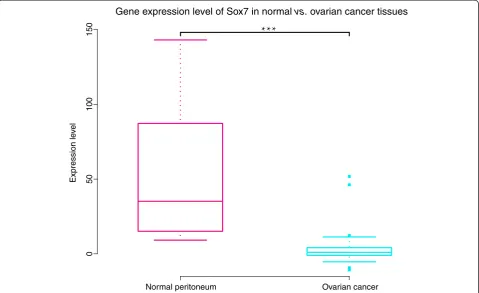
To investigate the correlation between SOX7 and the state of ovarian cancer, we compared the SOX7 gene expression profiles of forty-three serous ovarian cancers and ten peritoneum controls in GSE12470, with the false discovery rate (FDR) being controlled. We found that
the expression of SOX7 was reduced significantly in ovarian cancer tissues compared with normal controls (ovarian cancer mean: 3.50vs.normal peritoneum mean: 53.96, FDR =7.4e-07; Figure 1).
Correlation of reduced SOX7 expression with tumor progression
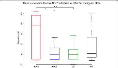
We assessed the expression of SOX7 in ovarian tissues of different tumor progression states. We used the gene expression data set GSE27651, which profiled six human ovarian surface epithelia (HOSE), eight serous borderline
HOSE SBOT LG HG
20
40
60
80
100
120
Expression le
ve
l
Gene expression level of Sox7 in tissues of different malignant state
Figure 2One-way ANOVA analysis of SOX7 mRNA expression levels among HOSE, SBOT, LG and HG.A significant correlation was found in HOSE relative to SBOT (p= 0.033) and HOSE compared with LG (p= 0.007).
Table 1 Biological processes enriched with genes dysregulated to a larger extent in ovarian cancer
Accession Term P-Values FDR
GO:0006414 translational elongation 4.692062542984977E-29 9.047961108371685E-26
GO:0006412 translation 2.04912911734554E-22 3.951447873919316E-19
GO:0006396 RNA processing 2.7244884076177707E-10 5.253780188674284E-7
GO:0022613 ribonucleoprotein complex biogenesis 1.3552603187281535E-9 2.6134227004703803E-6
GO:0016071 mRNA metabolic process 3.895055800131527E-8 7.51104669238778E-5
GO:0006397 mRNA processing 5.7556740309817435E-8 1.1098975527534805E-4
GO:0000375 RNA splicing, via transesterification reactions 3.5239047199996375E-7 6.795316527141715E-4
GO:0000377 RNA splicing, via transesterification reactions with bulged adenosine as nucleophile 3.5239047199996375E-7 6.795316527141715E-4
GO:0000398 nuclear mRNA splicing, via spliceosome 3.5239047199996375E-7 6.795316527141715E-4
GO:0042254 ribosome biogenesis 5.134416394199803E-7 9.900929787476365E-4
ovarian tumors (SBOT), thirteen low-grade serous ovar-ian carcinomas (LG), and twenty-two high-grade serous ovarian carcinomas (HG). As highly malignant cells are believed to arise from ovarian carcinoma of low malig-nancy, the expression value of SOX7 should be different among tissues of different malignancy. Differences of SOX7 gene expression were observed among the four groups (p=0. 012) by one-way analysis of variance (one-way ANOVA). Multiple comparisons showed significant down-regulation of SOX7 mRNA expression compared to HOSE in both SBOT (SBOT mean: 42.07 vs. HOSE mean: 81.98, student-t p= 0.033) and LG (LG mean: 40.85 vs. HOSE mean: 81.98, student-t p= 0.007). The amount of SOX7 expression in HG in comparison with HOSE was similar, although the difference was not statis-tically significant (Figure 2).
SOX7 as a negative regulator in Wnt/β-catenin pathway in ovarian cancer
It has been reported that SOX7, a member of the SOX family, functions as a transcriptional factor. To investigate
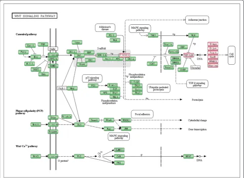
Figure 3Downstream genes chosen from KEGG annotated in Wnt/β-catenin pathway.Downstream genes in the Wnt/β-catenin pathway are denoted in red. The figure is created based on KEGG pathway. Only a part of the pathway is shown for clarity.
Table 2 The Pearson correlation coefficients between the expression levels of 13 genes and SOX7
Gene name r p
CCN-D1 −0.2753514639 0.0555052128
CCND2 −0.2083737906 0.1507783665
CCND3* −0.3789749206 0.0072460704
CTNNB1 −0.2298744109 0.1120735508
JUN* −0.328410818 0.0212317742
MMP7 −0.1436013727 0.3249317121
MYC* −0.3794897149 0.0071609446
PPARD(NR1C2) 0.0113166546 0.9384851238
TCF7* −0.4721329855 0.0006148219
TCF7L2* −0.5079689459 0.00019461
FOSL1 0.1976706188 0.1733712192
LEF1* −0.4230961749 0.0024547299
TCF7L1 0.014360968 0.9219826098
the mechanism by which SOX7 is involved in the onco-genesis and progression of ovarian cancers, we analyzed genes that were co-expressed with SOX7 and short-listed 7933 genes by Pearson correlation (FDR< 0.01) in GSE27651 (see Additional file 1: Table S1). Using DAVID with a 5% FDR control, we found that the 7933 genes were significantly overrepresented in eleven GO-biological processes involved mainly in transcription activ-ities (Table 1). As SOX7 has been reported to block the transcription of Wnt/β-catenin signaling pathway in various cancers, we postulated that it might have the same function in ovarian cancer. We chose thirteen genes from KEGG that were annotated in the Wnt/β-catenin pathway as down-stream or pivotal hub genes (Figure 3) and calculated the Pearson correlation coefficients of expression levels between
the 13 genes and SOX7 (Table 2). We found that the expres-sion of six of the 13 selected genes was significantly corre-lated with that of SOX7, all negatively (binomial distribution
p =0.0156). Additionally, a marginal correlation of cyclinD1 with SOX7 was observed (p =0.0555). Interestingly, as many as eleven of the 13 selected genes (except PPARD and TCF7L1) were negatively correlated with SOX7 (bino-mial distribution,p =0.0112), suggesting that SOX7 could be a negative regulator of the Wnt/β-catenin pathway.
Expression of SOX7, cyclin-D1 and COX2 proteins in normal ovarian tissues, borderline ovarian tumors and ovarian cancer
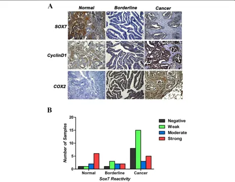
Eight of the ten analyzed normal ovarian tissue sections were positive for SOX7 immunoreactivity, with moderate
to strong staining intensity (Figure 4A), but the differences among the levels of staining intensity were not statistically significant. A minority of the ovarian cancer tissue sec-tions had positive SOX7 staining intensity, but negative or weak SOX7 staining intensity was seen in most of the malignant ovarian tissues (23/31; Figure 4B). SOX7 immu-noreactivity was nucleonic in every SOX7-positive ovarian tissue section. COX2 and cyclin-D1 are the target genes in the β-catenin signal pathway. The immuno-reactivity in COX2 and cyclin-D1 was crosscurrent compared to SOX7 in our research, which is consistent with the previous reports in other tissues (Figure 4A). For simultaneous comparison, each type of ovarian tissues used for photographing was from the same patient.
Relations between SOX7 and clinical or pathological characteristics in patients with epithelial ovarian carcinoma
We analyzed the correlation between SOX7 expression and clinical or pathological characteristics of the tissues. We observed that SOX7 was down-regulated mainly in serous cystadenocarcinoma (19/31) but did not find significant differences among the pathological types (i.e., serous cystadenocarcinoma, mucinous cystadenocarci-noma, and endometrioid carcinoma). On the other hand, we found that the down-regulation of SOX7 was signifi-cantly associated with the advanced stages (III-IV; 14/31,
p= 0.037), although no relationship was observed between SOX7 expression and other clinical features, such as path-ology grade or age (Table 3). Correlation analysis showed that SOX7 expression was negatively correlated with COX2 (rs=−0.618,p< 0.001) and cyclinD1 (rs=−0.583,
p< 0.001), two specific downstream targets in the Wnt-β-catenin pathway. The negative correlation of SOX7 with COX2 and cyclinD1 indicates a trend of inverse ex-pression pattern of SOX7 to COX2 and cyclin D1.
Discussion
Ovarian cancer remains to be a leading cause of death from gynecological malignancies. It is a huge challenge of current basic and clinical research to seek novel molecular markers for more accurate and efficient use in early diagnosis, treatment or prognosis of ovarian can-cer. In this study, we chose ovarian cancer to work on primarily due to the fact that this disease is so devastat-ing in females and that to date relatively little has been done on SOX7 in ovarian cancer. Our results obtained from different platforms indicate that the expression levels of SOX7 were significantly reduced in all types of ovarian cancers studied here, though at different extents. LG is the most malignant among ovarian cancer and may progress from SBOT, while HG, much less malignant than LG, likely develops from other kinds of precursors, such as normal epithelium of ovary or distal fallopian tube [24].
HG is a well-differentiated neoplasm and closely re-sembles normal tissues in many ways, so its prognosis is generally much better than LG [25]. As a result, the gene expression levels of SOX7 showed opposite tendencies to malignancy degrees. Based on the findings in this study, we propose that SOX7 is a key factor during ovarian cancer progression and is a useful prognostic marker.
Of great significance, we found that SOX7 was nega-tively correlated with Cyclin D1 and could be a negative regulator of the Wnt/β-catenin pathway. Cyclin D1, which controls the cell cycle, is the target gene ofβ-catenin and plays an important role in ovarian cancer. It is known that the Wnt/β-catenin signaling pathway is activated in epi-thelial ovarian cancer [26] and strongly involved in ovarian cancer development [27]. As SOX7 has been reported to block the transcriptional function of the Wnt/β-catenin signaling pathway and inhibit the activity of Wnt target genes including cyclin D1, c-Myc and COX-2 [15,17], we wondered whether there might be a correlation between SOX7 and these supposed target genes. Our results dem-onstrated that the expression levels of SOX7 and its
Table 3 Correlations of the SOX7 protein expression with the clinicopathological features of ovarian cancer and COX2/cyclinD1 Characteristic Total SOX7 expression P Over-expression Low-expression
All cases 31
Pathology type NS
Serous 26 7 19
Mucinous 2 0 2
Endometrioid 3 1 2
FIGO stage 0.037
Early (I-II) 16 7 9
Advance (III-IV) 15 1 14
Pathology grade NS
Low (1) 4 1 3
High (2 + 3) 27 7 20
Age (years) NS
<40 0 0 0
40-60 21 5 16
> 60 10 3 7
Cyclin D1 rs=−0.583,
p< 0.001
Over-expression 26 8 18
Low-expression 5 0 5
COX2 rs=−0.618,
p< 0.001
Over-expression 27 5 22
targets, COX-2 and cyclin D1, have an inverse relation-ship, further supporting our hypothesis that SOX7 is a negative regulator in the Wnt/β-catenin signaling pathway in ovarian cancer. Although our data showed a marginal correlation of cyclin D1 with SOX7 (p= 0.0555) at the mRNA level, the correlation at the protein level was strong, so both had similar trends.
SOX7, a member of the SOX-F subfamily encoding transcription factors that have a pivotal role in cardio-vascular development [8],has been implicated as a tumor suppressor in a variety of human cancers, e.g., colorectal, prostate, breast, liver and lung cancers [13,14,19,28]. Additionally, SOX7 may have potential usage as an inde-pendent prognostic marker in prostate and lung cancers as well as MDS patients [11,12,29]. Several lines of evi-dence indicate SOX7 as a negative regulator of the Wnt/ β-catenin signaling pathway [14,15]. Of great interest, a recent report based on whole genomic copy number analysis demonstrates SOX7 as a novel tumor suppres-sor, which is silenced in the majority of non-small cell lung cancer (NSCLC) samples [19]. Molecules modulat-ing SOX7 expression, such as miR-184 in hepatocellular carcinoma (HCC), have also been reported [28]. Another study published recently also showed that SOX7 could suppress HCC in vivoand in vitro[30].There are also contradicting reports to demonstrate that SOX7 mRNA was significantly up-regulated in several human cancer cells [20]. These results have prompted us to hypothesize that SOX7 might be a true tumor suppressor but behaves differently in different cancers.
Conclusions
Our work reported here suggests, for the first time, that SOX7 may play an important role as a tumor suppressor in ovarian cancer progression. Our results also revealed SOX7 as a negative regulator in the Wnt/β-catenin sig-naling pathway in ovarian cancer. Although further stud-ies are needed to elucidate the underlining mechanisms of interactions between SOX7 and the Wnt/β-catenin signaling pathway, our result demonstrates the suppres-sive function of SOX7 in the carcinogenic process of ovarian cancer.
Additional file
Additional file 1: Table S1.Short-listed 7933 genes were co-expressed with SOX7 by Pearson correlation (FDR< 0.01) in GSE27651.
Abbreviations
EOC:Epithelial ovarian cancer; NSCLC: Non-small cell lung cancers; GEO: Gene Expression Omnibus; KEGG: Kyoto Encyclopedia of Genes and Genomes; DAVID: Database for Annotation, Visualization and Integrated Discovery; BPs: Biological processes; GO: Gene Ontology; FDR: False discovery rate; HOSE: Human ovarian surface epithelia; SBOT: Serous borderline ovarian tumors; LG: Low-grade serous ovarian carcinomas; HG: High-grade serous ovarian carcinomas; HCC: Hepatocellular carcinoma.
Competing interests
The authors declare that they have no competing interests.
Authors’contributions
HL designed the study, BL carried out bioinformatics analysis and drafted the manuscript, HYL and ZQY participated in sample collection and experimental process, QS, SYY, JJK, DY and KF contributed reagents/materials/analysis tools. SLL finalized the manuscript. All authors read and approved the final manuscript.
Acknowledgements
The authors thank all the colleagues of genomics research center for comments on earlier versions of this manuscript. This work was supported by grants of the National Natural Science Foundation of China
(NSFC30970119, 81030029, 81271786, NSFC-NIH 81161120416).
Author details
1
Genomics Research Centre, Harbin Medical University, Harbin 150081, China.
2HMU-UCFM Centre for Infection and Genomics, Harbin, China.3Pathology
Department, The First Hospital of Qiqihaer City, Qiqihaer 161006, China.
4Department of Microbiology, Immunology and Infectious Diseases,
University of Calgary, Calgary, Canada.5Current address: Department of Biology, University of British Columbia, Vancouver, Canada.
Received: 2 July 2014 Accepted: 28 August 2014 Published: 5 September 2014
References
1. Jemal A, Siegel R, Xu J, Ward E:Cancer statistics, 2010.CA Cancer J Clin 2010,60(5):277–300.
2. Partridge EE, Barnes MN:Epithelial ovarian cancer: prevention, diagnosis, and treatment.CA Cancer J Clin1999,49(5):297–320.
3. Vrabie CD, Petrescu A, Waller M, Dina I:Clinical factors and biomarkers in ovarian tumors development.Rom J Morphol Embryol2008,49(3):327–338. 4. Ovarian cancer:A look at common myths and facts.Mayo Clin Womens
Healthsource2009,13(9):1–2.
5. Stovall DB, Cao P, Sui G:SOX7: From a developmental regulator to an emerging tumor suppressor.Histol Histopathol2013,29(4):439–445. 6. Long F, Wang X, Fang S, Xu Y, Sun K, Chen S, Xu R:A potential
relationship among beta-defensins haplotype, SOX7 duplication and cardiac defects.PLoS One2013,8(8):e72515.
7. Costa G, Mazan A, Gandillet A, Pearson S, Lacaud G, Kouskoff V:SOX7 regulates the expression of VE-cadherin in the haemogenic endothelium at the onset of haematopoietic development. Development2012,139(9):1587–1598.
8. Francois M, Koopman P, Beltrame M:SoxF genes: Key players in the development of the cardio-vascular system.Int J Biochem Cell Biol2010, 42(3):445–448.
9. Herpers R, van de Kamp E, Duckers HJ, Schulte-Merker S:Redundant roles for sox7 and sox18 in arteriovenous specification in zebrafish. Circ Res2008,102(1):12–15.
10. Takash W, Canizares J, Bonneaud N, Poulat F, Mattei MG, Jay P, Berta P: SOX7 transcription factor: sequence, chromosomal localisation, expression, transactivation and interference with Wnt signalling. Nucleic Acids Res2001,29(21):4274–4283.
11. Fan R, Zhang LY, Wang H, Yang B, Han T, Zhao XL, Wang W, Wang XQ, Lin GW:Methylation of the CpG island near SOX7 gene promoter is correlated with the poor prognosis of patients with myelodysplastic syndrome.Tohoku J Exp Med2012,227(2):119–128.
12. Li B, Ge Z, Song S, Zhang S, Yan H, Huang B, Zhang Y:Decreased expression of SOX7 is correlated with poor prognosis in lung adenocarcinoma patients.Pathol Oncol Res2012,18(4):1039–1045. 13. Stovall DB, Wan M, Miller LD, Cao P, Maglic D, Zhang Q, Stampfer MR,
Liu W, Xu J, Sui G:The regulation of SOX7 and its tumor suppressive role in breast cancer.Am J Pathol2013,183(5):1645–1653.
15. Chan DW, Mak CS, Leung TH, Chan KK, Ngan HY:Down-regulation of Sox7 is associated with aberrant activation of Wnt/b-catenin signaling in endometrial cancer.Oncotarget2012,3(12):1546–1556.
16. Clapper ML, Coudry J, Chang WC:beta-catenin-mediated signaling: a molecular target for early chemopreventive intervention.Mutat Res2004, 555(1–2):97–105.
17. Zhou X, Huang SY, Feng JX, Gao YY, Zhao L, Lu J, Huang BQ, Zhang Y: SOX7 is involved in aspirin-mediated growth inhibition of human colorectal cancer cells.World J Gastroenterol2011,17(44):4922–4927. 18. Zhang Y, Huang S, Dong W, Li L, Feng Y, Pan L, Han Z, Wang X, Ren G,
Su D, Huang B, Lu J:SOX7, down-regulated in colorectal cancer, induces apoptosis and inhibits proliferation of colorectal cancer cells.Cancer Lett 2009,277(1):29–37.
19. Hayano T, Garg M, Yin D, Sudo M, Kawamata N, Shi S, Chien W, Ding LW, Leong G, Mori S, Xie D, Tan P, Koeffler HP:SOX7 is down-regulated in lung cancer.J Exp Clin Cancer Res2013,32:17.
20. Katoh M:Expression of human SOX7 in normal tissues and tumors. Int J Mol Med2002,9(4):363–368.
21. Da Huang W, Sherman BT, Lempicki RA:Bioinformatics enrichment tools: paths toward the comprehensive functional analysis of large gene lists. Nucleic Acids Res2009,37(1):1–13.
22. Da Huang W, Sherman BT, Lempicki RA:Systematic and integrative analysis of large gene lists using DAVID bioinformatics resources. Nat Protoc2009,4(1):44–57.
23. Benjamini YHY:Controlling the false discovery rate: a practical and powerful approach to multiple testing.J R Stat Soc Ser B1995, 57:289–300.
24. King ER, Tung CS, Tsang YT, Zu Z, Lok GT, Deavers MT, Malpica A, Wolf JK, Lu KH, Birrer MJ, Mok SC, Gershenson DM, Wong KK:The anterior gradient homolog 3 (AGR3) gene is associated with differentiation and survival in ovarian cancer.Am J Surg Pathol2011,35(6):904–912.
25. McCluggage WG:Morphological subtypes of ovarian carcinoma: a review with emphasis on new developments and pathogenesis.Pathology2011, 43(5):420–432.
26. Arend RC, Londono-Joshi AI, Straughn JM Jr, Buchsbaum DJ:The Wnt/beta-catenin pathway in ovarian cancer: a review.Gynecol Oncol 2013,131(3):772–779.
27. Basu M, Roy SS:Wnt/beta-catenin pathway is regulated by PITX2 homeodomain protein and thus contributes to the proliferation of human ovarian adenocarcinoma cell, SKOV-3.J Biol Chem2013, 288(6):4355–4367.
28. Wu GG, Li WH, He WG, Jiang N, Zhang GX, Chen W, Yang HF, Liu QL, Huang YN, Zhang L, Zhang T, Zeng XC:Mir-184 post-transcriptionally regulates SOX7 expression and promotes cell proliferation in human hepatocellular carcinoma.PLoS One2014,9(2):e88796.
29. Zhong WD, Qin GQ, Dai QS, Han ZD, Chen SM, Ling XH, Fu X, Cai C, Chen JH, Chen XB, Lin ZY, Deng YH, Wu SL, He HC, Wu CL:SOXs in human prostate cancer: implication as progression and prognosis factors. BMC Cancer2012,12:248.
30. Wang C, Guo Y, Wang J, Min Z:The suppressive role of SOX7 in hepatocarcinogenesis.PLoS One2014,9(5):e97433.
doi:10.1186/s13048-014-0087-1
Cite this article as:Liuet al.:Reduced expression of SOX7 in ovarian cancer: a novel tumor suppressor through the Wnt/β-catenin signaling pathway.Journal of Ovarian Research20147:87.
Submit your next manuscript to BioMed Central and take full advantage of:
• Convenient online submission
• Thorough peer review
• No space constraints or color figure charges
• Immediate publication on acceptance
• Inclusion in PubMed, CAS, Scopus and Google Scholar
• Research which is freely available for redistribution