R E S E A R C H
Open Access
CD117
+
CD44
+
Stem T Cells Develop in the
Thymus and Potently Suppress T-cell
Proliferation by Modulating the CTLA-4
Pathway
Yang Wei
1,2†, Zhansheng Hu
1,3†, Wen Gu
1, Gang Liu
4, Bingyin Shi
1, Enqi Liu
5and Tie Liu
1*Abstract
Background:CD117 is expressed on double-negative (DN; CD4–CD8–) cells (Nat Rev Immunol 14:529–545; 2014), but whether it is expressed in other stages and its subsequent functions are unclear. We used an improved method of flow cytometry to analyze different populations of thymocytes (Sci Rep 4:5781; 2014). The expression of CD117 and CTLA-4 were directly assayed in the early stage of thymocytes.
Methods:Flow cytometry was used to analyze different populations of thymocytes, and T-cell proliferation assays, RT-PCR, and real-time RT-PCR were used to characterize the stem cells and examine the function of CD44+CD117+cells. Results:In DN cells, CD117 expression was greatest on CD44+CD25+cells (DN2), followed by CD44
+
CD25–(DN1), CD44– CD25+(DN3), and CD44–CD25–(DN4) cells. In thymocytes, CD117 expression was highest in DN cells, followed by single-positive (SP; CD4 or CD8) and double-positive (DP; CD4+CD8+) cells. Especially, CD117 expression was positively associated with CD44 and CTLA-4 expression. CTLA-4 expression was highest in DN cells, followed by SP and DP cells. CTLA-4 expression was positively associated with CD25, CD44, and Foxp3 expression. CD44+CD117+T cells expressed more CTLA-4, which suppressed T-cell proliferation and blocked CTLA-4 to cause antibody-induced T-cell proliferation. Conclusion:These results suggest that CD44+CD117+T cells are stem cells and a specific T-cell phenotype that initially develops in the thymus, but they do not progress through DN3and DN4stages, lack a DP stage, and potently suppress T-cell proliferation and modulate the CTLA-4 pathway.
Keywords:CD117, CTLA-4, CD25, CD44, Stem cell, Flow cytometry
Background
T-cell development in mice begins with hematopoietic progenitor cells that migrate from the bone marrow to the thymus [1, 2]. The earliest precursors in the T-cell lineage are found within thymocyte populations and lack CD4 and CD8 expression, so they are referred to as CD4−CD8− or double-negative (DN) thymocytes [3]. DN thymocytes consist of four subpopulations based on CD44 and CD25 expression [4]. DN cells that express CD4 and CD8 are CD4+CD8+ or double-positive (DP) thymocytes before
differentiating into single-positive (SP; CD4+ or CD8+) thymocytes [5].
Stems cells include embryonic stem (ES) cells, in-duced pluripotent stem (iPS) cells, adult tissue stem/ progenitor cells, and cancer stem cells. Mesenchymal stem cells (MSCs) have been shown to inhibit abnor-mally activated immune functions in dendritic cells, T cells, and B cells; however, preclinical efficacy studies to evaluate MSCs in autoimmune animal models have been contradictory [6–8].
Mammalian CD117 (c-kit) gene protein is a stem cell marker and a type III tyrosine kinase receptor [9, 10], widely expressed in hematopoietic stem cells (HSC), myeloid progenitor cells, as well as B cells and pro-T cells [11–13]. In the hematopoietic system this protein
* Correspondence:[email protected] †Equal contributors
1Immunology and Tumor Research Instituted, the First Affiliate Hospital, Xi’an Jiaotong University Health Science Center, Xi’an, Shaanxi 710061, People’s Republic of China
Full list of author information is available at the end of the article
contributes to cell growth, metabolism, proliferation, and survival [14, 15] during gametocyte development, pigmentation, and intestinal motility [16], and in the im-mune system contributes to inflammation [17]. The four DN subsets are defined by surface expression of c-kit, CD44, and CD25. Cells at the DN3stage are positive for
c-kitloCD44−CD25+. CD25 expression is downregulated as cells transition to the DN4cell stage [14, 18].
Nanog is an important transcription factor for main-taining self-renewal and pluripotency of ES cells and regulates the fate of inner cell mass of blastocysts in the embryo during early development. The homeoprotein Nanog is required for maintenance of pluripotency in mouse epiblast and ES cells [19].
Cytotoxic T lymphocyte antigen-4 (CTLA-4) is an in-hibitory relative of the T-cell costimulatory molecule, CD28. Whereas CD28 signaling promotes T-cell activa-tion, CTLA-4 serves an immunoregulatory function by suppressing the T-cell response [20–22]. CTLA-4 also has intrinsic functions for T-conv cells and regulates trafficking of self-reactive T cells [23, 24].
Flow cytometry can be used to characterize single cells in an efficient and expedient manner [25], and can be used to describe DN cells which are present in limited amounts in the mouse thymus. Previously, a highly re-producible multi-color flow cytometry method was de-veloped to increase thymocytes collected from 104 to more than 106 [26]. Using this method, we identified genes with low expression in DN cells and this permit-ted characterization of DN cell subsets.
In the present study, CD117, CTLA-4, CD4, CD8, CD25, and CD44 expression was measured in thymo-cytes and expression of CD117 and CTLA-4 was assayed directly in thymocytes and CD117+ stem T cells in the thymus and spleen. These data provide evidence of a re-lationship among CD117, CTLA-4, CD25, and CD44 cell surface markers on DN cells and other thymocytes.
Methods Animals
Female C57BL/6 mice (Taconic Farms, Germantown, NY, USA) were maintained under specific pathogen-free con-ditions and were studied at 4–6 weeks of age, according to protocols approved by the Institutional Animal Care and Use Committee at Xi’an Jiaotong University, China.
Flow cytometric analysis and cell sorting
Thymocytes and splenocytes were obtained from naive mice and suspended in PBS plus 1% fetal calf serum (FCS). To avoid nonspecific binding to mouse Fc-γ re-ceptors, cells were blocked with mouse CD16/CD32 mAb (0.25 μg/100 μl; BD Biosciences, Franklin Lakes, NJ, USA) for 15 min at room temperature. Cells were washed and then stained with FITC anti-CD4 (clone
RM4-5), PercP5.5-anti-CD8 (clone 53-6.7), APC-CY7-anti-CD44 (clone IM7), PE-CY7-anti-CD25 (clone PC61), PE-anti-CD117 (clone 2B8), FITC-CD117 (Clone 2B8), and APC-anti-CD3e (Clone 145-2C11) antibodies for 60 min at 4 °C. For some samples, surface-stained cells were fixed/permeabilized with a Cytofix/Cytoperm Kit (BD Biosciences) and stained with PE-anti-Foxp3 (clone FJK-16 s; eBioscience) and APC-anti-CTLA-4 (clone UC10-4B9; eBioscience) antibody for 45 min at 4 °C. The corresponding isotype controls (Armenian Hamster IgG, and Rat IgG2b,k) were purchased from eBioscience. Cells were sorted and analyzed using a FACSAria II, and data were analyzed using BD FACS-Diva software (BD Biosciences).
Culture medium and antibodies
RPMI 1640 medium containing 10% FCS was used for CD4 naive T-cell culture. Antibodies used for cell cul-ture were anti-mouse CD3 (145-2C11) and anti-mouse IL2 (300 u/ml) (BD BioSciences, San Jose, CA, USA).
Coculture system development
A coculture system was developed using a six-well Transwell plate (0.4μm pore size insert; Corning Incor-porated, Corning, NY, USA).
Naive T cell and CD44+CD117+cell sorting and
stimulation
Lymphocytes from the spleen and thymus were stained with fluorochrome-conjugated antibodies, CD4 (FITC), CD8 (PerCp5.5), CD44 (APC-CY7), and CD117 (PE) for 45 min at 4 °C. Cells were then washed twice and resus-pended at a density of 1 × 10⁸cells/ml. Naive CD4, CD4
+
CD44+CD117+, and CD8+CD44+CD117+ cells were sorted using BD FACS Aria II. Anti-CD3 antibody was coated onto 24-well flat-bottom plates at 1μg/ml in PBS overnight and then washed once with PBS. Naive T cells and CD4+CD44+CD117+ (CD8+CD44+CD117+) were then placed into the wells at a density of 1 × 10⁶cells/ml in culture. Cells were incubated at 37 °C containing 5% CO2for 7 days.
T-cell proliferation assays
Cell proliferation was quantified using a Cell Trace CFSE Cell Proliferation Kit (Invitrogen, Carlsbad, CA, USA). Splenic T cells were prepared by passing spleno-cytes through nylon-wool columns (108splenocytes were incubated in the column for 1 h at 37 °C in 5% CO2
reaction. CFSE-labeled cells (1 × 10⁶/well) were washed twice with RPMI 1640 culture medium and then cultured in complete RPMI 1640 medium with 2 mML-glutamine (Gibco), supplemented with 10 mM HEPES, 1% penicil-lin–streptomycin solution (Gibco), and 10% FBS (Gibco). A total 3μg/ml of ConA or IL-2 and anti-CD3 antibody was added for polyclonal stimuli. Samples not stimulated
with ConA or IL-2 and anti-CD3 were used as nega-tive controls. Cells were then incubated for 7 days and stained with CD4 and CD8. T-cell proliferation was measured by quantifying CFSE fluorescence in-tensity via flow cytometry.
CTLA-4 block assay
To block CTLA-4, 8μl anti-mouse CD152 (CTLA-4) or isotype Ctrl (Armenian Hamster IgG) antibody was added in medium before incubation during the T-cell proliferation assay.
RT-PCR and real-time RT-PCR
Total RNA was extracted from 106 sorted thymocytes from C57BL/6 mice, using TRIzol (Invitrogen). Chloro-form (0.2 ml; Sigma-Aldrich, USA) was added for every
Table 1Primers used for RT-PCR to detect CTLA-4, Nanog mRNAs β-actin sense GAA ATC GTG CGT GAC ATC AAA G
Antisense TGT AGT TTC ATG GAT GCC ACA G
CTLA-4 sense TCTGCAAGGTGGAACTCA
Antisense GCTAACTG CGACAAGGAT
Nanog sense ATCCCGAGAACTATTCTTG
Antisense GGTACTTCTGCTTCTGAAAC
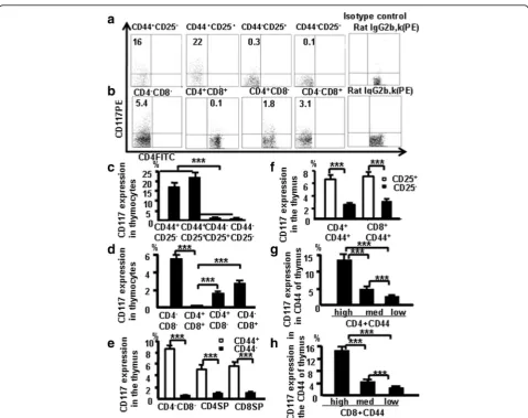
Fig. 1CD117 and CD44 expression in different thymocyte subpopulations.aCD117 expression in DN1, DN2, DN3, and DN4thymocytes.bCD117
expression in DN, DP, and SP thymocytes. DN thymocyte data (c) and thymocyte data (d) pooled from three independent experiments.eCD44 expression analyzed in the different subpopulations of mouse thymocytes.fCD117 expression evaluated on CD4+CD44+CD25+and CD8+CD44 +CD25+cells.gCD117 expression in CD44+CD4 SP cells, according to varying CD44 expression.hCD117 in CD44+CD8 SP cells, according to
1 ml of TRIzol used. Extracted RNA was shaken vigor-ously for 15 sec and incubated at room temperature for 2 min. After transferring the aqueous phase to a clean tube, isopropanol (Sigma-Aldrich) was added and the RNA was incubated at room temperature for 5 min. The RNA pellet was then washed with 1 ml of 75% ethanol, air-dried for 10 min, dissolved in 20μl of water, and incu-bated at 55 °C for 10 min. The OD ratio (λ 260 nm/ 280 nm) was between 1.9 and 2.0, and RNA samples were assessed with electrophoresis on 1.5% agarose gels and vi-sualized under UV light after ethidium bromide staining. RNA preparations were treated with DNase I to remove genomic DNA. cDNA was synthesized by incubating 20μl of mRNA in a sprint C1000 terminal cycler (Bio-Rad, USA). Negative controls contained the reaction mixture except for template DNA. For quantification, relative mRNA expression of specific genes was obtained by the 2−ΔΔCt method. The gene-specific primers (5′→3′) used are presented in Table 1. Data were normalized using transcripts for β-actin as controls, which were amplified with primers as presented in Table 1. Diluted cDNA (10μl) was mixed with 2μl of primer and 10μl IQ SYBR Green SuperMix and was assayed in triplicate using a
Stepone real-time system (ABI) under the following con-ditions: denaturation at 95 °C for 3 min, 40 cycles of 95 °C for 15 sec, and 60 °C for 1 min, followed by 30 sec of ex-tension at 72 °C. Each sample was analyzed in triplicate.
Statistical analysis
Mean and SD values were calculated with Microsoft Excel. At least three independent experiments were per-formed. A Tukey–Kramer post test was used to compare three or more means and a two-tailed unpaired Student’s
t test was used to compare two groups (p≤0.05 was considered statistically significant).
Results
CD117 and CD44 expression in different thymocyte subpopulations
CD117 plays an important role in the differentiation of T cells [9, 10]. T cells develop from CD3–CD4–CD8–c-kit+ cells through the modulation of cell surface molecules [27]. To confirm CD117 expression in the thymus during T-cell development, thymocytes were harvested from C57BL/6 mice, stained with CD4 (FITC), CD8 (PercP5.5), CD44 (APC-CY7), CD25 (PE-CY7), and CD117 (PE), and
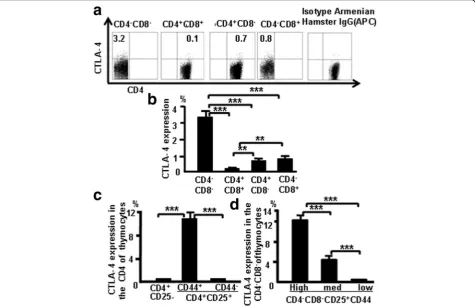
Fig. 2CTLA-4 expression correlates with CD25 and CD44 expression in different populations of thymocytes.aCTLA-4 expression in thymocytes of C57BL/6 mice.bCTLA-4 expression data pooled from three independent experiments.cCTLA-4 expression evaluated in CD4+CD25–and CD4 +CD25+cells.dCTLA-4 expression in CD25+DN cells, according to varying CD44 expression. Data are mean percent of total cells ± SD from three
analyzed by flow cytometry. This group of antibodies is now termed the “antibody cocktail.” Isotype Rat IgG2b,k was used as a negative control (CD117). DN2 (CD44 +
CD25+) cells had the highest frequency CD117 expres-sion, followed by DN1, DN3, and DN4 cells (Fig. 1a).
Pooled data from three independent experiments are shown in Fig. 1c. DN cells had more CD117-expressing cells in C57BL/6 mice followed by CD8+ SP cells, CD4+ SP cells, and DP cells (Fig. 1b); the pooled data from three independent experiments (Fig. 1d) indicate that CD117 expression was lowest in DP cells.
Thymocytes from naive mice were stained with antibody cocktail to confirm whether CD117 expression is associ-ated with CD44 expression in the other thymocyte subpop-ulations. In three experiments performed independently, more DN, CD4, and CD8 CD44+ cells expressed CD117 compared with DN, CD4 and CD8 CD44–cells (Fig. 1e). There was also a positive correlation between CD25 and CD117 expression among CD4+CD44+ cells and CD8+CD44+ cells (Fig. 1f ). Data (Fig. 1g, h) indicate that CD117 expressed in early stage T cells in the thymus positively correlates with CD44 in thymocytes.
CTLA-4 expression correlates with CD25 and CD44 expression in different thymocyte populations
CTLA-4 expression was examined in different subsets of thymocytes at various stages of T-cell development to
confirm CTLA-4 expression in T-cell development. More DN cells expressed CTLA-4 compared with CD4 SP, CD8 SP, and DP cells (Fig. 2a). The pooled data from three independent experiments are shown in Fig. 2b, and the data show that CTLA-4 expression is greater in early stages of T-cell development. More CD4+CD25
+
CD44+ cells produced CTLA-4 compared with CD4
+
CD25+CD44– and CD4+CD25– cells (Fig. 2c, d). Data show that surface expression of CD25 and CD44 appears to be positively correlated with CTLA-4 expression.
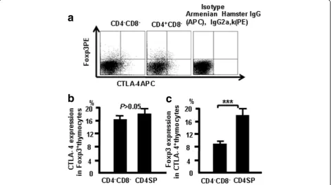
CTLA-4 correlates with Foxp3 expression in thymocytes
Thymocytes from naive mice were stained with CD4, CD8, CD44, CD25, CTLA-4 (APC), and Foxp3 (PE), and were analyzed by flow cytometry to study the relation-ship between CTLA-4 and Foxp3. More CTLA4 expres-sion was observed in Foxp3-positive CD4 SP cells compared with Foxp3-positive DN cells (Fig. 3). These data indicate that there is overlap of Foxp3 and CTLA-4 expression in some thymocytes.
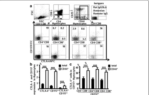
CTLA-4 expression in CD44+CD117+cells in the thymus
Thymocytes from naive mice were stained with the antibody cocktail and CTLA-4 (APC), we studied whether CD117 and CD44 expression correlates with CTLA-4 expression in the thymus (Fig. 4a). Data showed 0.5% of thymocytes were CD117+ cells, 0.7% of thymocytes were
CTLA-4+ cells, and 0.2% of thymocytes were CTLA-4
+
CD117+ cells. Furthermore, 2.9% of the CD44+ thymo-cytes were CD117+ cells, 3.2% of the CD44+ thymocytes were CTLA-4+ cells, and 0.6% of the CD44+ thymocytes were CTLA-4+CD117+cells (Fig. 4b). CTLA-4 expression was found in 19% of DN CD117+ cells, 18% of CD4
+
CD117+ cells, and 16% of CD8+CD117+ cells. Interest-ingly, CTLA-4 expression was also found in 34% of DN CD44+CD117+ cells, 36% of CD4+CD44+CD117+ cells, and 32% of CD8+CD44+CD117+ cells (Fig. 4c), demon-strating that CD44+and CD117+ expression is closely as-sociated with CTLA-4 in thymocyte stem T cells.
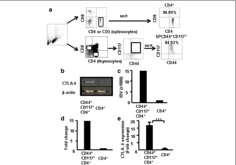
CD4+ SP cells and CD4+CD44+CD117+ cells were sorted from the thymocytes via FACS (Fig. 5a) to fur-ther verify whether CD44+CD117+ cells express higher levels of CTLA-4. The expression of CTLA-4 mRNA in CD44+CD117+ cells and CD4+ SP cells was compared by RT-PCR. Results showed that the level of CTLA-4 was higher in CD4+CD44+CD117+ cells, as determined by densitometry measurements of the gels (Fig. 5b). Data from three independent experi-ments were pooled (Fig. 5c, d). Furthermore, CD4+ SP and CD4+CD44+CD117+ cells were sorted from the thymocytes and the levels of CTLA-4 mRNA
were measured by real-time RT-PCR to further con-firm the aforementioned findings. As shown in Fig. 5e, CD4+CD44+CD117+ cells expressed higher levels of CTLA-4 than CD4+ SP cells (~14× higher). These re-sults corroborate the finding that CTLA-4 expression was much higher in CD4+CD44+CD117+ cells.
CTLA-4 expression correlates with CD44 expression in different populations of CD117-positive thymocytes
Thymocytes were stained with the combination of anti-bodies to CD4, CD8, CD44, CD25, CTLA-4 (APC), and CD117 (PE) and analyzed by flow cytometry, and data show that CTLA-4 expression correlates with thymic CD44 expression. Fig. 6a–c show these data which sug-gest that CD44 expression positively correlates with CTLA-4 expression in CD117-positive thymocytes.
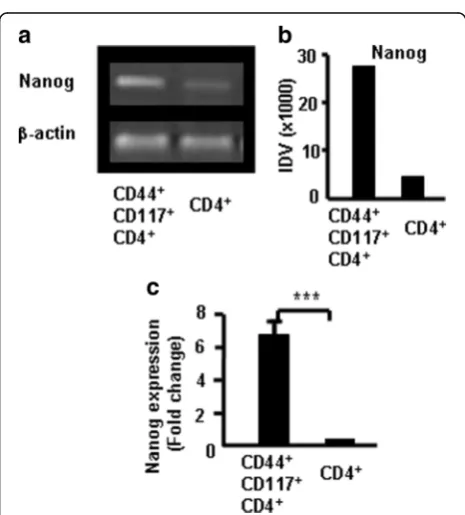
CD44+CD117+ cells expressed more Nanog in thymo-cytes. Nanog is an important transcription factor for maintaining the self-renewal and pluripotency of ESCs. It regulates the fate of the inner cell mass of blastocysts in the embryo during early development. The homeoprotein Nanog is required for maintenance of pluripotency in mouse epiblast and ES cells [19]. To characterize the CD44+CD117+ cells in the thymus, CD4+ SP cells and
Fig. 4CTLA-4 expression in CD44+CD117+cells in the thymus.aCD117 and CTLA-4 expression in the different population in the thymocytes. bCD117 and CTLA-4 in thymocytes and in the CD44+thymocytes.cCTLA-4 in CD117+thymocytes and in CD117+CD44+thymocytes. Data are
CD4+CD44+CD117+ cells were sorted from the thymo-cytes via FACS to verify whether CD44+CD117+ cells ex-press more Nanog. The Nanog mRNA exex-pression in CD44+CD117+ cells and CD4+ SP cells was compared using RT-PCR. Results showed that Nanog were greater in CD4+CD44+CD117+ cells than in CD4+SP cells as deter-mined by densitometric analysis of gels (Fig. 7a). Pooled data from three independent experiments are shown in Fig. 7b. CD4 SP and CD4+CD44+CD117+cells were sorted from thymocytes and Nanog mRNA expression was mea-sured using real-time RT-PCR to confirm previous find-ings. CD4+CD44+CD117+ cells expressed more Nanog than CD4+SP cells as shown in Fig. 7c. Data suggest that CD44+CD117+cells are stem cells.
CD44+CD117+stem T cells from the thymus and spleen
suppressed T-cell proliferation
CD117+ cells develop in the thymus and do not down-regulate CD44 and CD117 expression, which is measur-able in SP cells. We hypothesized that CD44+CD117+ T
cells are stem T cells, which are key to the development and function of the immune system, especially of T cells. To confirm whether CD44+CD117+ T cells affect T-cell proliferation, stem T-cell function was examined in vitro using a CFSE assay. CD4+CD117+CD44+ T cells from the thymus were sorted by FACSAria II and were cocul-tured with T cells (CD4+CD117+CD44+ T cells:CD4+ T cells) in the presence of Con A (3μg/ml). T-cell prolifer-ation was measured by flow cytometry to determine the CFSE (5 μM) fluorescence intensity, as shown in Fig. 8. Stem T cells from the thymus and T cells from the spleen were sorted by FACSAria and were incubated with cocultured T cells (stem T cells:T cells) at ratios of 1:20, 1:10, and 1:5 in the presence of Con A. T-cell pro-liferation was measured by flow cytometry to determine the CFSE fluorescence intensity (Fig. 8a, b). The percent-age of T-cell inhibition was 80%, 21%, and 5.1%, respect-ively. Further, both stem T cells and T cells from the spleen were sorted by FACSAria and were incubated with cocultured T cells (stem T cells:T cells) at ratios of
Fig. 5CTLA-4 expression in CD4+CD117+CD44+thymocytes by RT-PCR and real-time RT-PCR. CD4+CD117+CD44+and CD4+SP cells sorted from
1:20, 1:10, and 1:5 in the presence of Con A. The per-centage of cell inhibition was 84%, 26%, and 11%, re-spectively. The data from three independent experiments were pooled and are shown in Fig. 8c (stem cells from the thymus) and Fig. 8d (stem cells from the spleen). These results suggest that CD44+CD117+T cells significantly suppressed T-cell proliferation.
CTLA-4 suppressed T-cell proliferation and blocking CTLA-4 led to antibody-induced T-cell proliferation
CD44+CD117+ stem T cells from the thymus were sorted by FACSAria and incubated alone or cocul-tured (stem T cell:T cell) at a 1:2 ratio in the presence of anti-CD3 and IL-2 (300 u/ml) or blocking antibodies (1μg/100μl) to CTLA-4. Isotype Armenian Hamster IgG was used as a control. T-cell proliferation was measured by flow cytometry to determine the CFSE (2μM) fluores-cence intensity after 7-day incubation. The percentage of T-cell inhibition was 2.5% in T cells only (unstimulated) (Fig. 9Aa), 33% in T cells containing anti-CD3 + IL-2 (T cells only) (Fig. 9Ab), 5.3% in Isotype Armenian Ham-ster IgG (control, T cells were cultured with CD44+CD117+ T cells and Isotype Armenian Hamster IgG together) (Fig. 9Ac), 39% in T cells + CD44+CD117– T cells containing anti-CD3 + IL-2 (control, T cells were cultured with CD44+CD117– T cells) (Fig. 9Ad), 54% in the medium containing CTLA-4 broke antibody
Fig. 6CTLA-4 correlates with CD44 expression in different populations of CD117-positive thymocytes. Thymocytes were treated as indicated in Methods and analyzed by flow cytometry.aCTLA-4 expression in CD4+CD117+CD44+cells.bCTLA-4 expression in CD8+CD117+CD44+cells.cCTLA-4 expression in
DNCD117+CD44+cells. Data show mean percent of total cells ± SD from three independent experiments. *p <0.05, **p <0.01, ***p <0.001.DNdouble-negative
Fig. 7Nanog expression in CD117+CD44+cells measured with RT-PCR
(CD44+CD117+ T cells were cultured with CTLA-4 broke, after 1 h the cells were washed and cultured with T cells in the medium) (Fig. 9Ae), and 25% in CD44+CD117+ T cells and T cells in Transwell plate coculture system (CD44+CD117+ T cells and T cells were cultured in the Transwell system) (Fig. 9Af ). The data from three independent experiments were pooled and are shown in Fig. 9B. Data show that CD44+CD117+ stem T cells suppressed T-cell prolifer-ation by the interaction between the cells, and sup-pressive activity of stem T cells was reduced in the presence of anti-CTLA-4 antibody.
CD44+CD117+stem T cells can self-renew
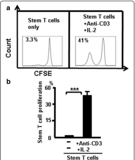
To confirm that CD44+CD117+ T cells can self-renew, thymic CD44+CD117+stem T cells were sorted with FAC-SAria and incubated alone or cocultured (stem T cell:T cell) at a 1:2 ratio in the presence of anti-CD3 and IL-2 (300 u/ ml). T-cell proliferation was measured by flow cytometry to measure CFSE (2μM) fluorescence intensity after 7-day in-cubation. T-cell proliferation was low in CD44+CD117+ stem T cells only (unstimulated), and this was greater in CD44+CD117+T cells containing anti-CD3 + IL-2 (Fig. 10). Thus, CD44+CD117+T cells can self-renew.
CD44+CD117+cells in blood and lymph nodes
To confirm CD44+CD117+ cells are exist in blood and lymph nodes, cells from naive mice were stained with the
combination of antibodies CD4 (FITC), CD8 (PerCp), CD25(PE-CY7), CD44 (APC-CY7), and CD117 (FITC) and analyzed by flow cytometry. The results showed 0.5% of CD4+CD117+ cells and 1.2% of CD4+CD117+ cells in blood and lymph nodes, respectively (Fig. 11). Further-more, 6.9% of CD4+ CD117+ cells and 14.6% of CD4+ CD117+ cells were present in CD44+ cells of blood and lymph nodes, respectively. These results suggest CD44
+
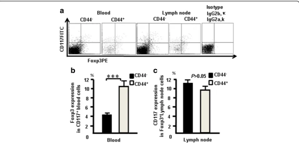
CD117+T cells are exist in blood and lymph nodes. CD44+CD117+ cells expressed Foxp3 in blood and lymph nodes. The cells from naive mice were stained with the following combination of antibodies: the antibody cocktail with Foxp3 (PE) and used to confirm whether CD117 and CD44 expression correlates with Foxp3 ex-pression in the blood and lymph nodes. Data show that 4.5% of CD4+CD117+ cells expressed Foxp3, 10.3% of CD4+CD44CD117+ cells expressed Foxp3+in blood cells, 11% of CD4+Foxp3+ cells expressed CD117, and 9.5% of CD4+CD44+ cells expressed Foxp3 in lymph nodes (Fig. 12a). The pooled data from three independent exper-iments are shown in Fig. 12b, c. These results suggested that CD44+CD117+cells and nTregcells are two different
populations of cells, although part of them overlap.
Discussion
Using an advanced flow cytometry method, we researched the characteristic of T-cell development in the thymus by measuring expression of CD117 and CTLA-4
Fig. 8CD4+CD117+cells from the thymus and spleen suppress T cells in vitro. T-cell proliferation was measured by flow cytometry to measure fluorescence intensity.aCD4+CD117+cells sorted from the thymus.bCD4+CD117+cells sorted from the spleen. Thymocyte data (c) and spleen
in thymocytes. Surprisingly, CD117+ stem T cells were found in mature thymocytes and CD117 and CTLA-4 ex-pression occurred at each stage of T-cell development. Changes in CD25, CD44, CD117, and CTLA-4 expression were examined in DN, DP, and SP cells using multi-color flow cytometry. CD117 was expressed in the early stages (DN1 and DN2) of T cells (Fig. 1a, c), and DN cells
expressed more CD117 than did DP and SP cells (Fig. 1b, d). CD117 expression was positively correlated with CD44 in thymocytes (Fig. 1e–h). CD44+CD117+T cells could be also found in blood and lymph nodes (Fig. 11). CD4SP and CD8SP cells contain many CD117+ cells, most of which are CD44+CD117+ cells. CD44+CD117+ cells have been used to label tumor stem cells [8, 10, 23, 28]. In the current study, CD117 and CD44 were used to label stem T cells and development stages were identified in the thymus.
Next, CTLA-4 expression was measured at different stages of T-cell development. CTLA-4 expression was higher in DN cells compared with SP and DP cells (Fig. 2a, b) and was positively correlated with CD25 and CD44 (Fig. 2c, d). CTLA-4 and Foxp3 overlapped in DN and CD4+cells (Fig. 3). CD44+CD117+ cells and nTreg cells are two different populations of cells,
Fig. 9Blocking CTLA-4 reduces the ability of CD4+CD117+CD44+
cells to suppress T-cell proliferation.aaT cells only;bT cells + anti-CD3 + IL-2;cCD44+CD117–T cells + anti-CD3 + IL-2;dCD44+CD117+ T cells + anti-CD3 + IL-2 + Isotype Armenian Hamster IgG;e(CD44
+CD117+T cells + CTLA-4 broke) + anti-CD3 + IL-2 + T cells; fCD44
+CD117+T cells cultured with T cells in a Transwell plate.BData
pooled from three independent experiments representing mean percent of totals ± SD. ***p <0.001
Fig. 10CD44+CD117+T cells have the ability to self renew.aCD4 +
CD44+CD117+T cells only and CD4+CD44+CD117+T cells + anti-CD3 + IL-2.bData pooled from three independent experiments representing mean percent ± SD. ***p <0.001
Fig. 11CD44+CD117+cells in blood and lymph nodes. Cells from
naive mice were stained with the combination of antibodies CD4 (FITC), CD8 (PerCp), CD25 (PE-CY7), CD44 (APC-CY7), and CD117 (FITC) and analyzed by flow cytometry.aCD117 expression and CD44+CD117+ cells in blood and lymph nodes.bData pooled
although parts of them overlap (Fig. 12). CD44
+
CD117+ cells had more CTLA-4 expression (Figs. 4 and 5). CD44 expression was positively correlated with CTLA-4 in CD117+ thymocytes (Fig. 6). CD117
+
CD44+ T cells are stem T cells that expressed more Nanog than other T cells in thymocytes (Fig. 7) and have the ability to self-renew (Fig. 10), confirming that CD44+CD117+ stem T cells suppressed T-cell pro-liferation and CD4+CD117+CD44+thymus cells had a po-tent suppressive function (Fig. 8). Because there is more CTLA-4 expression in stem T cells, we studied the effect of CTLA-4 on stem T cells. CD117+stem T cells were in-cubated with CTLA-4 blocking antibodies to confirm whether blocking CTLA-4 abrogated the suppressive mechanism of CD117+ stem T cells. CD44+CD117+ stem T cells and T cells proliferated, and when cultured separately this proliferation ceased. Blocking CTLA-4 abrogated the suppressive function of CD44+CD117+ stem T cells, and induced T-cell proliferation (Fig. 9). The suppressive activity of stem T cells was partly aborted by anti-CTLA-4 antibodies and the ability of CD44+CD117+cells to inhibit T cells was diminished in a Transwell plate, suggesting that this inhibition de-pends on cell-to-cell contact.
Stem T cells in the immune system differ from other stem cells with respect to development and why this is true is unclear. Research for study the effects of MSCs on lymphocyte proliferation and their immune modula-tion has provided invaluable data [29]. Stem T cells are analogous to MSCs; however, they highly express
abundant CTLA-4 and have a greater ability to modulate the immune system. How stem T cells interact with, and regulate, T cells, B cells, and natural killer cells, DCs, or neu-trophils is unclear, as is whether stem T cells alter lympho-cyte phenotype or induce development of T regulatory cells in vitro and/or in vivo, or whether stem T cells are anti-inflammatory in vivo.Tumor cells are associated with tumor stem cells [30], but CD44+CD117+stem T cells regulate T-cell proliferation. The difference between tumor stem T-cells and CD44+CD117+stem T cells is worthy of exploration.
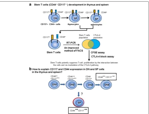
CD44+CD117+ stem T-cell development in the thymus and spleen. We sought to identify various gene/protein ex-pression changes at different stages of T-cell development. Many changes were identified, which are contradictory to the current theory of T-cell development. In this study, peak CD117 expression occurred in DN and CD117 cells and could still be detected in SP T cells in the thymus and spleen (Fig. 13). Thus, studies are needed to explain this phenomenon.
Previous work indicates that CD117 cells are present in DN1and DN2, but not in SP cells [31]. We observed that
CD117 cells such as NK T cells and Tregsdevelop in the
thymus, and do not follow the theory of cell or DN T-cell development. CD117 T-cell development may begin from CD44+DN cells and proceed directly to CD44+SP cells. It is unclear why NK T cells, Tregsand CD44+CD117+ cells
are T cells in phenotype but do not share the T-cell devel-opment pathway. We propose that CD44+CD117+ T cells may not have been derived from a common precursor as T cells, but rather pass through a different pathway.
Conclusions
In summary, CD44+CD117+ T-cell development oc-curred in the thymus and spleen. CD44+CD117+ T cells expressed abundant CTLA-4 and suppressed T-cell pro-liferation. Blocking CTLA-4 reduced the suppression of T-cell proliferation. CD44+CD117+ T cells are stem cells that expressed more Nanog and CTLA-4 and potently suppressed T-cell proliferation via modulating the CTLA-4 pathway. Finally, CD44+CD117+ T cells may have developed from CD4–CD8–CD44+CD25– T pro-genitor cells, without the involvement of DN3, DN4, and
CD4+CD8+(DP) stages in the thymus.
Abbreviations
CTLA-4:Cytotoxic T lymphocyte antigen-4; DN: Double-negative CD4–CD8–; DN1: Double-negative CD44+CD25–; DN2: Double-negative CD44+CD25+;
DN3: Double-negative CD44–CD25+; DN4: Double-negative CD44–CD25–;
DP: Double-positive CD4+CD8+; SP: Single-positive CD4 or CD8
Acknowledgements
The authors gratefully acknowledge The Xi'an Jiaotong University Cardiovascular Research Center for providing their flow cytometry services.
Funding
This study was supported by The First Affiliated Hospital, Xi'an Jiaotong University and Natural Science Foundation of Shaanxi Province of China (No. 2016JM8039).
Availability of data and materials
The data that support the findings of this study are available from the corresponding author on reasonable request.
Authors’contributions
TL contributed to the design of the study and writing the manuscript, performed experiments, and analyzed data. YW, ZSH, WG, GL, BYS, and EQL participated in the experimental design and manuscript preparation, performed some experiments, and analyzed data. All authors read and approved the final manuscript.
Competing interests
The authors declare that they have no competing interests.
Consent for publication Not applicable.
Ethics approval and consent to participate
All protocols were approved by the Institutional Animal Care and Use Committee at Xi’an Jiaotong University (Permit Number: 2016-278). All surgeries and sacrifices were performed under chloral hydrate anesthesia. Every effort was made to minimize animal suffering.
Author details
1Immunology and Tumor Research Instituted, the First Affiliate Hospital, Xi’an Jiaotong University Health Science Center, Xi’an, Shaanxi 710061, People’s Republic of China.2Core Research Laboratory, the Second Affiliated Hospital, Xi’an Jiaotong University Health Science Center, Xi’an, Shaanxi 710049, China. 3The First Affiliated Hospital, Jinzhou Medical University, Liaoning 121004, People’s Republic of China.4Clinical Research Center, Guangdong Medical Collage, Zhanjiang, Guangdong 524001, China.5The School of Medicine, Xi’an Jiaotong University Health Science Center, Xi’an, Shaanxi 710061, China.
Received: 12 August 2016 Revised: 10 December 2016 Accepted: 9 February 2017
References
1. Ritter MA, Boyd RL. Development in the thymus. it takes two to tango. Immunol Today. 1993;14:462–9.
2. Anderson G, Pongracz J, Parnell S, Jenkinson EJ. Notch ligand-bearing thymic epithelial cells initiate and sustain Notch signaling in thymocytes independently of T cell receptor signaling. Eur J Immunol. 2001;31:3349–54.
3. Shortman K. Cellular aspects of early T-cell development. Curr Opin Immunol. 1992;4:140–6.
4. Ceredig R, Rolink T. A positive look at double-negative thymocytes. Nat Rev Immunol. 2002;2:888–97.
5. Godfrey DI, Kennedy J, Suda T, Zlotnik A. A developmental pathway involving four phenotypically and functionally distinct subsets of CD3–CD4– CD8–triple-negative adult mouse thymocytes defined by CD44 and CD25 expression. J Immunol. 1993;150:4244–52.
6. Youd M, Blickarz C, Woodworth L, Touzjian T, Edling A, Tedstone J, Ruzek M, Tubo R, Kaplan J, Lodie T. Allogeneic mesenchymal stem cells do not protect NZBxNZW F1 mice from developing lupus disease. Clin Exp Immunol. 2010;161:176–86.
7. Schena F, Gambini C, Gregorio A, Mosconi M, Reverberi D, Gattorno M, Casazza S, Uccelli A, Moretta L, Martini A, et al. Interferon-gamma-dependent inhibition of B cell activation by bone marrow-derived mesenchymal stem cells in a murine model of systemic lupus erythematosus. Arthritis Rheum. 2010;62:2776–86.
8. Zhang S, Balch C, Chan MW, Lai HC, Matei D, Schilder JM, Yan PS, Huang TH, Nephew KP. Identification and characterization of ovarian cancer-initiating cells from primary human tumors. Cancer Res. 2008;68:4311–20. 9. Hombach-Klonisch S, Panigrahi S, Rashedi I, Seifert A, Alberti E, Pocar P,
Kurpisz M, Schulze-Osthoff K, Mackiewicz A, Los M. Adult stem cells and their trans-differentiation potential—perspectives and therapeutic applications. J Mol Med (Berl). 2008;86:1301–14.
10. Collins AT, Berry PA, Hyde C, Stower MJ, Maitland NJ. Prospective identification of tumorigenic prostate cancer stem cells. Cancer Res. 2005;65:10946–51.
11. Qiu FH, Ray P, Brown K, Barker PE, Jhanwar S, Ruddle FH, Besmer P. Primary structure of c-kit: relationship with the CSF-1/PDGF receptor kinase family—oncogenic activation of v-kit involves deletion of extracellular domain and C terminus. EMBO J. 1988;7:1003–11.
12. Yarden Y, Kuang WJ, Yang-Feng T, Coussens L, Munemitsu S, Dull TJ, Chen E, Schlessinger J, Francke U, Ullrich A. Human proto-oncogene c-kit. a new cell surface receptor tyrosine kinase for an unidentified ligand. EMBO J. 1987;6:3341–51.
13. Ray P, Krishnamoorthy N, Ray A. Emerging functions of c-kit and its ligand stem cell factor in dendritic cells: regulators of T cell differentiation. Cell Cycle. 2008;7:2826–32.
14. Plas DR, Thompson CB. Akt-dependent transformation. there is more to growth than just surviving. Oncogene. 2005;24:7435–42.
15. Keshet E, Lyman SD, Williams DE, Anderson DM, Jenkins NA, Copeland NG, Parada LF. Embryonic RNA expression patterns of the c-kit receptor and its cognate ligand suggest multiple functional roles in mouse development. EMBO J. 1991;10:2425–35.
16. Heissig B, Werb Z, Rafii S, Hattori K. Role of c-kit/Kit ligand signaling in regulating vasculogenesis. Thromb Haemost. 2003;90:570–6. 17. Metcalfe DD. Mast cells and mastocytosis. Blood. 2008;112(4):946–56. 18. Aifantis I, Mandal M, Sawai K, Ferrando A, Vilimas T. Regulation of T-cell
progenitor survival and cell-cycle entry by the pre-T-cell receptor. Immunol Rev. 2006;209:159–69.
19. Mitsui K, Tokuzawa Y, Itoh H, Segawa K, Murakami M, Takahashi K, Maruyama M, Maeda M, Yamanaka S. The homeoprotein Nanog is required for maintenance of pluripotency in mouse epiblast and ES cells. Cell. 2003;113:631–42.
20. Lenschow DJ, Walunas TL, Bluestone JA. CD28/B7 system of T cell costimulation. Annu Rev Immunol. 1996;14:233–58.
21. Gough SC, Walker LS, Sansom DM. CTLA4 gene polymorphism and autoimmunity. Immunol Rev. 2005;204:102–15.
22. Ise W, Kohyama M, Nutsch KM, Lee HM, Suri A, Unanue ER, Murphy TL, Murphy KM. CTLA-4 suppresses the pathogenicity of self antigen-specific T cells by cell-intrinsic and cell-extrinsic mechanisms. Nat Immunol. 2010;11:129–35.
23. Dalerba P, Dylla SJ, Park IK, Liu R, Wang X, Cho RW, Hoey T, Gurney A, Huang EH, Simeone DM, et al. Phenotypic characterization of human colorectal cancer stem cells. Proc Natl Acad Sci U S A. 2007;104:10158–63. 24. Jain N, Nguyen H, Chambers C, Kang J. Dual function of CTLA-4 in
regulatory T cells and conventional T cells to prevent multiorgan autoimmunity. Proc Natl Acad Sci U S A. 2010;107:1524–8.
25. Tung JW, Heydari K, Tirouvanziam R, Sahaf B, Parks DR, Herzenberg LA. Modern flow cytometry: a practical approach. Clin Lab Med. 2007;27:453–68. v.
26. Liu G, Li Z, Wei Y, Lin Y, Yang C, Liu T. Direct detection of FoxP3 expression in thymic double-negative CD4–CD8- cells by flow cytometry. Sci Rep 2014; 4:5781.
27. Di Santo JP, Radtke F, Rodewald HR. To be or not to be a pro-T? Curr Opin Immunol. 2000;12:159–65.
28. Fang D, Nguyen TK, Leishear K, Finko R, Kulp AN, Hotz S, Van Belle PA, Xu X, Elder DE, Herlyn M. A tumorigenic subpopulation with stem cell properties in melanomas. Cancer Res. 2005;65:9328–37.
29. Carrade DD, Borjesson DL. Immunomodulation by mesenchymal stem cells in veterinary species. Comp Med. 2013;63:207–17.
30. Hanahan D, Weinberg RA. Hallmarks of cancer: the next generation. Cell. 2011;144:646–74.
31. Bhandoola A, von Boehmer H, Petrie HT, Zúñiga-Pflücker JC. Commitment and developmental potential of extrathymic and intrathymic T cell precursors: plenty to choose from. Immunity. 2007;26:678–89.
• We accept pre-submission inquiries
• Our selector tool helps you to find the most relevant journal • We provide round the clock customer support
• Convenient online submission • Thorough peer review
• Inclusion in PubMed and all major indexing services • Maximum visibility for your research
Submit your manuscript at www.biomedcentral.com/submit