Published by Science and Education Publishing DOI:10.12691/jfnr-8-9-4
Addition of Opuntia ficus-indica Reduces Hypothalamic
Microglial Activation and Improves Metabolic
Alterations in Obese Mice Exposed to a High-fat Diet
Mercedes Victoria Urquiza-Martínez1, Héctor Eduardo Martínez-Flores2,*, Omar Guzmán-Quevedo3, Ana Elisa Toscano4, Raul Manhães de Castro4, Luz Torner2, Rosalío Mercado-Camargo2,
Rosa Elena Pérez-Sánchez2, María Carmen Bartolome-Camacho2
1
Programa Institucional de Doctorado en Ciencias Biológicas. Universidad Michoacana de San Nicolás de Hidalgo. Morelia, México 2
Facultad de Químico Farmacobiología, Universidad Michoacana de San Nicolás de Hidalgo, Morelia, México 3
Instituto Tecnológico Superior de Tacámbaro, Tacámbaro, Michoacán, México 4
Pós-Graduação em Neuropsiquiatria e Ciências do Comportamento, Universidade Federal de Pernambuco, Recife, Brasil 5
Centro de Investigación Biomédica de Michoacán, Instituto Mexicano del Seguro Social, Morelia 58341, Michoacán, México *Corresponding author: hector.martinez.flores@umich.mx
Received August 09, 2020; Revised September 10, 2020; Accepted September 18, 2020
Abstract
Opuntia ficus-indica consumption improves obesity and glucose and lipid metabolism, to name a few; however, the involved mechanism is poorly understood and attributed primarily to fiber. Obesity was recently shown to increase microglial cell activation in the hypothalamus. Here, we hypothesized that the addition of cactus flour (CF) to a high-fat diet (HFD) reduces hypothalamic inflammation, which is a key regulator of energy balance metabolism. Adult male C57Bl/6j mice underwent HFD (60% cal from fat) exposure over 12 weeks to develop obesity. The same type of HFD added with CF (17%) was administrated for 4 weeks. Bodyweight and food intake were recorded during the treatment, and glucose and insulin tolerance were evaluated at the end of the treatment. Additionally, the behavioral satiety sequence (BSS) was performed, and adiposity, along with microglia activation and density in the arcuate nucleus was determined. Herein, we found that CF normalizes body weight and adiposity without changes in absolute food intake. Moreover, CF modulated the cumulative caloric intake in a diet-dependent manner. Feed efficiency was decreased; glucose tolerance and insulin sensitivity were improved with CF treatment. BSS showed a decreased resting time and increased grooming for animals with an HFD-CF. Finally, CF consumption normalized microglia density in the arcuate nucleus of the hypothalamus in obese mice and significantly decreased their activation.This study shed new light on the understanding of the effects of cactus to reduce body weight, adiposity and improve glucose metabolism, and suggest they are mediated by the reduction of the microglial activation of the hypothalamic arcuate nucleus.Keywords
: obesity, energy metabolism, microglia, neuro-inflammation, insulinCite This Article:
Mercedes Victoria Urquiza-Martínez, Héctor Eduardo Martínez-Flores, Omar Guzmán-Quevedo, Ana Elisa Toscano, Raul Manhães de Castro, Luz Torner, Rosalío Mercado-Camargo, Rosa Elena Pérez-Sánchez, and María Carmen Bartolome-Camacho, “Addition of Opuntia ficus-indica Reduces Hypothalamic Microglial Activation and Improves Metabolic Alterations in Obese Mice Exposed to a High-fat Diet.” Journal of Food and Nutrition Research, vol. 8, no. 9 (2020): 473-483. doi: 10.12691/jfnr-8-9-4.1. Introduction
Obesity, defined as “a disease process characterized by excessive body fat accumulation with multiple organ-specific consequences” [1], is the result of an imbalance between energy consumed and expended. While mammals can keep a relatively constant body weight (BW) despite variations in day-to-day calorie intake/expenditure [2], studies from Milanski and Col [3] probe an acute hypothalamic inflammatory response after exposure to a HFD due to microglia activation, even before BW gain,
causing glucose metabolism alterations and adipocyte-derived hormone leptin resistance [3].
Hypothalamic regulation of energy balance (EB) relies on arcuate nucleus neuron’s production of some neurotransmitters and hormonal precursors to produce food intake stimulation (by Agouti related-peptide and Neuropeptide Y - inverse agonists of melanocortin 4 receptor-action) or inhibition - so called satiety - by means of pro-opiomelanocoritn (melanocortin receptor agonist)
eventual loss of pro-opiomelanocortin (POMC) neurons and gliosis characterized by activation of microglia cells, which produce several damaging cytokines and pro-oxidant compounds - making the weight loss very difficult to obese subjects [5].
This phenomenon is known as neuroinflammation, an event conducted by microglial activation. In this case, the insult is in charge of long-chain of saturated fats which act through TLR4 (Toll Like Receptor 4) in microglia inducing expression of inflammatory cytokines, activating inflammatory pathways - JNK and IKK - and recruiting bone-marrow-derived monocytes, which amplify and perpetuate hypothalamic inflammation [6]. These findings outline the important role of the hypothalamus in the onset of obesity - prior to adipose tissue increase - and its regulation on energy balance as a possible target of treatment [7].
Obesity studies in rodents like mice (C57Bl/6j) are well established in many important groups of investigation. This animal model provides a human-like response to a diet-induced obesity that includes the weight gain, glucose metabolism alterations, resistance to insulin and leptin, and the so called previously hypothalamic inflammation in arcuate nucleus [5,24] which provides a suitable obesity model to achieve the objectives of this study.
Opuntia ficus-indica provides a wide range of health benefits. Biomolecules including vitamins, minerals, antioxidants, and phenolic compounds make it a functional food due to its probed effects over blood glucose, lipids levels, body weight (BW), and inflammation, and as a neuroprotector [8,9,10]. However, the mechanisms underlying these mentioned benefits over a complex disease, like obesity, remain largely unknown. Since there is evidence supporting the anti-inflammatory effect of Opuntia extracts over microglia and nitric oxide - NO - production in vitro [11], we hypothesized that Opuntia consumption has a direct effect on the microglial cells in the arcuate nuclei of the hypothalamus of diet-induced obese mice, associated with improvement of metabolic parameters including BW, FI, glucose, insulin tolerance, adiposity, and feeding behavior. To test this hypothesis, we evaluated the activation state of the microglia in the hypothalamic arcuate nucleus in obese mice with and without Opuntia consumption. The primary focus was the density and activation of microglia, along with the metabolic parameters mentioned above. If the hypothesis is true, CF can be used as an aid to complement obesity treatment.
2. Materials and Methods
Twenty-eight male adult (8 weeks old) C57Bl/6j mice were purchased from the Institute of Neurobiology of the UNAM in Juriquilla, Querétaro, Mexico, and kept in light/dark cycles of 12h each, at a temperature of 22±2°C, with access to food and water ad libitum. Their initial average weight was 23.15±0.48 g. After 1week of habituation, they were housed in acrylic cages, 2 animals per cage. The experiments were designed and carried out under the Mexican official normativity for the production, care, and use of laboratory animals (NOM-062-ZOO-1999),
the general law of health of Mexico, and the American National Institutes of Health. This project was reviewed and accepted by the bioethics committee of the Universidad Michoacana de San Nicolás de Hidalgo (Rec. No. 01-19CE-FAC QFB).
2.1. Obesity Induction
A standard diet to keep the animals healthy (n=14) and HFD to induce obesity (n=14) were provided according to the AIN-93 [12] and the composition of a commercial HFD with 60% cal and fat was offered to the animals for 12 weeks. After this period, two groups of obese mice (n=7 each) received either HFD-CF or the HFD with fiber quantity enriched to be similar to the cactus added diet (cellulose and guar) (HFD group). Diet composition is given in detail on Table 1, the amount of vitamins and minerals were adjusted (reduced) on CF-diets, given the quantity of vitamin (~2.3%) and mineral (~16%) of reported data by Bensadon et al (2010) of Opuntia ficus-indica, to avoid an excess of any of them compared to the non-added CF diets.
Table 1. Diet ingredients and composition of the different diets used in the study. Depicted are the ingredients of the Standard Diet (SD), Standard Diet with Cactus Flour (SD-CF), High Fat Diet (HFD) and High Fat Diet with Cactus Flour (HFD-CF). Equivalence of 1kcal=4.184kJ
g/kg Standard Diet
Standard Diet + Cactus Flour
High-Fat Diet
High-Fat Diet + Cactus Flour Cornstarch 615 581 256 227 Sucrose 100 100 100 100 Casein 140 138 180 178 Cellulose 91 0 91 0
Guar 16 0 16 0
Soy oil/Lard 40 40 358 354 Mineral Mix 35 10 35 10 Vitamin Mix 10 7 10 7 Cactus Flour - 170 - 170 %kcal Protein 14.8 14.9 13.4 13.5 %kcal
Carbohydrate 75.6 73.5 26.5 24.8 %kcal
Lipid 9.5 9.7 60.0 60.4 KJoules/g 15.81 15.48 22.46 22.04 Kcal/g 3.78 3.70 5.37 5.27
Cactus was obtained from a local producer (Ziracuaretiro, Michoacán) and properly identified as
2.2. Body Weight Gain, Feed Consumption
and Feed Efficiency
BW gain and FI were recorded weekly (at the same hour) by weighing animals and food consumption using a digital scale (Sartorius®). The feed efficiency (FE) is used to measure the capability to transform ingested calories by an animal into body weight (g). The latter was calculated as follows: (body weight gained/caloric intake) X 100 [13].
2.3. Glucose and Insulin Tolerance Tests
To establish whether cactus flour consumption improves glucose and insulin tolerance, we performed glucose tolerance test (GTT) and insulin tolerance test (ITT). After 6h fasting (from 6.00 to 12.00 hours), basal glucose levels were determined in all the groups using a glucometer (One Touch Ultra 2®) with drop of blood from the tail. A solution of glucose (Meyer, México) (2g/kg body weight i.p. 30%w/v) or insulin NPH (AMSA, México) (1U/kg body weight i.p.) was injected and glucose levels were taken at 15, 30, 60, 90, and 120 min [14]. Both GTT and ITT were carried out in the same mice between the third and fourth week of exposure to cactus flour. To avoid any fatal outcomes, mice were given glucose or insulin at any probe of hypo/hyperglycemia (dizziness, asthenia, etc.).
2.4. Behavioral Satiety Sequence
To see whether cactus flour intake can modulate feeding behavior, a behavioral satiety sequence (BSS) was performed on the 25th day of the treatment. Mice fasted for 2 hours were offered a well-known amount of food and video-recorded (40 min) over the dark cycle, using the method described by Vickers [15], adapted from Halford
[16]. The 40 min video was divided into 8 periods of 5 min each to measure animal is the feeding, grooming, or sleeping time, as well as FI (g and kcal) at the end of the experiment.
2.5. Tissue Processing
At the end of the treatment (28th day), the animals were deeply anesthetized with pentobarbital (100mg/kg BW, i.p., Sedalphorte Salud y Bienestar Animal, Mexico) - lethal dose [17]. Then, retroperitoneal and epididymal adipose tissues were quickly removed and weighed to determine adiposity. Blood was extracted for further analysis with a direct heart puncture. Afterwards, the mice were transcardially perfused with cold phosphate buffered saline (PBS) and 4% paraformaldehyde and their brains were removed. The tissue was post-fixed overnight with 4% parafromaldehyde and crioprotected with 30% sucrose at 4°C [18].
2.6. Immunohistochemistry and
Morphological Analyses
To determine the effect of cactus flour consumption on hypothalamic inflammation, the density and activation of microglia in the arcuate nucleus were evaluated. To do this, brains were cut in coronal sections of 30µm, and
immunostained as described in Roque et al. (2016) [18]. Brain sections were incubated with the primary antibody anti-Iba1 (1:10000, FUJIFILM Wako Chemicals, USA), and followed by incubations with a biotinylated secondary antibody, and avidin-biotin complex solutions (ABC Elite Kit, Vector Laboratories, Burlingame, CA USA). Microglial cell counting and morphological analyses were performed by an examiner blind to the groups the samples belonged to. The arcuate nucleus was examined using a counting frame (0.0613±0.0017 mm2) to delimit the area and the number of microglial positive cells was divided by the area in mm2. Two fields per section were analyzed from 3 hypothalamic sections per mouse. The fields were observed at a 10X magnification, using an image analysis system (Axioskop 40 Zess® microscope, Zeiss). The total number of microglia was first considered from the total number of Iba1-immunoreactive cells in the counting frame. From these, Iba1-immunoreactive cells were classified as activated or non-activated microglia according to Roque et al (2016).
2.7. Statistical Analysis
All data are presented in means ± standard error of the mean (SEM). Artwork was created and data was analyzed with Graph Pad Prism 6 (La Jolla, California, USA) with T-student Test, one or two-way analysis of variance (ANOVA), followed by Bodnferroni’s post hoc tests. Values with p<0.05 (two-sided significance level) were considered significant. The sample size was calculated considering 90% test power using G*Power 3 (Heinrich-Heine University, Dusseldorf, Germany), [19].
3. Results
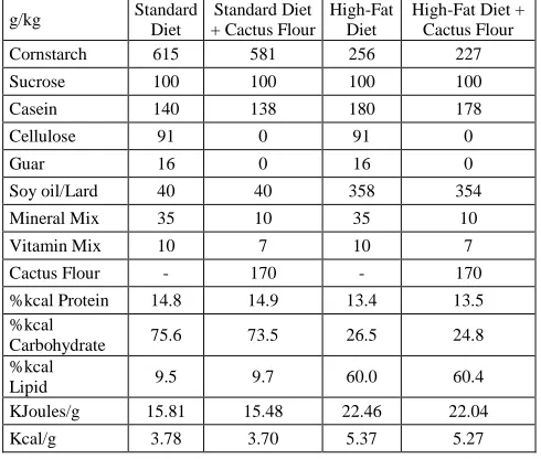
The HFD group exhibited 25.3% body weight gain after 12 weeks of feeding compared to the SD group (Figure 1A) (p=0.0001), confirming that it was a diet-induced obesity model [20].
Then, animals were fed for four weeks with cactus added diet or an adjusted fiber-diet. Cactus flour did not have any effect on BW during the treatment in SD, but completely normalized BW of obese mice from week two to the end of treatment (Figure 1B) (diet effect F[3, 120]=38.61, p<0.0001, time F[4, 120]=2.846, p=0.027 and
interaction F[12, 120]=1.205, p=0.28). The increased feed
efficiency observed in HFD was also blunted by adding cactus flour to the diet (Figure 1D) (F[3,20]=37.75, p<0.0001). SD-CF group exhibited an increase in both cumulative FI (F[3,24]=9.616, p=0.0002) and caloric intake
compared to the SD group; the HFD and HFD-CF groups ingested less food than SD group but there was no significant difference.
In terms of calories, HFD was higher than SD (Table 2) (F[3,24]=16.08, p<0.0001). The addition of cactus flour
significantly decreased caloric intake in HFD, contrasting with the effect observed when cactus flour was added to SD (Table 1 and Figure 1C).
when type 2 diabetes is suspected [21]. To know whether cactus consumption improves this parameter, we conducted GTT with all the groups. HFD-CF group had similar glucose levels (169.8±15.5 mg/dL) to SD group (153.4±22.2 mg/dL), contrary to HFD which maintains glucose levels up to 266.43±25.9 mg/dL (* vs SD). This difference is confirmed by the area under the curve (AUC) from the graph (diet effect F[3, 24]=5.525, p=0.005, time
F[5, 120]=86.45, p<0.0001 and interaction F[15, 120]=1.874, p=0.035) (Figure 2A and B). ITT serves to assess
peripheral tissue insulin sensitivity to introduce the circulating glucose efficiently into the cells. Like glucose tolerance, insulin sensitivity was also normalized by cactus consumption, when the mice were exposed to HFD (Figure 2C and D) (diet effect F[3, 16]=16.19, p<0.0001,
time F[5, 80]=86.09, p<0.0001 and interaction F[15, 80]=7.296, p<0.0001). Interestingly, neither glucose tolerance nor insulin sensitivity was affected by cactus consumption in SD mice, indicating that cactus flour only acts in high levels of energy (HFD).
Figure 1. Body weight over 12 weeks A) Diet-induced obesity in mice treated with a High Fat Diet (HFD) and the comparison with standard diet (SD), n=14 on each group. B) Weekly body weight change, C) Cumulative feed intake in g and kcal and D) Feed efficiency under cactus treatment of groups of mice fed with standard diet (SD), standard-diet with cactus flour (SD-CF), high fat diet (HFD) and high fat diet with cactus flour (HFD-CF); n=7 in each group. Two-way ANOVA (repeated measures for A and B) and one-way ANOVA (for C and D) followed by Bonferroni’s post hoc test. Data shown in means ± SEM. *P<0.05 vs SD
Table 2. Cumulative feed intake in grams and kilocalories of the diets used. Significant differences in food quantity (g) and energy (kcal) are shown for SD-CF diet, and differences only in energy are shown for HFD and SD-CF compared with SD diet. Also, SD-CF and HFD-CF present significant differences vs HFD diet
Groups Cumulative feed intake (g) Cumulative feed intake (kcal)
Mean SEM Mean SEM
Standard Diet 227.60 29.98 860.33 113.32
Standard Diet-Cactus flour 297.47* 22.10 1100.63* 81.76
High fat diet 181.25 5.49 971.50* 29.42
Figure 2. Blood glucose levels and area under the curve (AUC) of the experimental groups of mice. A) Glucose tolerance test (GTT) performed on the different groups of mice fed with standard diet (SD), standard-diet with cactus flour (SD-CF), high fat diet (HFD) and high fat diet with cactus flour (HFD-CF). B) AUC of GTT of the same groups. C) Insulin tolerance test (ITT) performed on all the groups. D) AUC of ITT of the groups. Analyzed with two-way ANOVA with a Bonferroni’s post hoc test, data shown in means ± SEM, n=7 *P<0.05 vs SD
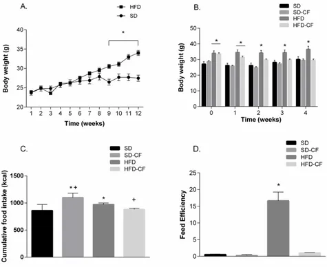
Feeding behavior, under constant environment features, can reflect the natural occurring of physiological processes involved in the regulation of feed intake, which commences with a period of feeding, after which the animal spends time grooming (liking and cleaning itself) and finally, rests/sleeps; the point of satiation is established in the intercept of feeding and resting curves
[16]. SD-CF mice showed a delayed point of satiation (period 6.6) (Figure 3B) compared to SD mice (period 3) (Figure 3A). In contrast, cactus flour did not affect the point of satiation in HFD-exposed mice. However, both HFD and HFD-CF groups exhibited a lower point of satiation (1.8 and 1.9, respectively) compared to the SD group (Figure 3A, C, and D). Although cactus consumption did not affect the point of satiation in HFD conditions, other beneficial effects were observed. Indeed, cactus flour decreased the time of rest in both SD and HFD mice (Figure 3A-D). In HFD-CF, an increase in grooming and feeding was observed, which explains the reduced resting time. Food intake showed significant
decrease on HFD group vs the others during the experiment, expressed in grams (F[3, 20]=3.236, p=0.044)
(Figure 3E) and kcal (F[3, 20]=3.939, p=0.0233) (Figure 3F).
Figure 3. Behavioral satiety sequence (BSS) of the different groups of mice. Time in seconds spent in the BSS of Standard Diet group (A), Standard Diet + Cactus Flour (B), High Fat Diet (C) and High Fat Diet + Cactus Flour (D). The vertical line shows the point of satiety where the feeding and resting line crosses. Food consumption is shown in grams (E) and in kcal (F) of the SD, SD-CF, HFD and HFD-CF groups of mice. One-way ANOVA followed by Bonferroni’s post hoc test was used. Data showed in means ± SEM, n=6, P≤0.05 vs *SD or +HFD
Next, we evaluated possible changes of the inflammatory state in the hypothalamic arcuate nucleus after cactus flour treatment. To do so, we analyzed the total density of microglia cells and the quantity of them that were activated (bigger and rounded) in mice under the different diet treatments. Photomicrographs (10X) representing each group of mice:
SD, SD-CF, HFD and HFD-CF are respectively shown in
Figure 5A-D. The total density of microglia (cells/mm2) was similar in SD, SD-CF and HFD-CF mice. Although not significantly, HFD had increased the total density of microglia (~28%), compared to the SD group as depicted in Figure 5E (F[3,24]=2.176, p=0.1171).
Figure 5. Micrography of hypothalamic coronal sections showing the Arcuate Nucleus (10X) of mice fed with A) Standard Diet (SD), B) Standard Diet with Cactus Flour (SD-CF) , C) High Fat Diet (HFD) and D) High Fat Diet with Cactus Flour (HFD-CF). 3V is the third ventricle. E) Total microglial density (cell number/mm2) in the arcuate nucleus and F) Activated microglial density (cell number/mm2) in the arcuate Nucleus of SD, SD-CF, HFD
Activated microglial cells (cells/mm2), characterized by a larger and rounded form, were selected, and quantified from the total population. HFD animals presented a significant 2-fold increase of activated microglia compared to SD and SD-CF groups. Treatment of HFD mice with cactus flour showed a significant reduction of the activated microglial density in the arcuate nucleus compared to HFD group, which was still higher than SD and SD-CF groups (Figure 5F) (F[3,24]=18.66, p<0.0001).
4. Discussion
Many of our results are consistent with previously published data of Opuntia ficus-indica health benefits
[10,23]. Besides that, diet-induced obesity models offer a similar response from what is observed in humans [24]. An inflammatory state of the hypothalamus, induced by a high-fat diet can alter the normal energy balance leading to many metabolic disturbances such as glucose intolerance and poor sensitivity to insulin and leptin action, altering satiety sensation in eating behavior, increasing feeding efficiency, lowering energy expenditure, and augmenting body weight gain at the expense of adipose tissue [25,26,27]. However, HFD also causes characteristic localized cell proliferation in the arcuate nucleus, particularly by an augmentation in microglial cell density with a proinflammatory phenotype
[28].
4.1. Body Weight Gain, Feed Intake and Feed
Efficiency
Natural compounds found in several food-derived products can reduce obesity-associated damage by modulating many metabolic aspects and probably by acting on the hypothalamic inflammatory state, such as the case of
Opuntia ficus-indica with probed effects over glucose metabolism [29,30], improvement on insulin secretion
[31], and weight loss [32]. Cactus can provide antioxidant biomolecules with complex functions over certain receptors involved in central nervous system inflammation induced by microglial cells [11].
Even though SD and HFD were adjusted to be similar in fiber (soluble and insoluble) quantity as the cactus added diets - SD-CF and HFD-CF - there were no significant differences to support the hypothesis that its health beneficial effects are due to the fiber content alone. A study performed on obese mice showed a comparison between a soluble (guar) vs insoluble (oat fiber) high-fat fiber-added diet during 45 weeks, showing that the difference between the groups on weight gain and insulin resistance was lower in the fiber insoluble group [33]. Consumption of fiber is associated with reduced diabetes risk, fecal energy loss by hindering macronutrient absorption, and slow gastric emptying, and it also promotes the release of cholecystokinin (CCK), Glucagon-like peptide 1 (GLP-1), and Peptide YY (PYY), gut hormone satiety promoters [34,35]. Fiber alone can be helpful in obesity management, but apparently requires long-term application to obtain such benefits.
4.2. Glucose and Insulin Effects
Alterations in glucose metabolism-glucose intolerance and Diabetes type 2 are common in obese subjects - HFD - that causes initial insulin resistance over muscle and liver and later over adipose tissue. Such alterations are linked to the protein kinase Akt signaling, however, there are independent Akt pathways involved in it as well, such as, defects on insulin receptor and Insulin Receptor Substrate (IRS) proteins [36].
Several diabetes and obesity studies with Opuntia spp
probed improvement in pancreatic islet integrity, β-cell proliferation, and insulin secretion induction by the hand of antioxidant, anti-inflammatory and circulating lowering lipid probed effects. The mechanisms proposed include a greater ratio of phosphorylatedAkt:Akt, higher expression of glucose transporter 2 (GLUT2) and peroxisome proliferator-activated receptor gamma (PPARγ); it also showed increased energy expenditure, reduced adipocyte size, increased ISR phosphorylation, and expression of genes related to fatty acid oxidation [32,37].
4.3. Satiety
The satiety cascade is a combination of physiological, behavioral, and psychological events that control appetite and are part of the eating process. Such cascade begins with hunger that leads to the initiation of eating, which later progresses into satiety for a period which ends, after which a new cycle commences. The brain integrates several signals from the processes to conjugate it with energy balance and reward pathways [38].
Satiety can be measured as the duration interval that separates eating from the next episode but can also be measured as the decrease in intake occurring in the next eating episode. While glucose level is a key factor that modulates hunger/satiety responses by means of eating carbohydrates, protein can also contribute with this phenomena showing even higher satiety potency, leaving fat at the end of hierarchy satiating power (protein>carbohydrates>fat). But micronutrients and non-nutrient food components can influence and promote satiety-like calcium, vitamins, fiber, and phenolic compounds, by increasing levels of anorexigenic hormones (PYY) and/or participating in peptide and neurotransmitter synthesis that control food intake [36]. Such effect could be related to the results obtained in our experiment, in which obese mice under treatment with cactus (HFD-CF) showed an earlier point of satiation possibly related to the micro and non-nutrient part of the flour rich in fiber (9.8g soluble and 54.45g insoluble/100g dry matter), polyphenols (2.69g Gallic Acid Equivalents/100g dry matter), calcium and vitamins
[8].
4.4. Microglial Proliferation and Activation
arcuate nucleus adjacent macrophagues - microglial cells - and promote inflammation by increasing production of interleukins (Il-6, Il-1β and TNFα) and iNOs- and POMC neuron loss [5]. In this study, we observed that cactus flour intake improves hypothalamic inflammation, suggesting that it is a mechanism involved in the metabolic benefits of cactus consumption. Cactus, rich in several phenolic compounds such as quercetin and kaempferol [40], can modulate energy metabolism on metabolic altered subjects without disturbing healthy ones, as they do not lose body weight beyond normal levels. Such effect can be exerted through the stimulation of thyroid hormone (T3) by increasing the quantity and duration of the iodothyronine deiodinase (D2) via protein kinase A activation [41]. Other mechanisms involved in energy balance restoration by polyphenols were suppression of dietary fat absorption, inflammatory reduction, sympathetic stimulation of thermogenesis, and modulating neuropeptides involved in feed intake and satiety [42,43,44]. Other possible mechanisms involving quercetin in this anti-inflammatory process promotes HO-1 enzyme expression in the lipid altered microglia, causing NF-κB interruption and resulting in an attenuation of oxidative stress, inflammation, and metabolic dysregulation [45].
Administration of different rich polyphenol extracts from other food sources exerted anti-obese effects acting on hypothalamic high fat-diet alterations. Polyphenols reduced the expression of inflammatory markers like interleukin 6 (IL-6) and IL-1β and negative modulators of leptin signaling (SOCS3 and PTP1B). The results obtained from these studies were the reduction of body weight, adipose mass depots, feed intake, glucose tolerance and insulin and leptin sensitivity [46,47,48].
Similarly, in this work, we found that the inflammatory state in the ARC (evidenced by microglia activation) was reduced by the administration of Opuntia flour, and that its effects could be exerted through its polyphenolic compounds. However, Opuntia administration could be slower to reach the desired effects over an important number of activated microglia due to its means of administration (oral) compared to the studies mentioned above which administrated the compounds intraperitoneally.
Cactus administration, even with a high-fat diet, can improve the inflammatory state on hypothalamic arcuate nucleus and thus cause reverse energy balance alterations. Effects of cactus administration are quite complete, as it serves to ameliorate glucose metabolism, improving energy spending, and reduce fat mass, and by its antioxidant properties, correct the inflammation state on central nervous system arcuate nucleus. Such beneficial effects cannot be attributed to fiber alone, as we know, the addition of fiber - on SD and HFD groups - by itself (cellulose and guar) did not have a health beneficial result (on HFD obese group) during this study; apparently it is the combination of bioactive molecules, e.g., the polyphenols present in the cactus flour that causes such an effect. There are more compounds than fiber in the cactus that function as metabolism regulators and allow the decrease of hypothalamic inflammation by reducing microglial cell density. Among the compounds found in it, several studies have found that phenolic compounds are able to restore energy balance disturbances and in doing
so, they correct the metabolic alterations characteristic of obesity.
Taken together, these data show an important effect of
Opuntia to improve obesity-related alteration including BW, adiposity, glucose tolerance, and insulin sensitivity. The treatment with this cactus most importantly reduced microglia density and activation in the arcuate nucleus, a key factor in the regulation of EB. In previous works, high density, and activation of microglia in this region have been linked with an inflammatory state responsible for obesity and obesity-related alteration [28]. In this context, this study suggests that Opuntia improves the metabolic phenotype by decreasing inflammation in the arcuate nucleus, which, in turn, reestablishes energy balance regulation. However, further studies are necessary to test that the CF in our model can modulate the expression of several neurotransmitters, inflammation markers (cytokines) and metabolism regulators (e.g. POMC, leptin, etc.). Also, an acute (hours) evaluation of the effect of CF on hypothalamic inflammation will serve to highlight its acute effect to revert inflammation at the CNS level. Additionally, despite the mice having over 85% genetic similarity with humans, clinical trials are necessary to confirm such a beneficial effect on humans from the perspective of using it as a strategy to treat the hypothalamic inflammation observed in obese humans.
5. Conclusions
In summary, our hypothesis is confirmed, in that
Opuntia ficus-indica reduces neuro-inflammation evidenced by the lower density of microglia and the reduction of its activation on arcuate nucleus of hypothalamus. These results can help to elucidate some part of the complex mechanisms involved in energy balance and its disruption by dietary factors. Thus, a gap for more investigation is opened to gather information (e. g. gene expression) that allows the development of proper and more effective treatment to obesity and its alterations.
Acknowledgements
To Ana Elisa Toscano’s team in Brazil and to Luz Torner team of IMSS who actively participate with capacitation and guidance. Declaration of Interest: none. Financial Support: This research was supported by the CONACYT founding under national and mixed grant agreement 286672/514268. Partial funding for this study was provided by a grant to L.T. from CONACyT (243419-2014).
Statement of Competing Interests
The authors have not competing interests.
List of Abbreviations
BSS Behavioral satiety sequence CCK Cholecystokinin
BW Body weight CF Cactus flour
D2 Iodothyronine deiodinase FE Feed efficiency
FI Feed intake
GLP-1 Glucagon-like peptide 1 GLUT2 Glucose transporter 2 GTT Glucose tolerance test HFD High fat diet
HFD-CF High fat diet and cactus flour
IKK The inhibitor of nuclear factor-κ (IκB) kinase Il Interleukins
i-NOS Inducible nitric oxide synthase IRS Insulin receptor substrate ITT Insulin tolerance test JNK c-Jun N-terminal kinases NF-κB Nuclear factor kappa B NO Nitric oxide
NPH Neutral protamine Hagedorn insulin POMC Pro-opiomelanocortin
PPARγ Peroxisome proliferator-activated receptor gamma
PTP1B Protein tyrosine phosphatase 1B PYY Peptide YY
SD Standard diet
SD-CF Standard diet and cactus flour SOCS3 Suppressor of cytokine signaling 3 T3 Thyroid hormone
TLR4 Toll like receptor 4
TNFα Tumor necrosis factor alpha
References
[1] Scottish Intercollegiate Guidelines Network (SIGN). Clinical management of obesity: a national clinical guideline No 15 Feb. 2010.
[2] Ravussin, Y., Leibel, R.L. and Ferrante Jr, A.W., “A missing link in body weight homeostasis: the catabolic signal of the overfed state” Cell Metab 20(4), 565-72, 2014.
[3] Milanski, M., Degasperi, G., Coope, A., Morari, J., Denis, R., Cintra, D.E., Tsukumo, D.M.L., Anhe, G., Amaral, M.E., Takahashi, H.K., Curi, R., Oliveira, H.C., Carvalheira, J.B.C., Bordin, S., Saad, M.J. and Velloso, L.A., “Saturated fatty acids produce an inflammatory response predominantly through the activation of TLR4 signaling in hypothalamus: implications for the pathogenesis of obesity”. J Neurosci, 29(2), 359-70, 2009.
[4] Gautron, L., Elmquist, J.K. and Williams, K.W., “Neural control of energy balance: translating circuits to therapies”. Cell, 161(1), 133-45. 2015.
[5] Berkseth, K.E., Guyenet, S.J., Melhorn, S.J., Donghoon, L., Thaler, J.P., Schur, E.A. and Schwartz, M.W., “Hypothalamic gliosis associated with high-fat diet feeding is reversible in mice: a combined immunohistochemical and magnetic resonance imaging study”, Endocrinol, 155(8), 2858-67, 2014.
[6] Dragano, N.R., Haddad-Tovolli, R. and Velloso, L.A., “Leptin, neuroinflammation and obesity. Endocrine Immunology, Karger Pub., 48, 84-96, 2017.
[7] Cota, D., Proulx, K. and Seeley, R.J., “The role of CNS fuel sensing in energy and glucose regulation”, Gastroenterol, 132(6), 2158-68, 2007.
[8] Bensadón, S., Hervert-Hernández, D., Sáyago-Ayerdi, S.G. and Goñi, I., “By-products of Opuntia ficus-indica as a source of antioxidant dietary fiber”, Plant Foods Human Nutr, 65(3), 210-16, 2010.
[9] Valencia-Sandoval, K., Brambila-Paz, J.D.J. and Mora-Flores, J.S., “Evaluación del nopal verdura como alimento funcional mediante opciones reales”, Agrociencia, 44(8), 955-63, 2010.
[10] Osuna-Martínez, U., Reyes-Esparza, J. and Rodríguez-Fragoso, L., “Cactus (Opuntia ficus-indica): a review on its antioxidants properties and potential pharmacological use in chronic diseases”, Nat Prod Chem Res, 2, 153.
[11] Lee, M.H., Kim, J.Y., Yoon, J.H., Lim, H.J., Kim, T.H., Jin, C., Kwak, W-J, Han, C-K. and Ryu, J-H-. “Inhibition of nitric oxide synthase expression in activated microglia and peroxynitrite scavenging activity by Opuntia ficus indica var. saboten”, Phytother Res, 20(9), 742-47, 2006.
[12] Reeves, P.G., Nielsen, F.H., Fahey Jr, G.C., “AIN-93 purified diets for laboratory rodents: final report of the American Institute of Nutrition ad hoc writing committee on the reformulation of the AIN-76A rodent diet”, J Nutr, 123(11), 1939-51, 1993.
[13] Parekh, P.I., Petro, A.E., Tiller, J.M., Feinglos, M.N., and Surwit, R.S., “Reversal of diet-induced obesity and diabetes in C57BL/6J mice”, Metabolism, 47(9), 1089-96, 1998.
[14] Andrikopoulos, S., Blair, A.R., Deluca, N., Fam, B.C., Proietto, J., “Evaluating the glucose tolerance test in mice”. Am J Physiol-Endoc Metab, 295(6), E1323-32, 2008.
[15] Vickers, S.P., Clifton, P.G., Dourish, C.T. and Tecott, L.H., “Reduced satiating effect of d-fenfluramine in serotonin 5-HT2C receptor mutant mice”, Psychopharmacol, 143(3), 309-14, 1999.
[16] Halford, J.C., Wanninayake, S.C. and Blundell, J.E., “Behavioral satiety sequence (BSS) for the diagnosis of drug action on food intake”, Pharmacol Biochem Behav, 61(2), 159-68, 1998.
[17] Lamanna, C. and Hart, E.R. “Relationship of lethal toxic dose to body weight of the mouse”, Toxicol Appl Pharm, 13(3), 307-15, 1968.
[18] Roque, A., Ochoa-Zarzosa, A. and Torner, L., “Maternal separation activates microglial cells and induces an inflammatory response in the hippocampus of male rat pups, independently of hypothalamic and peripheral cytokine levels, Brain Behav Immun, 55, 39-48, 2016.
[19] Faul, F., Erdfelder, E., Lang, A.-G. and Buchner, A. “G*Power 3: A flexible statistical power analysis program for the social, behavioral, and biomedical sciences”, Behav Res Methods, 39, 175-91, 2007.
[20] Hariri, N. and Thibault, L., “High-fat diet-induced obesity in animal models”, Nutr Res Rev, 23(2), 270-299, 2010.
[21] Vinué, Á. and González-Navarro, H., “Glucose and Insulin Tolerance Tests in the Mouse”, Methods Mol Biol, 1339, 247-54, 2015.
[22] Mann, A., Thompson, A., Robbins, N. and Blomkalns, A.L., “Localization, identification, and excision of murine adipose depots”, J Vis Exp, (94), e52174.
[23] El-Mostafa, K., El Kharrassi, Y., Badreddine, A., Andreoletti, P., Vamecq, J., Kebbaj., M.S.E., Latruffe, N., Lizard, G., Nasser, B. and Cherkaoui-Malki, M., “Nopal cactus (Opuntia ficus-indica) as a source of bioactive compounds for nutrition, health and disease”, Mol, 19(9), 14879-901, 2014.
[24] Thaler, J.P., Yi, C.X., Schur, E.A., Guyenet, S.J., Hwang, B.H., Dietrich, M.O., Zhao, X., Sarruf, D.A, Izgur, V., Maravilla, K.R., Nguyen, H.T., Fischer, J.D., Matsen, M.E., Wisse, B.E., Morton, G.J., Horvath, T.L., Baskin, D.G., Tschöp, M.H. and Schwartz, M.W., “Obesity is associated with hypothalamic injury in rodents and humans”, J Clin Invest, 122(1), 153-162, 2012.
[25] Wang, C.Y. and Liao, J.K., “A mouse model of diet-induced obesity and insulin resistance”, Methods Mol Biol, 821,421-33, 2012.
[26] Jais, A. and Brüning, J.C., “Hypothalamic inflammation in obesity and metabolic disease”, J Clin Invest, 127(1), 24-32, 2017.
[27] Dorfman, M.D. and Thaler, J.P., “Hypothalamic inflammation and gliosis in obesity”, Curr Opin Endocrinol, 22(5), 325, 2015.
[28] André, C., Guzman-Quevedo, O., Rey, C., Rémus-Borel, J., Clark, S., Castellanos-Jankiewicz, A., Ladeveze, E., Leste-Lasserre, T., Nadjar, A., Abrous, D.N., Laye, S. and Cota D., “Inhibiting microglia expansion prevents diet-induced hypothalamic and peripheral inflammation”, Diabetes, 66(4), 908-919, 2017.
[29] Luo, C., Zhang, W., Sheng, C., Zheng, C., Yao, J. and Miao, Z., “Chemical composition and antidiabetic activity of Opuntia Milpa Alta extracts”, Chem Biodivers, 7, 2869-79, 2010.
oral glucose before and after exercise in healthy men”, Int J Sport Nutr Exerc Metab, 22, 284-91, 2012.
[31] López-Romero, P., Pichardo-Ontiveros, E., Avila-Nava, A., Vázquez-Manjarrez, N., Tovar, A.R., Pedraza-Chaverri, J. and Torres, N., “The effect of nopal (Opuntia ficus indica) on postprandial blood glucose, incretins, and antioxidant activity in Mexican patients with type 2 diabetes after consumption of two different composition breakfasts”, J Acad Nutr Diet, 114(11), 1811-18, 2014.
[32] Rodríguez-Rodríguez, C., Torres, N., Gutiérrez-Uribe, J.A., Noriega, L.G., Torre-Villavalzo, I., Leal-Díaz, A.M., Antunes-Ricardo, M., Márquez-Mota, C., Ordaz, G., Chavez-Santoscoy, R.A., Serna-Saldivar, S.O., Tovar, A.R., “The effect of isorhamnetin glycosides extracted from Opuntia ficus-indica in a mouse model of diet induced obesity”, Food Func, 6(3), 805-15, 2015.
[33] Isken, F., Klaus, S., Osterhoff, M., Pfeiffer, A. and Weickert, M., “Effects of long-term soluble vs. insoluble dietary fiber intake on high-fat diet-induced obesity in C57BL/6J mice”, J Nutr Bioch, 21(4), 278-84, 2010.
[34] Halford, J.C. and Harrold, J.A., “Satiety-enhancing products for appetite control: science and regulation of functional foods for weight management”, Proc Nutr Soc, 71(2), 350-62, 2012.
[35] van der Klaauw, A.A. and Farooqi, I.S. “The hunger genes: pathways to obesity”, Cell, 161(1), 119-32, 2015.
[36] Czech, M.P. “Insulin action and resistance in obesity and type 2 diabetes”, Nat Med, 23(7), 804, 2017.
[37] Angulo-Bejarano, P.I., Martínez-Cruz O. and Paredes-López, O., “Phytochemical content, nutraceutical potential and biotechnological applications of an ancient Mexican plant: nopal (Opuntia ficus-indica)”, Curr Nutr Food Sci, 10(3),196-217, 2014.
[38] Amin, T. and Mercer, J.G. “Hunger and satiety mechanisms and their potential exploitation in the regulation of food intake”, Curr Obes Rep, 5(1), 106-12, 2016.
[39] Tremblay, A. and Bellisle, F., “Nutrients, satiety, and control of energy intake” Appl Physiol Nutr Metab 40(10), 971-79, 2015.
[40] Saleem, M., Kim, H.J., Han, C.K., Jin, C. and Lee, Y.S., “Secondary metabolites from Opuntia ficus-indica var. saboten”, Phytochem, 67(13), 1390-94, 2006.
[41] da-Silva, W.S., Harney, J.W., Kim, B.W., Li, J., Bianco, S.D.C., Crescenzi, A., Christoffolete, M.A., Huang, S.A., Bianco, A.C., “The small polyphenolic molecule kaempferol increases cellular energy expenditure and thyroid hormone activation”, Diabetes, 56(3), 767-76, 2007.
[42] Panickar, K.S. “Effects of dietary polyphenols on neuroregulatory factors and pathways that mediate food intake and energy regulation in obesity”, Mol Nutr Food Res, 57(1), 34-47, 2013.
[43] Magrone, T., Perez de Heredia, F. and Jirillo, E., “Functional foods and nutraceuticals as therapeutic tools for the treatment of diet-related diseases”, Can J Physiol Pharmacol, 91(6), 387-96, 2013.
[44] Mohamed, S., “Functional foods against metabolic syndrome (obesity, diabetes, hypertension and dyslipidemia) and cardiovasular disease”, Trends Food Sci Technol, 35(2), 114-28, 2014.
[45] Yang, J, Kim, C.S., Tu, T., Kim, M-S., Goto, T., Kawada, T., Choi, M-S., Park, T., Sung, M-K., Yun, J.W., Choe, S-Y., Lee, J.H., Joe, Y., Choi, H-S., Back, S.H., Chung, H.T. and Yu, R., “Quercetin protects obesity-induced hypothalamic inflammation by reducing microglia-mediated inflammatory responses via HO-1 induction”, Nutrients, 9(7), 650, 2017.
[46] Okuda, M.H., Zemdegs, J.C.S., de Santana, A.A., Santamarina, A.B., Moreno, M.F., Hachul, A.C.L., dos Santos, B., Oller, C.M., do Nascimento, C.M.O., Ribeiro, E.B. and Oyama, L.M., “Green tea extract improves high fat diet-induced hypothalamic inflammation, without affecting the serotoninergic system”, J Nutr Biochem, 25(10), 1084-89, 2014.
[47] Wu, Y., Yu, Y., Szabo, A., Han, M. and Huang, X-F., “Central inflammation and leptin resistance are attenuated by ginsenoside Rb1 treatment in obese mice fed a high-fat diet”, PloS one,9(3), e92618, 2014.
[48] Khare, P., Jagtap, S., Jain, Y., Baboota, R.K., Mangal, P., Boparai, R.K., Bhutani, K.K., Sharma, S.S., Premkumar, L.S., Kondepudi, K.K., Chopra, K. and Bishnoi, M., “Cinnamaldehyde supplementation prevents fasting-induced hyperphagia, lipid accumulation, and inflammation in high-fat diet-fed mice”, Biofactors, 42(2), 201-11, 2016.