R E V I E W
Considerations in designing systems for large
scale production of human cardiomyocytes from
pluripotent stem cells
Allen Chen, Sherwin Ting, Jasmin Seow, Shaul Reuveny and Steve Oh
*Abstract
Human pluripotent stem cell (hPSC)-derived cardiomyocytes have attracted attention as an unlimited source of cells for cardiac therapies. One of the factors to surmount to achieve this is the production of hPSC-derived cardiomyocytes at a commercial or clinical scale with economically and technically feasible platforms. Given the limited proliferation capacity of differentiated cardiomyocytes and the difficulties in isolating and culturing committed cardiac progenitors, the strategy for cardiomyocyte production would be biphasic, involving hPSC expansion to generate adequate cell numbers followed by differentiation to cardiomyocytes for specific applications. This review summarizes and discusses up-to-date two-dimensional cell culture, cell-aggregate and microcarrier-based platforms for hPSC expansion. Microcarrier-based platforms are shown to be the most suitable for up-scaled production of hPSCs. Subsequently, different platforms for directing hPSC differentiation to cardiomyocytes are discussed. Monolayer differentiation can be straightforward and highly efficient and embryoid body-based approaches are also yielding reasonable cardiomyocyte efficiencies, whereas microcarrier-based approaches are in their infancy but can also generate high cardiomyocyte yields. The optimal target is to establish an integrated scalable process that combines hPSC expansion and cardiomyocyte differentiation into a one unit operation. This review discuss key issues such as platform selection, bioprocess parameters, medium development, downstream processing and parameters that meet current good manufacturing practice standards.
* Correspondence:[email protected]
Bioprocessing Technology Institute, 20 Biopolis Way, Centros #06-01, Singapore 138668, Singapore
Introduction
Cardiovascular disease is the leading cause of death globally, accounting for 244.8 per 100,000 deaths in 2008 [1]. Although novel drugs and devices have enhanced the quality of life for patients with cardiovascular disease, they have not necessarily decreased morbidity or mortality [2]. Human adult cardiomyocytes have a turnover rate of less than 1% per year [3], indicating a limited regenerative capacity of the human adult heart. Resident cardiac stem cells and cardiac progenitor cells have been reported in the heart [4,5] and they have the ability to differentiate into all the constituent cell lineages of the myocardium, therefore participating in the repair process of a myocardial injury [6]. However, these cells cannot restore very large infarcts and an external therapeutic intervention is needed to compensate the heart’s inadequate intrinsic repair ability. As such, heart transplantation currently remains the only definitive treatment for end-stage patients. Unfortunately, donor hearts are critically deficient; thus, new therapeutic paradigms for heart failure are warranted.
A potential cure for heart failure can be achieved through cardiovascular cell therapy, which aims to repopulate damaged myocardium with new contractile cells and re-store the heart. Pluripotent stem cells have nearly unlimited self-renewal capabilityin vitroand have the ability to differ-entiate into all three germ layers, thus giving rise to all cell types of the human body [7]. Since the initial demonstra-tion that contracting cardiomyocytes can be generated from both human embryonic stem cells (hESCs) [8] and human induced pluripotent stem cells (hiPSCs) [9], stem cell technology has raised hopes for a source of unlimited numbers of human cardiomyocytes to rebuild the heart. In experimental animal models of acute myocardial infarction, transplantation of hESC-derived cardiomyocytes to the injury site has been shown to benefit heart function [10-12]. It was shown that the functional improvement of the heart is transient and presumably due to paracrine contributions of transplanted hESC-derived cardiomyocytes
© 2014 Chen et al.; licensee BioMed Central Ltd. The licensee has exclusive rights to distribute this article, in any medium, for 12 months following its publication. After this time, the article is available under the terms of the Creative Commons Attribution License (http://creativecommons.org/licenses/by/2.0), which permits unrestricted use, distribution, and reproduction in any medium, provided the original work is properly cited.
that led to increased vascularization [13]. Nevertheless, results presented so far are heartening because they present a prospect for survival and maturation of cardiomyocytes [14]. In cases of myocardial infarction, one billion cells potentially need to be replaced [15], emphasizing the need for reproducible and high yield differentiation protocols.
Besides their significance in regenerative medicine, cardi-omyocytes generated in vitro are also needed for cardiac safety pharmacology testing. Unforeseen cardiotoxicity is one of the most common causes of late-stage clinical attri-tion [16]. Many drugs in the market have been withdrawn due to unexpected drug-induced electrophysiological alter-ations of the heart [17]. An example is the well-known case of rofecoxib, which was withdrawn from the market due to concerns about increased risk of cardiotoxicity and stroke associated with long-term, high dosage use. The early detection of any drug side effects can halt the process of futile and cost-intensive drug development and, more importantly, safeguard the health of patients. However, physiologically relevant in vitrocardiac models are limited as no current immortalized human cell lines accurately re-semble functional cardiomyocytes of the heart for assessing preclinical cardiotoxic responses of drugs. Current cardiac models are typically animal models and in vitro assays, which lack cross-species translation due to differences in biological pathways and pharmacokinetic properties. Stud-ies have already shown that hiPSC-derived cardiomyocytes will react to cardioactive drugs with expected responses, supporting the concept of large scale human cell-based predictive toxicology screens [18]. The use of human pluri-potent stem cell (hPSC)-derived cardiomyocytes offers the pharmaceutical industry a precious tool for accelerating clinical introduction, reduce drug development costs and, most importantly, improving drug safety. Currently, GE Healthcare (GE Healthcare PTE Ltd., Singapore, Singapore) is providing cryopreserved hPSC-derived cardiomyocytes (Cytiva™Cardiomyocyte). GE Healthcare has demonstrated that these cardiomyocytes can be used to develop a multi-plexed cell imaging-based cardiotoxicity assay with fluores-cent probes measuring plasma membrane integrity, cellular calcium levels, cell number and mitochondria status.
A third application of hPSC-derived cardiomyocytes is drug discovery. Over the years, many animal models, par-ticularly mouse, have been generated and widely used. Al-though these models have shed light on our understanding of the onset and progression of cardiac diseases, they do not always recapitulate the phenotype seen in patients. Several studies have demonstrated the usage of hiPSC-derived cardi-omyocytes as models for human cardiac disease [19-21]. Leveraging on models of cardiomyopathies, hPSC-derived cardiomyocytes can thus serve as a platform for screening existing therapies, testing experimental drug combinations and developing new ones. As high-content screens usually require more than 108purified cells [22], challenges to
overcome are scalable differentiation methods and purification of cell fractions of interest.
The immense potential of hPSC-derived cardiomyocytes in regenerative medicine, cardiotoxicity pharmacology test-ing and drug discovery is indeed tantaliztest-ing. To effectively serve these applications, however, a reproducible, efficient and cost-effective platform for in vitro cardiomyocyte generation must be developed.
In this review, we examine the progress in development of large scale cardiomyocyte differentiation platforms from hPSCs. We describe the performance of existing reported systems and summarize the main obstacles for further research and optimization.
Expansion of human pluripotent stem cells and cardiogenesis
The limited proliferating capacity of differentiated cardio-myocytes dictates the design of the production process. It is possible to isolate and cultivate cardiac progenitors as shown in several studies using fluorescence-activated cell sorting of the KDR+/PDGFRA- population [23] or genetic-ally modified NKX2-5 reporter cell line [24]. Even though these progenitors have the potential to be used directly for cell therapy and as the source for cardiomyocyte gener-ation [25], their isolgener-ation, maintenance requirements and expansion capacity provide several challenges. Wang and colleagues [26] have comprehensively reviewed the use of cardiac progenitors for cell therapy. Further, the differenti-ation step of hPSCs to cardiomyocytes has a limited cell expansion of two- to five-fold [27,28]. Thus, a scalable, large scale platform for cardiomyocyte production would have to emphasize hPSC expansion rather than cardio-myocyte expansion.
Expansion of human pluripotent cells
Prior to establishment of a working cell bank and expan-sion method, it is important to select robust hPSC lines for growth ability and cardiomyocyte differentiation. Especially during the screening of hiPSC clones, it would be ideal to include parameters used in the later expansion as part of the selection criteria. For example, if microcarriers are chosen for hiPSC expansion, then suitable hiPSC clones should also be tested on microcarriers for cell growth and expression of pluripotent markers. In our group, we have observed variability in expansion capability of hPSCs as well as cardiomyocyte differentiation efficiency. Moreover, the mode of cell expansion (for example, feeder cells or extra-cellular matrix (ECM) coating) can affect cardiomyocyte differentiation efficiency. Hence, it is important to verify these effects before large scale cardiomyocyte generation.
As shown in Figure 1, three hPSC expansion platforms are available. To evaluate these different hPSC expansion platforms for cardiomyocyte generation, we will examine
Chenet al. Stem Cell Research & Therapy2014,5:12 Page 2 of 13
the technical issues involved in each and their implications on subsequent cardiomyocyte differentiation.
Platforms for human pluripotent stem cell expansion Two-dimensional tissue culture
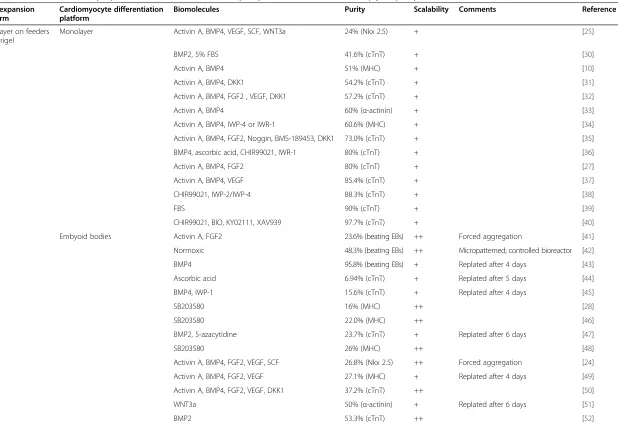
Conventional adherent monolayer culture has been widely utilized for hPSC expansion on either feeder cells or ECM (for example, Matrigel, BD Biosciences PTE Ltd., Singapore, Singapore) coated plates in conditioned medium or commercial serum-free medium such as mTeSR™1 (Stemcell Singapore PTE Ltd., Singapore, Singapore) and StemPro®hESC SFM (Life Technologies, Invitrogen Singapore PTE Ltd., Singapore, Singapore) (Figure 1a,c). The majority of reported cardiomyocyte differentiation pro-tocols utilized either hPSCs co-cultured with feeder cells or on Matrigel with feeder cell-conditioned medium (Table 1).
Expansion of hPSCs can be scaled up by using multi-layer flasks (Figure 1d). These multi-layer plates are currently utilized as the cell expansion platform according to good manufacturing practice for supporting cell therapy-related clinical trials [29]. Rowley and colleagues [29] estimated that up to 240 billion hPSCs can be produced with manual handling of 36 multilayer plates. For larger scales, suspension cultures were recommended (for example, microcarrier culture; step 1 in Figure 1g,h) [29]. Another drawback of hPSC expansion in these tissue culture flasks is the lack of representative sampling and online process monitoring or controls. This can undermine the consistency and the quality of the cells. Moreover, the production process requires extensive manual handling and large clean room facilities, which make large scale produc-tion expensive.
Figure 1Bioprocessing of human pluripotent stem cell-derived cardiomyocytes.Illustrations showing phase contrast images of(a)two-dimensional human embryonic stem cell colony culture in mTeSR™1 medium and(b)human embryonic stem cell-derived beating cardiomyocytes (encircled by the red line). Scale bars are 200μm. For small scale production, human pluripotent stem cells (hPSCs) can be expanded from(c)single layer tissue culture flasks to(d)multilayer flasks (Corning HYPERFlask®) with minimal controls. For clinical and commercial applications, hPSCs can be expanded and differentiated in suspension as(e)cell aggregates or on(f)microcarriers in bioreactors(g,h)with automated controls. 2D, two-dimensional; EB, embryoid body; qRT-PCR, quantitative reverse transcriptase-polymerase chain reaction.
Chenet al. Stem Cell Research & Therapy2014,5:12 Page 3 of 13
Table 1 Methods for cardiomyocyte differentiation from human pluripotent stem cells, ranked by purity of yields
hPSC expansion platform
Cardiomyocyte differentiation platform
Biomolecules Purity Scalability Comments Reference
Monolayer on feeders or Matrigel
Monolayer Activin A, BMP4, VEGF, SCF, WNT3a 24% (Nkx 2.5) + [25]
BMP2, 5% FBS 41.6% (cTnT) + [30]
Activin A, BMP4 51% (MHC) + [10]
Activin A, BMP4, DKK1 54.2% (cTnT) + [31]
Activin A, BMP4, FGF2 , VEGF, DKK1 57.2% (cTnT) + [32]
Activin A, BMP4 60% (α-actinin) + [33]
Activin A, BMP4, IWP-4 or IWR-1 60.6% (MHC) + [34]
Activin A, BMP4, FGF2, Noggin, BMS-189453, DKK1 73.0% (cTnT) + [35]
BMP4, ascorbic acid, CHIR99021, IWR-1 80% (cTnT) + [36]
Activin A, BMP4, FGF2 80% (cTnT) + [27]
Activin A, BMP4, VEGF 85.4% (cTnT) + [37]
CHIR99021, IWP-2/IWP-4 88.3% (cTnT) + [38]
FBS 90% (cTnT) + [39]
CHIR99021, BIO, KY02111, XAV939 97.7% (cTnT) + [40]
Embyoid bodies Activin A, FGF2 23.6% (beating EBs) ++ Forced aggregation [41]
Normoxic 48.3% (beating EBs) ++ Micropatterned; controlled bioreactor [42]
BMP4 95.8% (beating EBs) + Replated after 4 days [43]
Ascorbic acid 6.94% (cTnT) + Replated after 5 days [44]
BMP4, IWP-1 15.6% (cTnT) + Replated after 4 days [45]
SB203580 16% (MHC) ++ [28]
SB203580 22.0% (MHC) ++ [46]
BMP2, 5-azacytidine 23.7% (cTnT) + Replated after 6 days [47]
SB203580 26% (MHC) ++ [48]
Activin A, BMP4, FGF2, VEGF, SCF 26.8% (Nkx 2.5) ++ Forced aggregation [24]
Activin A, BMP4, FGF2, VEGF 27.1% (MHC) + Replated after 4 days [49]
Activin A, BMP4, FGF2, VEGF, DKK1 37.2% (cTnT) ++ [50]
WNT3a 50% (α-actinin) + Replated after 6 days [51]
BMP2 53.3% (cTnT) ++ [52]
Chen
et
al.
Stem
Cell
Research
&
Therapy
2014,
5
:12
Page
4
o
f
1
3
http://stemc
ellres.com
/content/5/1
Table 1 Methods for cardiomyocyte differentiation from human pluripotent stem cells, ranked by purity of yields(Continued)
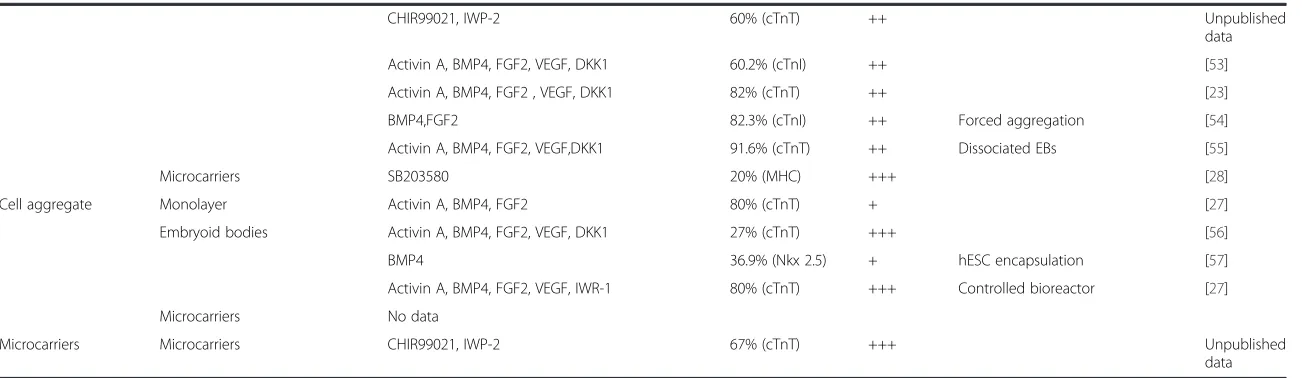
CHIR99021, IWP-2 60% (cTnT) ++ Unpublished
data
Activin A, BMP4, FGF2, VEGF, DKK1 60.2% (cTnI) ++ [53]
Activin A, BMP4, FGF2 , VEGF, DKK1 82% (cTnT) ++ [23]
BMP4,FGF2 82.3% (cTnI) ++ Forced aggregation [54]
Activin A, BMP4, FGF2, VEGF,DKK1 91.6% (cTnT) ++ Dissociated EBs [55]
Microcarriers SB203580 20% (MHC) +++ [28]
Cell aggregate Monolayer Activin A, BMP4, FGF2 80% (cTnT) + [27]
Embryoid bodies Activin A, BMP4, FGF2, VEGF, DKK1 27% (cTnT) +++ [56]
BMP4 36.9% (Nkx 2.5) + hESC encapsulation [57]
Activin A, BMP4, FGF2, VEGF, IWR-1 80% (cTnT) +++ Controlled bioreactor [27]
Microcarriers No data
Microcarriers Microcarriers CHIR99021, IWP-2 67% (cTnT) +++ Unpublished
data
BMP, bone morphogenetic protein;cTnI, cardiac troponin I;cTnT, cardiac troponin T;EB, embryoid body;FBS, fetal bovine serum;FGF, fibroblast growth factor;hESC, human embryonic stem cell;hPSC, human pluripotent stem cell;MHC, myosin heavy chain;SCF, stem cell factor;VEGF, vascular endothelial growth factor.
Chen
et
al.
Stem
Cell
Research
&
Therapy
2014,
5
:12
Page
5
o
f
1
3
http://stemc
ellres.com
/content/5/1
Cell aggregate culture
Cell aggregate culture has been developed as a scalable ex-pansion platform for hPSCs, which utilizes the characteris-tic of hPSCs to form aggregates in suspension. hPSCs in single cell suspension usually undergo apoptosis or anoikis. In order to prevent these detrimental effects and establish a viable hPSC suspension culture, hPSCs are partially dissoci-ated in order to form cell aggregates and/or ROCK-Myosin signaling pathway inhibitors (Y27632 or Blebbistatin) are added to improve survival of dissociated hPSCs. Despite significant cell loss during the initial cell aggregate forma-tion (28 to 76%), several groups have obtained cell concen-trations of 1 to 2 × 10⁶ cells/ml with 12.5- to 17.7-fold expansion using various hPSC lines [56,58-60]. Since hPSC aggregation is usually associated with spontaneous differen-tiation, maintaining pluripotency can be demanding. Exces-sive cell aggregation can induce spontaneous differentiation or necrosis due to nutrient diffusion limitation. The maintenance of aggregate culture is usually carried out by limiting aggregate size to under 500μm by constant disag-gregation of the culture. The process is done manually and can affect culture viability and reproducibility. Designing a scalable bioprocess for aggregate culture may require the integration of reproducible, automated processes for repeti-tive dissociation and formation of uniform cell aggregates. hPSC aggregate cultures were recently reviewed by O'Brien and Laslett [61].
Microcarrier culture
The development of the microcarrier platform for hPSC expansion has attracted interest for generating cardiomyo-cytes in a scalable manner. hPSCs cultured on microcarriers exhibited higher cell density and expansion than two-dimensional tissue or aggregate cultures, while maintaining pluripotency and karyotypic stability [62]. A recent study by Bardy and colleagues [63] reported the highest cell concen-tration of 6.1 × 10⁶ cells/ml with 20-fold expansion in spinner flasks in serum-free medium. The microcarrier-expanded cells exhibited the capacity to differentiatein situ to neural progenitors [63], endoderm progeny [64], and car-diomyocytes [62,65]. However, in order to establish a viable hPSC microcarrier culture, several factors have to be consid-ered, such as microcarrier coating and type, bioreactor design, operating parameters and feeding of medium. These factors can have significant impacts on cell growth, pluripo-tency and lineage commitments [66]. Compared to other classical microcarrier cultures (for example, Vero cells and mesenchymal stem cells) that form monolayers of cells on microcarriers, hPSCs form multi-layered cell-microcarrier aggregates during propagation. Several groups have screened a variety of microcarriers and coatings for initial cell attach-ment and hPSC expansion and examined their impact on prolonged cultivation [64,65,67,68]. A study by Chen and colleagues [67] examined in detail the effects of shape, size,
surface charge, and coatings of microcarriers on hPSC culti-vation. Microcarrier shape and size affect the compactness of cell-microcarrier aggregates and consequently cell growth. hPSCs cultured on spherical microcarriers (Ø >180 μm) exhibited more open structures whereas compact cell-microcarrier aggregates were observed in cylindrical microcarriers [67]. The compactness of cell-microcarrier aggregates increased with the decrease in microcarrier diameter and this had a negative impact on cell growth, likely due to limited nutrient diffusion for the cells within the aggregates [67]. Matrigel coating has been shown to be critical for hPSC cultivation on microcarriers. Recently, it was shown that laminin and vitronectin can replace Matrigel as defined coatings on microcarriers without compromising cell growth [69]. ROCK inhibitors (Y27632 or Blebbistatin) were later shown to enable hPSC growth on ECM coating-free microcarriers [70].
Scaling up of hPSC microcarrier cultures has been demonstrated by several groups using spinner flasks. Serra and colleagues [71] carried out hPSC microcarrier culturing in bioreactors (300 ml) with automated process control. The authors achieved 2.17 × 10⁶cells/ml in 11 days with a seeding density of 1.5 × 10⁵cells/ml (15-fold expansion). A comprehensive review of the bioprocess parameters for hPSC microcarrier culture was recently published [66]. It is important to note that hPSCs propagated on microcarriers can undergo changes in their phenotype. hPSCs cultured on microcarriers exhibited longer doubling time and a gen-erally lower lactic acid specific production rate than those cultured on a tissue culture plate in serum-free media [72]. Leung and colleagues [73] reported different sensitivities of hPSC lines to shear stress. hPSCs expanded on micro-carriers in spinner flasks were able to differentiate efficiently to neural progenitors [63] and endoderm progeny [64]. However, this was not the case for cardiac differentiation. We have observed that hPSCs expanded in agitated micro-carrier cultures and differentiated to cardiomyocytes by the p38 MAP kinase protocol [28,74] exhibited lower yields of cardiomyocytes when compared to cells expanded in static microcarrier culture (unpublished results). We hypothesized that the shear effect caused subtle changes in hPSCs that reduced their cardiac differentiation propensity. Hence, it is important to establish quality controls to monitor these sub-tle changes in hPSC culture before cardiac differentiation.
Cardiogenesis
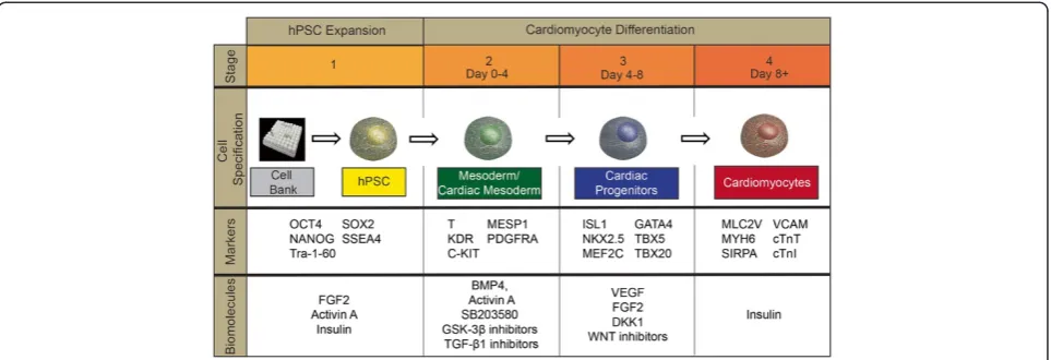
Cardiomyocyte differentiation is based on the recapitulation of in vivo cardiogenesis, which depends on a series of complex molecular signaling pathways [75,76]. The differen-tiation efficiency of these methods depends on the biomole-cules used (growth factors or small molecule inhibitors), hPSC culture expansion conditions and the timely activation or deactivation of molecular signals necessary to guide the differentiation toward cardiac lineages (Figure 2) [77].
Chenet al. Stem Cell Research & Therapy2014,5:12 Page 6 of 13
Moreover, in all protocols, the concentrations and duration of growth factors or small molecule inhibitor treatments depend on the platform (embryoid body (EB), two-dimensional tissue culture or microcarrier cultures) and the hPSC line.
Current cardiac differentiation protocols use either small molecule inhibitors or growth factors to induce the signals for cardiac differentiation. In both cases, the signaling leads to a cascade of three sequential stages: mesodermal induc-tion, cardiac progenitor and cardiomyocyte generation and maintenance [78] (Figure 2). Mesodermal induction, which is monitored by the expression levels of KDR and PDGFR-α [23,53], takes place during the first 3 to 4 days of cardio-myocyte differentiation. This step is usually induced with the growth factors, bone morphogenetic protein (BMP)4 and Activin A, which activate the transforming growth factor (TGF)-βsignaling pathway, which is crucial for meso-derm differentiation [79]. Mesomeso-derm induction can be also achieved by the addition of small molecules, such as GSK-3β inhibitors (CHIR99021 or BIO) [38,40]. These inhibitors increase endogenous levels of BMP2/4, thus indir-ectly activating the TGF-βsignaling pathway [38].
Cardiac progenitor induction is achieved by removal of the TGF-β pathway activators and addition of the growth factors, fibroblast growth factor-2 and/or vascular endothe-lial growth factor, which activate the ERK signaling pathway [80], or by small molecules that inhibit WNT signaling (for example, KY02111, XAV939, IWP-2 and IWR-1). As these factors drive mesodermal cells towards the cardiac progeni-tor lineage, they inhibit the development of smooth muscle and endothelial cell lineages [32,50]. A common finding in this stage is that the addition of insulin inhibits the cardiac progenitor differentiation process [49].
The final stage of cardiomyocyte production (day 8 and later) focuses on maturation of cells to cardiomyocytes and their maintenance. Differentiated cardiomyocytes can be
maintained in simple serum-free medium, which can minimize outgrowth of fibroblasts and maintain cardio-myocyte purity. Prolonged cultivation of cardiocardio-myocytes has been shown to increase the amount of mature ventricu-lar phenotype [38].
Current platforms for generating cardiomyocytes from human pluripotent stem cells
After having established a scalable hPSC expansion method, a suitable cardiac differentiation process should be developed. The variety of protocols used by research groups indicates the complexity of the differentiation process and hence the difficulty of applying universally efficient differen-tiation protocols to the different hPSC lines and culture conditions (Table 1). To date, cardiomyocyte differentiation platforms can be divided into three categories, namely monolayer, EB and microcarrier cultures. Selecting a suit-able platform for cardiomyocyte production may depend on several factors, including type of intended application (for example, cell therapy, disease modeling, cardiac toxicology study).
Two-dimensional tissue culture
In the two-dimensional tissue culture differentiation plat-form, hPSCs expanded in monolayer can be directly differ-entiated to cardiomyocytes by a simple change of hPSC growth medium to cardiac differentiation medium. The first efficient directed differentiation protocol was reported by co-culturing hESCs with mouse endoderm-like cells (END-2), which generated 85% cells exhibiting ventricular-like action potentials [81]. Differentiation protocols were further refined by using more defined conditions with known growth factors, extracellular matrices and eliminat-ing the need for END-2 cells. As shown in Table 1, a simple protocol of using serum-free medium supplemented with Activin A and BMP4 can direct the differentiation to
Figure 2Schematic of human pluripotent stem cell expansion and differentiation to cardiomyocytes.Biomolecules (growth factors and small molecules) play important roles in human pluripotent stem cell (hPSC) expansion and differentiation to cardiomyocytes. Intracellular and cell surface markers associated with each of the three main stages can be used to monitor the progression of differentiation.
Chenet al. Stem Cell Research & Therapy2014,5:12 Page 7 of 13
cardiomyocytes. In brief, Activin A was added to confluent hPSC monolayer culture for 1 day, then BMP4 for 4 days followed by the removal of growth factors and maintenance in serum-free medium afterward [10]. The protocol was further improved with additional growth factors or small molecules to accommodate interline variability that usually exists among hPSC lines. Recently, small molecules target-ing TGF-β, BMP and WNT signaling have shown the potential to replace growth factors in directing hPSC to car-diomyocytes [38]. In particular, small molecules targeting WNT signaling were the most promising. The sequential activation of WNT via GSK-3β inhibitors (for example, CHIR99021 and BIO) for 1 day, followed by WNT inhib-ition (for example, KY02111, XAV939, IWP-2 and IWR-1) at day 3 and removal of small molecules after day 5 achieved cardiomyocyte purity of up to 98% at day 14 [38]. Prolonged cultivation or using the novel WNT inhibitor KY02111 generated more ventricular cardiomyocytes [38,40]. These molecules have been applied successfully in cardiomyocyte differentiation using hPSCs cultured on synthetic ECM peptide coated plates [38].
Embryoid body culture
In the EB differentiation platform, cells from two-dimensional cell cultures are usually dissociated into single cells followed by re-aggregation in order to generate homo-geneous EB cultures with uniform sizes that are further dif-ferentiated to cardiomyocytes (Figure 1e). In general, this dissociation process can lead to significant cell death [82]. Differentiating hPSCs as EBs was first employed in order to accurately recapitulate the complex assembly of cell adhe-sion and intracellular signaling of early embryogenesis [82]. hPSCs undergo spontaneous differentiation when cultured in suspension as EBs, forming cells of the three lineages. Without guidance towards cardiomyocyte differentiation and control of the culture conditions, the efficiency of cardiomyocyte differentiation was usually low (<1%) [30].
Significant progress has been made to improve EB-based cardiomyocyte differentiation efficiency with the identifica-tion of growth factors and pathways associated with cardiac differentiation (Table 1). Most of the reported EB cultures use hPSCs expanded in two-dimensional tissue cultures on feeder cells, in particular mouse embryonic fibroblasts. Upon cell dissociation, the residual feeder cells in the cell suspension provide attachment matrices for the generation of EBs. These EBs achieve higher cell viability and aggregate stability compared to EBs generated from two-dimensional tissue cultures without feeder layers (for example, Matrigel) [83].
EB differentiation protocols are more complicated than those used in two-dimensional tissue cultures due to their three-dimensional shape and the effects of the EB micro-environment [82]. Specifically, the size of EBs has been shown to influence cell differentiation towards different
lineages [84]. In addition, concentrations of small molecules or growth factors used for differentiation had to be adjusted as these soluble factors have to diffuse through a multi-layered cell environment [82]. As cells differentiate toward the cardiac lineage, the formation of excessive aggregate size as a result of cell proliferation or attachment of several aggregates into one (agglomeration) could cause necrosis and consequently reduce cardiomyocyte yield [74]. In order to overcome these problems, several groups have placed single dissociated cells into V- or U-bottom micro-wells coupled with centrifugation to generate uniform cell-aggregates with cell numbers ranging from 200 to 1,000 cells per aggregate [53,58]. Others have tried encapsulation of hESCs and differentiation to cardiomyocytes [57]. We have tested the use of inert dextran beads as a means to separate cell aggregates from each other. In brief, dextran beads were added to culture wells containing EBs to fill spaces between aggregates, forming two to three layers of beads. Without these inert beads separating the aggregates, frequent manual manipulation of cell aggregates is required in order to prevent agglomeration [74]. In some cases, 4-day-old EBs can be plated on ECM-coated tissue culture plates (for example, gelatine), forming beating cell layers afterward. Under these culture conditions, the yield and purity of the cardiomyocytes improved significantly, achieving 64 to 89% cardiac troponin T (cTnT)-positive cardiomyocytes with a variety of hPSC lines [54].
Furthermore, hPSCs can be expanded as cell aggregates in suspension without microcarriers as discussed previ-ously. Two studies showed that these aggregate cultures ex-panded in a spinner flask can be subsequently differentiated
in situto cardiomyocytes as EBs [56,58]. Matsuura and col-leagues [27] reported 80% cTnT-positive cardiomyocytes generated in a controlled bioreactor. Even though the efficiency of cardiomyocyte differentiation was lower than some of the reported EB-based methods (Table 1), cell ag-gregate expansion followed byin situdifferentiation is more scalable than monolayer expansion.
Microcarrier platform
Microcarriers can be applied in two ways for cardiomyocyte differentiation. Firstly, they can be used to assist EB forma-tion, stabilization and prevention of agglomeration. Lecina and colleagues [28] used five different microcarriers to examine the effect of microcarrier type, size, shape and concentration on the efficiency of EB-based cardiomyocyte differentiation using SB203580, a mitogen-activated protein kinase inhibitor. Only the small positively charged Tosoh-10 beads of ØTosoh-10μm, which did not support cell expansion [67], were able to stabilize EB structures and achieve effi-cient cardiomyocyte differentiation. Using these microcar-riers, a cardiomyocyte yield of 90% beating aggregates and 17% cells expressing cardiomyocyte markers (myosin heavy chain and alpha-smooth muscle actin) were achieved in
Chenet al. Stem Cell Research & Therapy2014,5:12 Page 8 of 13
agitated cultures. In these cultures, two- to three-fold higher efficiency (0.28 to 0.62 cardiomyocytes generated per hESC seeded) than the EB-based method (0.13 to 0.22 cardiomyocyte generated per hESC seeded) was achieved [28,74]. Conventional larger microcarriers (Cytodex1 (Ø190
μm) and DE-53 (length 130μm × diameter 35μm)) that ex-hibited higher hPSC expansion capability were not efficient in generation of beating cell-microcarrier aggregates during the cardiomyocyte differentiation process.
In a second approach, hPSC microcarrier aggregates gen-erated during cell expansion can be directly differentiated to cardiomyocytes [62,65]. Reported experimental results using this approach are still limited. However, our group has applied microcarrier expanded hPSC cells for cardio-myocyte differentiation and obtained high cell density of 8 × 10⁶cells/ml (40.4-fold cell expansion) with high cardio-myocyte purity of 67% cTnT and yields of 268 cardiomyo-cytes per hPSC seeded (Figure 1f; unpublished data). Furthermore, we have observed that the method of hPSC microcarrier culture expansion (static versus agitated con-dition in spinner flask) can affect the efficiency of cardio-myocyte differentiation. The cause for this phenomenon is still under investigation.
Considerations for scaling up cardiomyocyte production
We have analyzed three hPSC expansion platforms (two-dimensional, cell-aggregate and microcarrier cultures) for their potential to be coupled with various differentiation platforms. These platforms can be applied using a variety of media, culture methods and conditions. In the following section we discuss critical criteria that should be considered in selection of conditions for designing a scalable cardio-myocyte production system.
Selection of cardiomyocyte production platform
The selection of cardiomyocyte production platform is dictated by the quantity of cardiomyocytes required for a particular application. Three methods of hPSC expansion and seven options for cardiomyocyte differentiation are avail-able for cardiomyocyte production (Tavail-able 1). The initial se-lection of hPSC expansion platform would be based on the amount of cardiomyocytes and the purity needed. The monolayer culture platform can be considered the most straightforward method when compared to suspension-based systems, achieving relatively high differentiation efficiency (Table 1). For large scale production purposes, cell-aggregate and microcarrier-based systems should be considered. Furthermore, process monitoring and control can be easily applied in suspension platforms to ensure process consistency and reproducibility (Figure 1g,h). We be-lieve the integration of hPSC expansion and cardiomyocyte differentiation as a one unit operation in suspension would be the best approach to scale up cardiomyocyte production.
Bioprocess parameters for cardiomyocyte production in suspension
hPSC expansion in suspension with automated online process monitoring and controls has been reported [71]. Serra and colleagues [71] examined the effects of oxygen and operation mode (perfusion versus semi-continuous) in hPSC microcarrier cultures [71]. Oxygen was preferable at 30% air saturation instead of 5%. hPSCs in perfusion mode (dilution rate of 0.5 day-1) showed lower lactate production and shorter lag phase than semi-continuous culture (50% medium exchange per day) [71]. The perfusion system might be ideal for stage-specific cardiomyocyte differenti-ation compared to current processes with constant fluctua-tions in metabolite and growth factor concentrafluctua-tions. Such a system with optimized medium feed can also provide greater efficiencies, replacing current empirical media feeding schemes for hPSC expansion and cardiomyocyte differentiation.
Maintaining a homogeneous suspension culture is critical in process monitoring and controls, but can be challenging considering the size variations of cell aggregate and cell-microcarrier aggregate cultures. Stirring can also induce shear stress responses. Recently, we have observed that agitation-induced shear stress in spinner microcarrier cultures reduced cardiomyocyte differentiation efficiency. Applying agitation during the first 3 days of cardiomyocyte differentiation (for both microcarrier- and EB-based methods) suppressed cardiomyocyte differentiation. This inhibition could be alleviated when intermittent agitation was employed. The literature reports that shear stress affects the TGF-βpathway, which is vital in cardiomyocyte differentiation [85,86].
Medium development
Medium development is critical in the development of hPSC expansion and differentiation processes. In most papers, hPSCs were expanded in conditioned medium on Matrigel-coated plates or mitotically inactivated feeder cells (Table 1). Medium components like serum replacement (Knockout Serum Replacement (KOSR, Life Technologies)), feeder cells and Matrigel for coating of plates or microcarriers pose risks of pathogen contamination. Therefore, defined xeno-free conditions for hPSC expansion have been developed. Lately, it was reported that hPSCs can be cultured in a simple xeno-free, defined medium with eight components on recombinant vitronectin coated plates [87]. This defined and cheaper medium has significant impact on future hPSC clinical applications where the high medium cost has been one of the bottlenecks for studying large scale process devel-opment. This medium can also be adapted for hPSC expan-sion in aggregate or microcarrier cultures. Significant progress has also been made in establishing defined media for each stage of cardiomyocyte differentiation. Media used by various groups are relatively defined and include known
Chenet al. Stem Cell Research & Therapy2014,5:12 Page 9 of 13
growth factors or small molecules with trace amounts of serum albumin (bovine or human) added. Serum-supplemented medium has been used by several groups in generating stable EBs (Table 1). To tackle this issue, Ng and colleagues [88] developed a defined medium, denoted as APEL, for EB formation and differentiation. To address the cost efficiency, small molecules that target WNT signaling transduction pathways have been found to be suitable replacements for costly growth factors.
Downstream processing
Studies on development and optimization of downstream processing of hPSC-derived cardiomyocytes are very lim-ited. The process of harvesting cardiomyocytes from mono-layer, EB or microcarrier cultures has been demonstrated for small scale research purposes. Monolayer cultures, EBs or cell-microcarrier aggregates are treated with dissociation enzymes followed by passing through a sieve to remove undissociated cell aggregates and microcarriers [74]. These processes are usually not optimized and can become more problematic during scaling up. Enzyme concentration, treatment time, mode of mixing, and sieve loading capacity can affect cell viability and harvest efficiency. Hence, there is a need for further investigation into establishing a scalable and efficient cell harvesting process.
Another aspect of downstream processing is the purifica-tion of cardiomyocytes from mixed cell populapurifica-tions [31,89]. Considering the amount of cells required for cell therapy, the effects of unwanted cells in cell replacement therapy are largely unknown. Tumorigenicity related to undifferen-tiated hPSCs remains one of the main concerns, which can be addressed by two approaches. Choo and colleagues [90] used cytotoxic monoclonal antibodies against hPSCs to eliminate undifferentiated hPSCs. Others reported the use of magnetic activated micro-beads coupled to multiple anti-bodies to remove undifferentiated hPSCs and enrich cardi-omyocytes from the heterogeneous cell population after differentiation [31,89,91,92]. However, the antibody-based approach might not be economically viable when scaled up due to the quantity of antibodies needed and their associated costs. Alternatively, Tohyama and colleagues [93] showed a novel approach based on cardiomyocyte metabolic properties, in which cardiomyocytes (up to 99% purity) were selected from mixed cell populations using glucose-depleted medium containing high lactate. This method is inexpensive, scalable and could be easily integrated into a cardiomyocyte production platform. In addition, hPSC-derived cardiomyocyte cultures contain three sub-phenotypes (atrial, ventricular and nodal). The ratio between these subtypes can be altered by modulating signaling pathways during the differentiation [78]. It was reported that activation of neuregulin/ErbB signaling can increase the nodal fraction within the cardiomyocyte popu-lation [94]. Overexpression of microRNA 499, decreased
retinoid signaling, electrical stimulation, prolonged cultiva-tion and use of the WNT inhibitor KY02111 were shown to increase the ventricular subtype [35,38,40,95,96]. Thus, isolation and purification of the required sub-phenotype could be further investigated in order to improve efficiency. Specifically, enriched nodal cells could potentially form a biological pacemaker whereas ventricular cardiomyocytes could be used for cell therapy after ventricular myocardial infarction.
Lastly, the purified cardiomyocytes can be either cryo-preserved or applied directly for tissue engineering. Pro-gress has been made showing the functionality of cryopreserved and dissociated cardiomyocytes. A study by Xu and colleagues [97] showed that cardiomyocytes can be dissociated into single cells, cryopreserved and thawed without significant loss (70-77% recovery). These cells could be used for transplantation, comparable to freshly isolated cells [97].
Meeting current good manufacturing practice requirements Production of cardiomyocytes at clinical commercial scales that follows the current good manufacturing practice (cGMP) standard will be challenging as most of the current processes are carried out for research purposes without any consideration for cGMP require-ments. A defined xeno-free integrated process of expansion, differentiation and downstream purification within a closed system with appropriate monitoring and control systems would be the most suitable for cGMP manufacturing. To characterize the cardiomyocytes produced, quality control assays (step 3 in Figure 1) should be standardized to include cardiomyocyte gene (for example, quantitative RT-PCR) and protein marker (flow cytometry and immunocytochemistry) expression analysis, characterization of electrophysiological properties (multi-electrode array and patch clamp), structural property and organization (electron microscopy) analysis, and determination of calcium signaling (calcium imaging) [98]. Automated high-throughput imaging-based assays developed recently can be advantageous for providing reliable, non-invasive, multi-parameter, real-time monitor-ing of cardiomyocytes in suspension as EBs or on microcarriers. In cases where downstream purification of cardiomyocytes to subtypes is performed, quality control of cell identity should be done by using the whole cell patch clamping technique, high content immunocyto-chemistry and flow cytometry [78]. Patch clamping measures the action potential of individual cells and can be both time consuming and low throughput. Flow cytometry analysis using antibodies against these subtypes has been utilized as a high-throughput method. Anti-bodies against myosin light chain 2 (MLC2) isoforms, namely MLC2a and MLC2v, are widely used to determine ventricular and atrial-like cardiomyocytes.
Chenet al. Stem Cell Research & Therapy2014,5:12 Page 10 of 13
Conclusions and future directions
Cardiac differentiation is an extremely delicate and dynamic process that involves the activation and inhibition of multiple signaling pathways at different time points. Due to this complexity, the development of a protocol that can effi-ciently differentiate hPSCs to cardiomyocytes in a scalable platform has still yet to be developed and optimized. In this review, current platforms for hPSC expansion have been reviewed for their propensity to be adapted into a scalable bioprocess and their efficiency in terms of cardiomyocyte differentiation. The different protocols developed over the past few years have focused on generation of high purity cardiomyocytes without considering issues involved in scaling up the processes. Establishment of a scalable cardio-myocyte production platform requires a more holistic approach integrating parameters related to scaling up hPSC expansion, cardiomyocyte differentiation and downstream purification into a one unit operation.
In summary, current demands from regenerative medi-cine, drug testing, and disease modeling require develop-ment of cardiomyocyte production processes that have to meet a variety of requirements (for example, serum-free media, cGMP requirements, cost of production, quality con-trol and downstream bioprocessing). Beyond these issues, several others have to be considered in future applications. For example, cardiomyocytes produced according to the current methods are immature in their marker expression and electrical and mechanical functionality [77]. This poses problems for the applications mentioned previously as im-mature cardiomyocytes may not be the most ideal models of adult cardiomyocytes. Moreover, for cell therapy an ap-propriate cell delivery method should be developed [77,99]. These challenges call for multidisciplinary efforts that adapt current cardiomyocyte differentiation protocols to develop a cost-effective, scalable and cGMP compliant process, and that resolve issues of downstream purification and quality control as well as cell maturation and delivery systems.
Abbreviations
BMP:Bone morphogenetic protein; cGMP: current good manufacturing practice; cTnT: cardiac troponin T; EB: Embryoid body; ECM: Extracellular matrix; hESC: human embryonic stem cell; hiPSC: human induced pluripotent stem cell; hPSC: human pluripotent stem cell; MLC2: Myosin light chain 2; RT-PCR: Reverse transcriptase-polymerase chain reaction; TGF: Transforming growth factor.
Competing interests
The authors declare that they have no competing interests.
Published: 21 January 2014
References
1. Roger VL, Go AS, Lloyd-Jones DM, Benjamin EJ, Berry JD, Borden WB, Bravata DM, Dai S, Ford ES, Fox CS, Fullerton HJ, Gillespie C, Hailpern SM, Heit JA, Howard VJ, Kissela BM, Kittner SJ, Lackland DT, Lichtman JH, Lisabeth LD, Makuc DM, Marcus GM, Marelli A, Matchar DB, Moy CS, Mozaffarian D, Mussolino ME, Nichol G, Paynter NP, Soliman EZ,et al:Executive summary: heart disease and stroke statistics - 2012 update: a report from the American Heart Association. Circulation2012,125:188–197.
2. Nugent HM, Edelman ER:Tissue engineering therapy for cardiovascular disease.Circ Res2003,92:1068–1078.
3. Bergmann O, Bhardwaj RD, Bernard S, Zdunek S, Barnabe-Heider F, Walsh S, Zupicich J, Alkass K, Buchholz BA, Druid H, Jovinge S, Frisen J:Evidence for cardiomyocyte renewal in humans.Science2009,324:98–102.
4. Anversa P, Kajstura J, Leri A, Bolli R:Life and death of cardiac stem cells: a paradigm shift in cardiac biology.Circulation2006,113:1451–1463. 5. Beltrami AP, Barlucchi L, Torella D, Baker M, Limana F, Chimenti S, Kasahara H,
Rota M, Musso E, Urbanek K, Leri A, Kajstura J, Nadal-Ginard B, Anversa P:Adult cardiac stem cells are multipotent and support myocardial regeneration. Cell2003,114:763–776.
6. Anversa P, Leri A, Kajstura J:Cardiac regeneration.J Am Coll Cardiol2006, 47:1769–1776.
7. Ameen C, Strehl R, Bjorquist P, Lindahl A, Hyllner J, Sartipy P:Human embryonic stem cells: current technologies and emerging industrial applications.Crit Rev Oncol Hematol2008,65:54–80.
8. Itskovitz-Eldor J, Schuldiner M, Karsenti D, Eden A, Yanuka O, Amit M, Soreq H, Benvenisty N:Differentiation of human embryonic stem cells into embryoid bodies compromising the three embryonic germ layers.Mol Med2000,6:88–95.
9. Zwi L, Caspi O, Arbel G, Huber I, Gepstein A, Park IH, Gepstein L:Cardiomyocyte differentiation of human induced pluripotent stem cells.Circulation2009, 120:1513–1523.
10. Laflamme MA, Chen KY, Naumova AV, Muskheli V, Fugate JA, Dupras SK, Reinecke H, Xu C, Hassanipour M, Police S, O'Sullivan C, Collins L, Chen Y, Minami E, Gill EA, Ueno S, Yuan C, Gold J, Murry CE:Cardiomyocytes derived from human embryonic stem cells in pro-survival factors enhance function of infarcted rat hearts.Nat Biotechnol2007,25:1015–1024.
11. Leor J, Gerecht S, Cohen S, Miller L, Holbova R, Ziskind A, Shachar M, Feinberg MS, Guetta E, Itskovitz-Eldor J:Human embryonic stem cell transplantation to repair the infarcted myocardium.Heart2007,93:1278–1284.
12. Caspi O, Huber I, Kehat I, Habib M, Arbel G, Gepstein A, Yankelson L, Aronson D, Beyar R, Gepstein L:Transplantation of human embryonic stem cell-derived cardiomyocytes improves myocardial performance in infarcted rat hearts. J Am Coll Cardiol2007,50:1884–1893.
13. van Laake LW, Passier R, den Ouden K, Schreurs C, Monshouwer-Kloots J, Ward-van Oostwaard D, van Echteld CJ, Doevendans PA, Mummery CL: Improvement of mouse cardiac function by hESC-derived cardiomyocytes correlates with vascularity but not graft size.Stem Cell Res2009,3:106–112. 14. van Laake LW, Passier R, Monshouwer-Kloots J, Verkleij AJ, Lips DJ, Freund C, den Ouden K, Ward-van Oostwaard D, Korving J, Tertoolen LG, van Echteld CJ, Doevendans PA, Mummery CL:Human embryonic stem cell-derived cardiomyocytes survive and mature in the mouse heart and transiently improve function after myocardial infarction.Stem Cell Res2007,1:9–24. 15. Laflamme MA, Murry CE:Regenerating the heart.Nat Biotechnol2005,
23:845–856.
16. Kola I, Landis J:Can the pharmaceutical industry reduce attrition rates? Nat Rev Drug Discov2004,3:711–715.
17. Braam SR, Tertoolen L, van de Stolpe A, Meyer T, Passier R, Mummery CL: Prediction of drug-induced cardiotoxicity using human embryonic stem cell-derived cardiomyocytes.Stem Cell Res2010,4:107–116.
18. Davis RP, van den Berg CW, Casini S, Braam SR, Mummery CL:Pluripotent stem cell models of cardiac disease and their implication for drug discovery and development.Trends Mol Med2011,17:475–484. 19. Carvajal-Vergara X, Sevilla A, D'Souza SL, Ang YS, Schaniel C, Lee DF, Yang L,
Kaplan AD, Adler ED, Rozov R, Ge Y, Cohen N, Edelmann LJ, Chang B, Waghray A, Su J, Pardo S, Lichtenbelt KD, Tartaglia M, Gelb BD, Lemischka IR:Patient-specific induced pluripotent stem-cell-derived models of LEOPARD syndrome. Nature2010,465:808–812.
20. Moretti A, Bellin M, Welling A, Jung CB, Lam JT, Bott-Flugel L, Dorn T, Goedel A, Hohnke C, Hofmann F, Seyfarth M, Sinnecker D, Schomig A, Laugwitz KL: Patient-specific induced pluripotent stem-cell models for long-QT syndrome.N Engl J Med2010,363:1397–1409.
Note:This article is part of a thematic series onCardiovascular
regeneration edited by Ronald Li. Other articles in the series can
be found online at http://stemcellres.com/series/cardiovascular.
Chenet al. Stem Cell Research & Therapy2014,5:12 Page 11 of 13
21. Novak A, Barad L, Zeevi-Levin N, Shick R, Shtrichman R, Lorber A, Itskovitz-Eldor J, Binah O:Cardiomyocytes generated from CPVTD307H patients are arrhythmogenic in response to beta-adrenergic stimulation.J Cell Mol Med2012,16:468–482.
22. Braam SR, Passier R, Mummery CL:Cardiomyocytes from human pluripotent stem cells in regenerative medicine and drug discovery.Trends Pharmacol Sci
2009,30:536–545.
23. Kattman SJ, Witty AD, Gagliardi M, Dubois NC, Niapour M, Hotta A, Ellis J, Keller G: Stage-specific optimization of activin/nodal and BMP signaling promotes cardiac differentiation of mouse and human pluripotent stem cell lines. Cell Stem Cell2011,8:228–240.
24. Elliott DA, Braam SR, Koutsis K, Ng ES, Jenny R, Lagerqvist EL, Biben C, Hatzistavrou T, Hirst CE, Yu QC, Skelton RJ, Ward-van Oostwaard D, Lim SM, Khammy O, Li X, Hawes SM, Davis RP, Goulburn AL, Passier R, Prall OW, Haynes JM, Pouton CW, Kaye DM, Mummery CL, Elefanty AG, Stanley EG:NKX2-5(eGFP/w) hESCs for isolation of human cardiac progenitors and cardiomyocytes.Nat Methods2011,8:1037–1040. 25. Wong SS, Bernstein HS:Cardiac regeneration using human embryonic stem
cells: producing cells for future therapy.Regen Med2010,5:763–775. 26. Wang WE, Chen X, Houser SR, Zeng C:Potential of cardiac stem/progenitor
cells and induced pluripotent stem cells for cardiac repair in ischaemic heart disease.Clin Sci2013,125:319–327.
27. Matsuura K, Wada M, Shimizu T, Haraguchi Y, Sato F, Sugiyama K, Konishi K, Shiba Y, Ichikawa H, Tachibana A, Ikeda U, Yamato M, Hagiwara N, Okano T: Creation of human cardiac cell sheets using pluripotent stem cells. Biochem Biophys Res Commun2012,425:321–327.
28. Lecina M, Ting S, Choo A, Reuveny S, Oh S:Scalable platform for hESC differentiation to cardiomyocytes in suspended microcarrier cultures. Tissue Eng Part C Methods2010,16:1609–1619.
29. Rowley J, Abraham E, Campbell A, Brandwein H, Oh S:Meeting lot-size challenges of manufacturing adherent cells for therapy.BioProcess Int2012,10:16–22. 30. Laflamme MA, Chen KY, Naumova AV, Muskheli V, Fugate JA, Dupras SK,
Reinecke H, Xu C, Hassanipour M, Police S, O'Sullivan C, Collins L, Chen Y, Minami E, Gill EA, Ueno S, Yuan C, Gold J, Murry CE:Cardiomyocytes derived from human embryonic stem cells in pro-survival factors enhance function of infarcted rat hearts.Nat Biotechnol2007,25:1015–1024.
31. Uosaki H, Fukushima H, Takeuchi A, Matsuoka S, Nakatsuji N, Yamanaka S, Yamashita JK:Efficient and scalable purification of cardiomyocytes from human embryonic and induced pluripotent stem cells by VCAM1 surface expression.PLoS ONE2011,6:e23657.
32. Woll PS, Morris JK, Painschab MS, Marcus RK, Kohn AD, Biechele TL, Moon RT, Kaufman DS:Wnt signaling promotes hematoendothelial cell development from human embryonic stem cells.Blood2008,111:122–131.
33. Melkoumian Z, Weber JL, Weber DM, Fadeev AG, Zhou Y, Dolley-Sonneville P, Yang J, Qiu L, Priest CA, Shogbon C, Martin AW, Nelson J, West P, Beltzer JP, Pal S, Brandenberger R:Synthetic peptide-acrylate surfaces for long-term self-renewal and cardiomyocyte differentiation of human embryonic stem cells. Nat Biotechnol2010,28:606–610.
34. Hudson J, Titmarsh D, Hidalgo A, Wolvetang E, Cooper-White J:Primitive cardiac cells from human embryonic stem cells.Stem Cells Dev2012,21:1513–1523. 35. Zhang Q, Jiang J, Han P, Yuan Q, Zhang J, Zhang X, Xu Y, Cao H, Meng Q,
Chen L, Tian T, Wang X, Li P, Hescheler J, Ji G, Ma Y:Direct differentiation of atrial and ventricular myocytes from human embryonic stem cells by alternating retinoid signals.Cell Res2011,21:579–587.
36. Cao N, Liang H, Huang J, Wang J, Chen Y, Chen Z, Yang HT:Highly efficient induction and long-term maintenance of multipotent cardiovascular progenitors from human pluripotent stem cells under defined conditions. Cell Res2013,23:1119–1132.
37. Ye L, Zhang S, Greder L, Dutton J, Keirstead SA, Lepley M, Zhang L, Kaufman D, Zhang J:Effective cardiac myocyte differentiation of human induced pluripotent stem cells requires VEGF.PLoS ONE2013,8:e53764. 38. Lian X, Hsiao C, Wilson G, Zhu K, Hazeltine LB, Azarin SM, Raval KK, Zhang J,
Kamp TJ, Palecek SP:Robust cardiomyocyte differentiation from human pluripotent stem cells via temporal modulation of canonical Wnt signaling.Proc Natl Acad Sci U S A2012,109:E1848–E1857.
39. Moon SH, Ban K, Kim C, Kim SS, Byun J, Song MK, Park IH, Yu SP, Yoon YS: Development of a novel two-dimensional directed differentiation system for generation of cardiomyocytes from human pluripotent stem cells. Int J Cardiol2013,168:41–52.
40. Minami I, Yamada K, Otsuji TG, Yamamoto T, Shen Y, Otsuka S, Kadota S, Morone N, Barve M, Asai Y, Tenkova-Heuser T, Heuser JE, Uesugi M, Aiba K, Nakatsuji N:A small molecule that promotes cardiac differentiation of human pluripotent
stem cells under defined, cytokine- and xeno-free conditions.Cell reports2012, 2:1448–1460.
41. Burridge PW, Anderson D, Priddle H, Barbadillo Munoz MD, Chamberlain S, Allegrucci C, Young LE, Denning C:Improved human embryonic stem cell embryoid body homogeneity and cardiomyocyte differentiation from a novel V-96 plate aggregation system highlights interline variability.Stem Cells2007, 25:929–938.
42. Niebruegge S, Nehring A, Bar H, Schroeder M, Zweigerdt R, Lehmann J: Cardiomyocyte production in mass suspension culture: embryonic stem cells as a source for great amounts of functional cardiomyocytes.Tissue Eng Part A
2008,14:1591–1601.
43. Takei S, Ichikawa H, Johkura K, Mogi A, No H, Yoshie S, Tomotsune D, Sasaki K: Bone morphogenetic protein-4 promotes induction of cardiomyocytes from human embryonic stem cells in serum-based embryoid body development. Am J Physiol Heart Circ Physiol2009,296:H1793–H1803.
44. Cao N, Liu Z, Chen Z, Wang J, Chen T, Zhao X, Ma Y, Qin L, Kang J, Wei B, Wang L, Jin Y, Yang HT:Ascorbic acid enhances the cardiac differentiation of induced pluripotent stem cells through promoting the proliferation of cardiac progenitor cells.Cell Res2012,22:219–236.
45. Ren Y, Lee MY, Schliffke S, Paavola J, Amos PJ, Ge X, Ye M, Zhu S, Senyei G, Lum L, Ehrlich BE, Qyang Y:Small molecule Wnt inhibitors enhance the efficiency of BMP-4-directed cardiac differentiation of human pluripotent stem cells.J Mol Cell Cardiol2011,51:280–287.
46. Graichen R, Xu X, Braam SR, Balakrishnan T, Norfiza S, Sieh S, Soo SY, Tham SC, Mummery C, Colman A, Zweigerdt R, Davidson BP:Enhanced cardiomyogenesis of human embryonic stem cells by a small molecular inhibitor of p38 MAPK. Differentiation2008,76:357–370.
47. Gai H, Leung EL, Costantino PD, Aguila JR, Nguyen DM, Fink LM, Ward DC, Ma Y:Generation and characterization of functional cardiomyocytes using induced pluripotent stem cells derived from human fibroblasts. Cell Biol Int2009,33:1184–1193.
48. Gaur M, Ritner C, Sievers R, Pedersen A, Prasad M, Bernstein HS, Yeghiazarians Y: Timed inhibition of p38MAPK directs accelerated differentiation of human embryonic stem cells into cardiomyocytes.Cytotherapy2010,12:807–817. 49. Willems E, Spiering S, Davidovics H, Lanier M, Xia Z, Dawson M, Cashman J,
Mercola M:Small-molecule inhibitors of the Wnt pathway potently promote cardiomyocytes from human embryonic stem cell-derived mesoderm.Circ Res2011,109:360–364.
50. Yang L, Soonpaa MH, Adler ED, Roepke TK, Kattman SJ, Kennedy M, Henckaerts E, Bonham K, Abbott GW, Linden RM, Field LJ, Keller GM: Human cardiovascular progenitor cells develop from a KDR+embryonic-stem-cell-derived population.Nature2008,453:524–528.
51. Tran TH, Wang X, Browne C, Zhang Y, Schinke M, Izumo S, Burcin M: Wnt3a-induced mesoderm formation and cardiomyogenesis in human embryonic stem cells.Stem Cells2009,27:1869–1878.
52. Pal R, Mamidi MK, Das AK, Bhonde R:Comparative analysis of cardiomyocyte differentiation from human embryonic stem cells under 3-D and 2-D culture conditions.J Biosci Bioeng2013,115:200–206.
53. Sa S, McCloskey KE:Stage-specific cardiomyocyte differentiation method for H7 and H9 human embryonic stem cells.Stem Cell Rev2012,8:1120–1128. 54. Burridge PW, Thompson S, Millrod MA, Weinberg S, Yuan X, Peters A,
Mahairaki V, Koliatsos VE, Tung L, Zambidis ET:A universal system for highly efficient cardiac differentiation of human induced pluripotent stem cells that eliminates interline variability.PLoS ONE2011,6:e18293. 55. Shinozawa T, Furukawa H, Sato E, Takami K:A novel purification method of
murine embryonic stem and human-induced pluripotent stem cell-derived cardiomyocytes by simple manual dissociation.J Biomol Screen
2012,17:683–691.
56. Chen VC, Couture SM, Ye J, Lin Z, Hua G, Huang HI, Wu J, Hsu D, Carpenter MK, Couture LA:Scalable GMP compliant suspension culture system for human ES cells.Stem Cell Res2012,8:388–402.
57. Jing D, Parikh A, Tzanakakis ES:Cardiac cell generation from encapsulated embryonic stem cells in static and scalable culture systems.Cell Transplant
2010,19:1397–1412.
58. Singh H, Mok P, Balakrishnan T, Rahmat SNB, Zweigerdt R:Up-scaling single cell-inoculated suspension culture of human embryonic stem cells. Stem Cell Res2010,4:165–179.
59. Olmer R, Haase A, Merkert S, Cui W, Paleček J, Ran C, Kirschning A, Scheper T, Glage S, Miller K, Curnow EC, Hayes ES, Martin U:Long term expansion of undifferentiated human iPS and ES cells in suspension culture using a defined medium.Stem Cell Res2010,5:51–64.
Chenet al. Stem Cell Research & Therapy2014,5:12 Page 12 of 13
60. Amit M, Laevsky I, Miropolsky Y, Shariki K, Peri M, Itskovitz-Eldor J:Dynamic suspension culture for scalable expansion of undifferentiated human pluripotent stem cells.Nat Protocols2011,6:572–579.
61. O'Brien C, Laslett AL:Suspended in culture - human pluripotent cells for scalable technologies.Stem Cell Res2012,9:167–170.
62. Oh SKW, Chen AK, Mok Y, Chen X, Lim U-M, Chin A, Choo ABH, Reuveny S: Long-term microcarrier suspension cultures of human embryonic stem cells. Stem Cell Res2009,2:219–230.
63. Bardy J, Chen A, Lim YM, Wu S, Wei S, Weiping H, Chan K, Reuveny S, Oh SKW: Microcarrier suspension cultures produce high yields of neural progenitor cells from human pluripotent stem cells.Tissue Eng Part C Methods2013,19:166–180. 64. Lock LT, Tzanakakis ES:Expansion and differentiation of human embryonic
stem cells to endoderm progeny in a microcarrier stirred-suspension culture. Tissue Eng Part A2009,15:2051–2063.
65. Phillips BW, Horne R, Lay TS, Rust WL, Teck TT, Crook JM:Attachment and growth of human embryonic stem cells on microcarriers.J Biotechnol2008, 138:24–32.
66. Chen A, Reuveny S, Oh SK:Application of human mesenchymal and pluripotent stem cell microcarrier cultures in cellular therapy: achievements and future direction.Biotechnol Adv2013,31:1032–1046.
67. Chen AK-L, Chen X, Choo ABH, Reuveny S, Oh SKW:Critical microcarrier properties affecting the expansion of undifferentiated human embryonic stem cells. Stem Cell Res2011,7:97–111.
68. Nie Y, Bergendahl V, Hei DJ, Jones JM, Palecek SP:Scalable culture and cryopreservation of human embryonic stem cells on microcarriers. Biotechnol Prog2009,25:20–31.
69. Heng BC, Li J, Chen AK-L, Reuveny S, Cool SM, Birch WR, Oh SK-W:Translating human embryonic stem cells from 2-dimensional to 3-dimensional cultures in a defined medium on laminin- and vitronectin-coated surfaces. Stem Cells Dev2012,21:1701–1715.
70. Medicinal and other products and human and animal transmissible spongiform encephalopathies: memorandum from a WHO meeting. Bull World Health Organ1997,75:505–513.
71. Serra M, Brito C, Sousa MFQ, Jensen J, Tostões R, Clemente J, Strehl R, Hyllner J, Carrondo MJT, Alves PM:Improving expansion of pluripotent human embryonic stem cells in perfused bioreactors through oxygen control. J Biotechnol2010,148:208–215.
72. Chen X, Chen A, Woo TL, Choo ABH, Reuveny S, Oh SKW:Investigations into the metabolism of two-dimensional colony and suspended microcarrier cultures of human embryonic stem cells in serum-free media.Stem Cells Dev
2010,19:1781–1792.
73. Leung HW, Chen A, Choo A, Reuveny S, Oh S:Agitation can induce differentiation of human pluripotent stem cells in microcarrier cultures. Tissue Eng Part C Methods2010,17:165–172.
74. Ting S, Lecina M, Reuveny S, Oh S:Differentiation of human embryonic stem cells to cardiomyocytes on microcarrier cultures.Curr Protocols Stem Cell Biol2012. Chapter 1:Unit1D 7.
75. Wagner M, Siddiqui MA:Signal transduction in early heart development (I): cardiogenic induction and heart tube formation.Exp Biol Med (Maywood)2007, 232:852–865.
76. Wagner M, Siddiqui MA:Signal transduction in early heart development (II): ventricular chamber specification, trabeculation, and heart valve formation. Exp Biol Med (Maywood)2007,232:866–880.
77. Burridge PW, Keller G, Gold JD, Wu JC:Production of de novo cardiomyocytes: human pluripotent stem cell differentiation and direct reprogramming. Cell Stem Cell2012,10:16–28.
78. Xu C:Differentiation and enrichment of cardiomyocytes from human pluripotent stem cells.J Mol Cell Cardiol2012,52:1203–1212.
79. Watabe T, Miyazono K:Roles of TGF-beta family signaling in stem cell renewal and differentiation.Cell Res2009,19:103–115.
80. Chen Y, Amende I, Hampton TG, Yang Y, Ke Q, Min JY, Xiao YF, Morgan JP: Vascular endothelial growth factor promotes cardiomyocyte differentiation of embryonic stem cells.Am J Physiol Heart Circ Physiol2006,291:H1653–H1658. 81. Mummery C, Ward-van Oostwaard D, Doevendans P, Spijker R, van den
Brink S, Hassink R, van der Heyden M, Opthof T, Pera M, de la Riviere AB, Passier R, Tertoolen L:Differentiation of human embryonic stem cells to cardiomyocytes: role of coculture with visceral endoderm-like cells. Circula-tion2003,107:2733–2740.
82. Bratt-Leal AM, Carpenedo RL, McDevitt TC:Engineering the embryoid body microenvironment to direct embryonic stem cell differentiation. Biotechnol Prog2009,25:43–51.
83. Denning C, Allegrucci C, Priddle H, Barbadillo-Munoz MD, Anderson D, Self T, Smith NM, Parkin CT, Young LE:Common culture conditions for maintenance and cardiomyocyte differentiation of the human embryonic stem cell lines, BG01 and HUES-7.Int J Dev Biol2006,50:27–37.
84. Peerani R, Rao BM, Bauwens C, Yin T, Wood GA, Nagy A, Kumacheva E, Zandstra PW:Niche-mediated control of human embryonic stem cell self-renewal and differentiation.EMBO J2007,26:4744–4755. 85. Wang W, Ha CH, Jhun BS, Wong C, Jain MK, Jin ZG:Fluid shear stress
stimulates phosphorylation-dependent nuclear export of HDAC5 and mediates expression of KLF2 and eNOS.Blood2010,115:2971–2979. 86. Dimmeler S, Assmus B, Hermann C, Haendeler J, Zeiher AM:Fluid shear
stress stimulates phosphorylation of Akt in human endothelial cells: involvement in suppression of apoptosis.Circ Res1998,83:334–341. 87. Chen G, Gulbranson DR, Hou Z, Bolin JM, Ruotti V, Probasco MD, Smuga-Otto K,
Howden SE, Diol NR, Propson NE, Wagner R, Lee GO, Antosiewicz-Bourget J, Teng JM, Thomson JA:Chemically defined conditions for human iPSC derivation and culture.Nat Methods2011,8:424–429.
88. Ng ES, Davis R, Stanley EG, Elefanty AG:A protocol describing the use of a recombinant protein-based, animal product-free medium (APEL) for human embryonic stem cell differentiation as spin embryoid bodies.Nat Protoc2008, 3:768–776.
89. Van Hoof D, Dormeyer W, Braam SR, Passier R, Monshouwer-Kloots J, Ward-van Oostwaard D, Heck AJ, Krijgsveld J, Mummery CL:Identification of cell surface proteins for antibody-based selection of human embryonic stem cell-derived cardiomyocytes.J Proteome Res2010,9:1610–1618.
90. Choo AB, Tan HL, Ang SN, Fong WJ, Chin A, Lo J, Zheng L, Hentze H, Philp RJ, Oh SKW, Yap M:Selection against undifferentiated human embryonic stem cells by a cytotoxic antibody recognizing podocalyxin-like protein-1. Stem Cells2008,26:1454–1463.
91. Dubois NC, Craft AM, Sharma P, Elliott DA, Stanley EG, Elefanty AG, Gramolini A, Keller G:SIRPA is a specific cell-surface marker for isolating cardiomyocytes derived from human pluripotent stem cells.Nat Biotechnol2011,29:1011–1018. 92. Schriebl K, Satianegara G, Hwang A, Tan HL, Fong WJ, Yang HH, Jungbauer A,
Choo A:Selective removal of undifferentiated human embryonic stem cells using magnetic activated cell sorting followed by a cytotoxic antibody. Tissue Eng Part A2012,18:899–909.
93. Tohyama S, Hattori F, Sano M, Hishiki T, Nagahata Y, Matsuura T, Hashimoto H, Suzuki T, Yamashita H, Satoh Y, Egashira T, Seki T, Muraoka N, Yamakawa H, Ohgino Y, Tanaka T, Yoichi M, Yuasa S, Murata M, Suematsu M, Fukuda K:Distinct metabolic flow enables large-scale purification of mouse and human pluripotent stem cell-derived cardiomyocytes.Cell Stem Cell2013,12:127–137. 94. Zhu WZ, Xie Y, Moyes KW, Gold JD, Askari B, Laflamme MA:Neuregulin/ErbB
signaling regulates cardiac subtype specification in differentiating human embryonic stem cells.Circ Res2010,107:776–786.
95. Chan YC, Ting S, Lee YK, Ng KM, Zhang J, Chen Z, Siu CW, Oh SK, Tse HF: Electrical stimulation promotes maturation of cardiomyocytes derived from human embryonic stem cells.J Cardiovasc Transl Res2013,6:989–999. 96. Fu JD, Rushing SN, Lieu DK, Chan CW, Kong CW, Geng L, Wilson KD,
Chiamvimonvat N, Boheler KR, Wu JC, Keller G, Hajjar RJ, Li RA:Distinct roles of microRNA-1 and -499 in ventricular specification and functional maturation of human embryonic stem cell-derived cardiomyocytes.PLoS ONE2011,6:e27417. 97. Xu C, Police S, Hassanipour M, Li Y, Chen Y, Priest C, O'Sullivan C, Laflamme MA,
Zhu WZ, Van Biber B, Hegerova L, Yang J, Delavan-Boorsma K, Davies A, Lebkowski J, Gold JD:Efficient generation and cryopreservation of cardiomyo-cytes derived from human embryonic stem cells.Regen Med2011,6:53–66. 98. Mordwinkin NM, Burridge PW, Wu JC:A review of human pluripotent stem
cell-derived cardiomyocytes for high-throughput drug discovery, cardiotoxi-city screening, and publication standards.J Cardiovasc Transl Res2013,6:22–30. 99. Buikema J, van der Meer P, Sluijter JP, Domian IJ:Engineering myocardial
tissue: the convergence of stem cells biology and tissue engineering technology.Stem Cells2013. in press.
doi:10.1186/scrt401
Cite this article as:Chenet al.:Considerations in designing systems for large scale production of human cardiomyocytes from pluripotent stem cells.Stem Cell Research & Therapy20145:12.
Chenet al. Stem Cell Research & Therapy2014,5:12 Page 13 of 13