Acad J Surg,
Vol.
2
, No.
1-2
(201
5
)
ORIGINAL REPORT
The Effects of Sex Hormones on Liver Regeneration after Liver Trauma in
Animal Model
Shirvan Salaminia1, Saman Nikeghbalian2, Farzaneh Dehghani3, Babak Sabet4,
Amir Ali Mafi5, Seyed Ali Malek-Hosseini2, Nader Tanideh6, Farid Moradian7
1 Department of Surgery, Yasuj University of Medical Sciences, Yasuj, Iran
2 Shiraz Organ Transplantation Center, Namazee Hospital, Shiraz University of Medical Sciences, Shiraz, Iran 3 Department of Anatomy and Histology, Morphometric Stereology Research Laboratory, Shiraz University of Medical
Sciences, Shiraz, Iran
4 Department of General Surgery, Shahid Beheshti University of Medical Sciences, Tehran, Iran
5 Shahid Modarres Clinical Research and Development Center, Shahid Beheshti University of Medical Sciences, Tehran, Iran 6 Laboratory Animals Research Center, Shiraz University of Medical Sciences, Shiraz, Iran
7 Department of General Surgery, Shiraz University of Medical Sciences, Shiraz, Iran
Received: 5 Jul. 2014; Received in revised form: 1 Aug. 2014; Accepted: 22 Sep. 2014
Abstract
Background: The surgical management of liver injuries remains a great challenge for the traumatologists and general surgeons. We hypothesized that administration of 17 β-estradiol, a female sex hormone, improves hepatocellular healing after liver trauma.
Methods: In an experimental model, 60 rats were divided into six subgroups: A (male control), B (male and estradiol), C (castrated male and estradiol), D (female control), F (female and estradiol), and G (oopherectomized female). After inducing liver trauma, estradiol subgroups received 3 doses of intravenous 17 β-estradiol (1 mg/kg) every 8 hours. 2 weeks post trauma, animals were sacrificed and hepatocellular regeneration was measured with the help of stereologic parameters of regeneration. Hepatocellular healing was compared between previous left lobe samples and the new post-traumatic right lobe samples.
Results: Stereological parameters of rats receiving 17 β-estradiol after trauma was much better regarding mean angiogenesis point counting and volume density, compared with non-receiver groups after 2 weeks of trauma (P < 0.005). There was no significant difference for hepatocyte nucleus, hepatocyte point counting and volume density between estradiol receiver and non-receiver groups. In a comparison between subgroups, female sex had the same effect as giving estradiol. Oopherectomized female rats had more fibrogenesis but less angiogenesis (P < 0.005). Fibrogenesis was more in groups that were estradiol non-receiver (P < 0.005). In an explicit comparison of control females and males, estradiol infused males and females, and castrated male or oopherectomized female groups showed that stereological parameters of hepatocyte and hepatocyte nucleus were lower in female subgroups, but angiogenesis was better for female groups except for oopherectomized females. Conclusions: This study did support the administration of exogenic female hormone as an approach to augment the angiogenesis as a good index of regeneration for traumatic liver in rats.
© 2015 Tehran University of Medical Sciences. All rights reserved.
Citation: Salaminia Sh, Nikeghbalian S, Dehghani F, Sabet B, Mafi AA, Malek Hosseini SA, et al. The Effects of Sex Hormones on Liver Regeneration after Liver Trauma in Animal Model. Acad J Surg, 2015; 2(1-2): 2-6.
Keywords: Liver diseases, Liver regeneration, Hepatocytes, Gonadal steroid hormones, Estradiol, Hormone replacement therapy, Models, Animal, Rats
Introduction
Despite the relatively protected location, liver is the most commonly injured organ following penetration and blunt abdominal trauma (1,2). Although, the mortality rates from liver trauma have reduced over the
female sex hormones on trauma and hemorrhage and improvement of liver function after trauma-hemorrhage (3-5). Recently, Feliciano and Pachter suggested that any blunt liver trauma regardless of its magnitude should be managed non-operatively if the patient is hemodynamically stable (2). Regeneration of the liver is affected by internal milieu of cellular and hormonal factors. Some of the suggested factors are as follows: Norepinephrine, epidermal growth factor, tissue necrosis factor, prostaglandins, steroid, sex hormones, interleukin-6, hepatocyte growth factor, insulin, glucagon, epinephrine, chemokines, etc (6,7). We
hypothesized that administration of 17 β-estradiol, a
female sex hormone, improves hepatocellular healing after liver trauma and this study was prepared to evaluate the effect of sex hormones after liver trauma.
Materials and Methods
Process of inducing liver trauma
In an animal model, 60 rats (30 males and 30 females) weighing 180-270 g in the animal lab of Shiraz University of Medical Sciences, were selected for this study. Female and male rats were divided into six subgroups, as follow: Subgroup A: Control males, which had neither been castrated, nor received estradiol; Subgroup B: Males that received estradiol; Subgroup C: Males that had been castrated 3 months prior to the study and received estradiol; Subgroup D: Female control, which had not been oopherectomized and no estradiol was administered; Subgroup E: Female group that received estradiol; Subgroup F: female group that had been oopherectomized 3 months prior to the study but no estradiol was given.
Each case fasted the night before the procedure. However, they were allowed to drink a limited amount of water. Keflin 100 mg/kg (IV) was administered before the procedure. At the time of the procedure, each case was sedated and anesthetized by Ketamine-Xylaxine. Through a midline abdominal incision of about 5 cm, we entered the abdominal cavity. Afterwards by applying a determined crushing clamp within 2 cm of its arms, a crushing injury was induced in the right liver lobe in a standard and measurable manner at three separate points (about 1 cm apart from each other), for 10 minutes and the crushed area demarcated with a proline stitch. In this method, we were able to induce equal liver injury in all cases. Simultaneously, a small sample from the left liver lobe was taken and sent for stereological study and compared with the traumatized samples. Toward the
end of the procedure, a single dose of 17 β-estradiol (1
mg/kg) was injected intravenously (subgroup B, C and
E) and the abdominal incision was closed in multiple
layers. Afterwards, two extra doses of 17 β-estradiol
with interval of 8 hours were administered. The cases were followed under close observation for 2 weeks.
Later, all rats were scarified and samples of the right lobe of liver (the site of trauma) were sent for stereological study. Each equal sample sectioned by an
electrical microtome into ten thin slices (5 µm) and
stained by hematoxylin and eosin (H and E) stain, and
studied by light microscopy with 40 (400)
magnification power. Hepatocellular healing was compared between previous left lobe samples and the new post-traumatic right lobe samples based on stereological findings of cellular healing and also between subgroups by using, point counting and measurement of proportional volume of different elements of tissue and also angiogenesis response. The
stereologic parameters of cell healing and
angiogenesis, which were measured include:
hepatocyte point counting and volume density (proportional volume), hepatocyte nucleus volume density and point counting, angiogenesis point counting and volume density and fibrous tissue volume density and point counting. Data was analyzed with ANOVA and also t-test of SPSS for Windows (version 20; IBM Corp., Armonk, NY, USA). P < 0.050 was considered significant.
Stereologic parameters and analysis
The liver biopsy fragments were taken and analyzed considering the hepatocytes and its nucleus. Each fragment fixed and embedded into paraffin blocks.
Several sections were cut in 5 µm thickness and stained
with H and E solution. 15 random fields were studied blindly moving the stage of microscope in each animal sample.
In this study tissues were examined on a video-microscope system (E-200, Nikon, Japan) connected to a Sony LV-474UB VHS video recorder (Sony, Japan) and a 21-inch LG flatiron LG-M2262A (LG, South Korea) color monitor. Slides were viewed with objective lens. A Weibel stereology graticule (38 points) was fitted inside the 2.5-connecting lens to project the image of the graticule onto the monitor. Therefore, overlay of the image of any slide placed on the microscope stage. Morphometric examination was done blindly using the point counting method of Weibel at a magnification of ×400. Volume fraction of each liver tissue compartment on each of the ten histological sections was calculated by placing the liver section on the microscope stage, which the grid points were superimposed onto the sectional image. Each point was classified as overlying the bile duct,
parenchyma (hepatocyte and sinusoids),
non-parenchymal vessels or other structures within the portal fields. The points that hit the biliary and portal system were not evaluated in this study. These “other structures” consisted mainly of connective tissue and inflammatory cells (fibrosis). Volume fractions were calculated as the fraction of points overlying any given compartment over the total number of points counted.
The proportional volume fraction (calculated as a
percentage) occupied by each liver tissue compartment (×) of liver regeneration was then determined as in Eq. (1), where Vpx = volume fraction of liver × component (e.g hepatocyte nucleus), Psx = the number of points occupied by liver × component within the field, and Pstx = total number of points on the field.
Vpx=Psx/Pstx.
Results
A comparison of point-counting stereological data and volume density of pre and post-trauma of male and female rat groups is given in table 1. There were significant differences (P < 0.050) between quantitative data derived from the animal liver samples before and after trauma. This data showed that the means of
different measured stereological parameters were increased significantly compared to before trauma (P < 0.001).
The conversion of liver fraction data to numerical density data and comparison with each of different sex provided some different results on the most part (Table 2). There were significant differences (P < 0.050) in the counting point numbers and also volume density or fraction between males and females in all of the stereological parameters. However, There were no significant differences between these parameters in liver samples before the trauma (P > 0.050).
The mean of the stereological parameters of liver samples after trauma again were significantly different
between those animals groups with 17 β-estradiol
infusions compared without infusion (Table 3).
Table 1. Comparison of stereolgic parameters before and after trauma
Stereologic parameter Before trauma Mean ± SD After traumaMean ± SD P value
Hepatocyte point counting 244.0.7 ± 37.7 266.6 ± 16.62 0.001
Nucleus point counting 74.12±4.75 85.0 ± 17 0.001
Angiogenesis point counting 27.45 ± 26.1 115.67 ± 58.48 0.001
Fibrous tissue point counting 27.45 ± 26.1 79.05 ± 41.74 0.001
Fibrous volume density 6.32 ± 6.12 15.22 ± 8.41 0.001
Angiogenesis volume density 6.32 ± 6.13 21.8 ± 10.42 0.001
Nucleus volume density 19.02 ± 2.89 16.2 ± 2.94 0.001
Hepatocyte volume density 46.66 ± 6.7 68.34 ± 9.65 0.001
SD: Standard deviation
Table 2. Comparison of stereologic parameters consider in gender
Stereologic parameter MeanMale Female P value
± SD Mean ± SD
Hepatocyte point counting 268.24 ± 34.47 218.42 ± 19.48 0.001
Nucleus point counting 94.34 ± 16.08 74.73 ± 11.33 0.001
Angiogenesis point counting 94.97 ± 39.85 138.77 ± 64.50 0.005
Fibrous tissue point counting 68.14 ± 15.60 91.23 ± 62.24 0.039
Fibrous volume density 12.99 ± 31.16 17.70 ± 11.38 0.037
Angiogenesis volume density 18.02 ± 7.30 26.00 ± 11.83 0.004
Nucleus volume density 17.93 ± 2.52 14.26 ± 2.03 0.001
Hepatocyte volume density 51.12 ± 5.53 41.69 ± 3.69 0.001
SD: Standard deviation
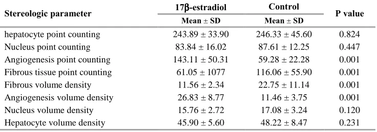
Table 3. Comparison of stereologic parameters considering 17 β-estradiol injection Stereologic parameter 17ββββ-estradiol Mean Control P value
± SD Mean ± SD
hepatocyte point counting 243.89 ± 33.90 246.33 ± 45.60 0.824
Nucleus point counting 83.84 ± 16.02 87.61 ± 12.25 0.447
Angiogenesis point counting 143.11 ± 50.31 59.28 ± 22.28 0.001
Fibrous tissue point counting 61.05 ± 1077 116.06 ± 55.90 0.001
Fibrous volume density 11.56 ± 2.34 22.75 ± 11.14 0.001
Angiogenesis volume density 26.83 ± 8.77 11.46 ± 3.75 0.001
Nucleus volume density 15.76 ± 2.72 17.08 ± 3.24 0.120
Analysis of stereological data of both pre and post-trauma of animal samples with ANOVA post-hoc Benferroni test showed that there is no different within and between all of the subgroups from either sex before the trauma. Volume fractions of hepatocytes, hepatocyte nucleuses, and all of their related point counting are significantly higher in male than in females, and also are higher in groups that not given 17
β-estradiol (P < 0.050). Angiogenesis in all of female
groups and groups with 17 β-estradiol were
significantly higher and this is also certified by the lack of female hormone sex in the oopherectomized female rats that showed decreased angiogenesis and increased fibrosis significantly (P < 0.050). Fibrosis is seen more significantly in male groups and females with the lack of any female sex hormones (P < 0.050).
Discussion
There were significant differences between quantitative data derived from the animal liver samples before and after trauma in the counting point numbers and volume density or fraction between males and females in all of stereological parameters of hepatocyte, hepatocyte nucleus, angiogenesis and fibrogenesis. Certainly when we consider angiogenesis as an index of proliferation,
then in our study all groups with 17 β-estradiol shows
better angiogenesis and also this parameter was also better in female groups except the oopherectomized
female group with no 17 β-estradiol. However, in this
group fibrosis was seen more significantly compared to other group. Results of our study indicated that all of the stereologic parameters had significant difference after trauma, considering sex and groups.
If we consider that more volume density and point counting of hepatocytes and hepatocyte nucleus is an evidence for more liver proliferation, then in this study we can conclude that the estradiol effect has no effect on liver regeneration after trauma. Therefore, exogenous estradiol in castrated rat had had not any positive effect on liver regeneration and also did not affect female rats compare to the oopherectomized cases, and male rats had more liver regeneration. The same finding was achieved by Tsukamoto and Kojo (8)
in which liver regeneration was significantly
suppressed in rats who received glucocorticoids or indomethacin. On the contrary, administration of
dehydroepiandrosterone, a male sex hormone,
following trauma-hemorrhage has been reported to restore hepatocellular function and reduce hepatic damage that was observed in ovariectomized female rats under such conditions (9,10).
Contrarily with hepatocyte stereological
parameters, we found that administration of 17
β-estradiol can help to enhance angiogenesis and
decrease fibrosis after liver trauma in this animal model. Multiple lines of evidence suggest that estrogen
directly modulates angiogenesis via effects on endothelial cells. Despite these consistent observations,
the mechanisms by which estrogen regulates
angiogenesis under physiological and pathological circumstances have not been defined. Angiogenesis is a critical event in wound healing, tumor growth, and the inflammatory vasculitides. In addition, animal studies have shown that estrogen increases the frequency of cancer (breast, cervix, vagina, kidney, and liver), and there is evidence that estrogen may increase the risk of various cancers in humans and this effect is related to angiogenic effect of this hormone (11). Fibrosis as another clue to inflammation and difficult regeneration is seen more significantly in male groups and females with lack of any female sex hormones. Many studies have shown a beneficial effect of estradiol on liver
function in trauma-hemorrhage shock models.
Mizushima et al. demonstrated that estradiol has salutary effects on depressed hepatocellular functions following trauma-hemorrhage in male animals. Administration of estradiol significantly improved hepatocellular function (i.e., maximal velocity and overall efficiency of in vivo indocyanine green clearance) (4,12). In previous literature, controversial and sometimes opposite statements regarding the effects of male and female sex hormones under stress conditions have been mentioned. Androgens have been implicated as the causative factor for the post-injury dysfunction in males (13). Some surveys showed that depressed splenic and peritoneal immune response after trauma-hemorrhage can be normalized by single
dose of 17 β-esteradiol (14,15). In addition, female sex
steroids seem to be protective after trauma-hemorrhage and severe blood loss, as the administration of estrogen prevents the androgen-induced immunosuppression in
castrated male mice. Nonetheless the precise
underlying mechanisms for these immunomodulatory effects of sex hormone steroids after shock remain unknown (16).
Considering the validity of stereological parameters for evaluating this problem, Similar methods have already been utilized in several studies about liver morphology and structure for different issues by applying morphometric and stereological parameters. In one study, stereologic study suggested that estrogenic effects are associated with liver peroxisome proliferation. However, none of these studies evaluated tissue healing and regeneration after liver trauma (17). There are also several studies that evaluated the application of stereological parameters in either radiologic or histologic quantification (18). Using point counting stereology evaluated the effect of telmisartan on liver fibrosis in an animal in one study and showed decreased liver fibrogenesis for this drug after using in rat with diabetes mellitus (19). Thus, stereologic study seem to be a reliable quantitative measurement that we applied in our study.
Liver regeneration and shortening of its recovery time always has been the central aim of many studies. All previous studies on the effects of estrogens and sex hormones on acute trauma and hemorrhage were based on an evaluation of some temporary parameters such as blood flow, perfusion status, inflammatory and immunomodulatory effects of sex steroids. In addition, they were done by high-tech methods (e.g.: laser Doppler US, biologic dye studies and radionuclide
measurements) and also were expensive and
uncommon. However, our study assessed the final effects of sex steroids on liver trauma and gave more objective and measurable information that are applicable at anytime and anywhere.
In conclusion, the administration of 17 β-estradiol
augment the regeneration angiogenesis of traumatic liver in this animal model and angiogenesis, a good index of regeneneration and healing was better with estradiol. Further studies are recommended to clarify the exact liver regeneration process based on clinical and stereological parameters and evaluate the effect of sex hormones on liver regeneration after liver trauma in a human model.
References
1. Parks RW, Chrysos E, Diamond T. Management of liver trauma. Br J Surg. 1999; 86(9): 1121-35.
2. Feliciano DV, Pachter HL. Hepatic trauma revisited. Curr Probl Surg. 1989; 26(7): 453-524.
3. Kuebler JF, Jarrar D, Toth B, Bland KI, Rue L, III, Wang P, et al. Estradiol administration improves splanchnic perfusion following trauma-hemorrhage and sepsis. Arch Surg. 2002; 137(1): 74-9.
4. Mizushima Y, Wang P, Jarrar D, Cioffi WG, Bland KI, Chaudry IH. Estradiol administration after trauma-hemorrhage improves cardiovascular and hepatocellular functions in male animals. Ann Surg. 2000; 232(5): 673-9. 5. Remmers DE, Wang P, Cioffi WG, Bland KI, Chaudry
IH. Testosterone receptor blockade after trauma-hemorrhage improves cardiac and hepatic functions in males. Am J Physiol. 1997; 273(6 Pt 2): H2919-H2925. 6. Mangnall D, Bird NC, Majeed AW. The molecular
physiology of liver regeneration following partial hepatectomy. Liver Int. 2003; 23(2): 124-38.
7. Francavilla A, Vujanovic NL, Polimeno L, Azzarone A, Iacobellis A, Deleo A, et al. The in vivo effect of
hepatotrophic factors augmenter of liver regeneration, hepatocyte growth factor, and insulin-like growth factor-II on liver natural killer cell functions. Hepatology. 1997; 25(2): 411-5.
8. Tsukamoto I, Kojo S. Effect of glucocorticoid on liver regeneration after partial hepatectomy in the rat. Gut. 1989; 30(3): 387-90.
9. Kuebler JF, Jarrar D, Wang P, Bland KI, Chaudry IH. Dehydroepiandrosterone restores hepatocellular function and prevents liver damage in estrogen-deficient females following trauma and hemorrhage. J Surg Res. 2001; 97(2): 196-201.
10. Banta S, Yokoyama T, Berthiaume F, Yarmush ML. Effects of dehydroepiandrosterone administration on rat hepatic metabolism following thermal injury. J Surg Res. 2005; 127(2): 93-105.
11. Losordo DW, Isner JM. Estrogen and angiogenesis: A review. Arterioscler Thromb Vasc Biol. 2001; 21(1): 6-12. 12. Kawasaki T, Chaudry IH. The effects of estrogen on
various organs: therapeutic approach for sepsis, trauma, and reperfusion injury. Part 2: liver, intestine, spleen, and kidney. J Anesth. 2012; 26(6): 892-9.
13. Schneider CP, Schwacha MG, Samy TS, Bland KI, Chaudry IH. Androgen-mediated modulation of macrophage function after trauma-hemorrhage: central role of 5alpha-dihydrotestosterone. J Appl Physiol (1985). 2003; 95(1): 104-12.
14. Angele MK, Schwacha MG, Ayala A, Chaudry IH. Effect of gender and sex hormones on immune responses following shock. Shock. 2000; 14(2): 81-90.
15. Ba ZF, Shimizu T, Szalay L, Bland KI, Chaudry IH. Gender differences in small intestinal perfusion following trauma hemorrhage: the role of endothelin-1. Am J Physiol Gastrointest Liver Physiol. 2005; 288(5): G860-G865. 16. Murashov AK, Pak ES, Hendricks WA, Tatko LM.
17beta-Estradiol enhances neuronal differentiation of mouse embryonic stem cells. FEBS Lett. 2004; 569(1-3): 165-8.
17. Ortiz-Zarragoitia M, Cajaraville MP. Effects of selected xenoestrogens on liver peroxisomes, vitellogenin levels and spermatogenic cell proliferation in male zebrafish. Comp Biochem Physiol C Toxicol Pharmacol. 2005; 141(2): 133-44.
18. Sahin B, Emirzeoglu M, Uzun A, Incesu L, Bek Y, Bilgic S, et al. Unbiased estimation of the liver volume by the Cavalieri principle using magnetic resonance images. Eur J Radiol. 2003; 47(2): 164-70.