CrossMark
Published by DiscoverSys
ABSTRACT
Background: Segmental bone defects resulting from traumatic injuries are complicated problems with significant long-term morbidity. Vascularized bone grafts, by definition, are placed with their vascularity intact, and thus are immediately viable.
Case: A 46 years old man referred to Sanglah Hospital after roadway accident and subsequently fell to the valley. Open wound Ø 5x2 cm (already stitched), bone loss 13 cm length, tenderness over the arm and elbow, radial artery and ulnar artery palpable, O2 saturation 98%, no paresthesia, no active ROM elbow flexion and extension, active ROM wrist 80/70 , active ROM MCP-IP 90/45, and patient can do a thumb extension. Already done the free vascularized fibular graft, and stabilized with 3.5 mm reconstruction plate and screwing for intercondylar fracture. Then checked with angiography for vascular viability.
Discussion: Management of bone defects after severe open fractures of the distal humerus encompasses many technical difficulties. In
these cases, appropriate osteosynthesis may not always feasible, and bone grafting should be considered for the restoration of normal elbow anatomy. Vascularized bone transfers are more efficient than conventional corticocancellous interposition grafting for the management of massive bone loss (>6 cm). The free osteocutaneous fibula flap is a composite tissue transfer suitable to address combined defects. Clinical monitoring and early detection of anastomoses failure can be done within hours after the surgery by evaluating the skin paddle viability.
Conclusion: The surgeon should decide elbow reconstruction in severe injuries that compromise several structures and create tissue defects by careful evaluation. The kind of lesion and mechanism of trauma are not necessarily the most important factors for planning treatment. The free osteocutaneous fibular graft should be further considered as a reconstructive option for the treatment of metaphyseal or juxta-articular complex defects of the elbow joint.
Keywords: humeral bone defect, bone loss, free vascularized fibular graft
Cite This Article: Artha, K.Y.W., Asmara, A.A.G.Y. 2018. Reconstruction of a severe open distal humerus fracture and intercondylar fracture with complete loss of 13 cm humeral bone by using a free vascularized fibular graft: A case report. Bali Medical Journal 7(2): 415-419. DOI:10.15562/ bmj.v7i2.1058
Reconstruction of a severe open distal humerus
fracture and intercondylar fracture with complete
loss of 13 cm humeral bone by using a free
vascularized fibular graft: A case report
Kadek Yuris Wira Artha,1* AA Gede Yuda Asmara2
INTRODUCTION
Reconstruction of large skeletal deficiencies presents a challenging problem to the orthopedic surgeon; methods have evolved significantly during the past decades. Several studies have demonstrated the clinical usefulness of autogenous cortical bone and allograft reconstruction, but resorption of bone and fractures of the graft are possible complications.1
Vascularized bone grafts, by more rapid healing and hypertrophy, may tolerate mechanical loading better and reduced the incidence of resorption and stress fracture. Although several clinical studies have shown the usefulness of vascularized grafts concerning union and resistance to infection, we have found no studies of bone dynamics in vascu-larized grafts.1
The traditional solution for reconstruction of segmental bone defects has included autoge-nous cortical cancellous bone obtained from the
segments without vessel anastomoses, i.e., fibula; allograft bone transplants; and custom prostheses. Autogenous bone grafts are considered as the gold standard for bone replacement, mainly because they offer minimum immunological rejection, complete histocompability and provide the best osteoconductive, osteogenic, and osteoinductive properties.2,3,4 However, clinical benefit is not guar-anteed and there is an associated 8-39% complica-tion.5 Each of these techniques possesses problems which frequently compromise the desired objec-tives of long-term stability and viability required for functional reconstruction of an extremity.6
In autografts of cortical bone or whole bones, the majority of osteocytes die, requiring the eventual replacement of the dead bone by creeping substi-tution process in which dead bone is ultimately replaced by living bone. In massive allografts, 1Orthopaedic and Traumatology
Resident, Medical Faculty Udayana University, Sanglah General Hospital, Denpasar, Bali, Indonesia; 2Orthopaedic and Traumatology Consultant, Medical Faculty Udayana University, Sanglah General Hospital, Denpasar, Bali, Indonesia
*Correspondence to:
Kadek Yuris Wira Artha, Orthopaedic and Traumatology Resident, Medical Faculty Udayana University, Sanglah General Hospital, Denpasar, Bali, Indonesia
yurisartha@gmail.com
Received: 2018-02-06 Accepted: 2018-2-25
Volume No.: 7
Issue: 2
First page No.: 415
P-ISSN.2089-1180
E-ISSN.2302-2914
which is replaced with viable bone from the recip-ient bed. Complications of segmental autografts and allografts include nonunion between the graft and the recipient bone, resorption, collapse of particular segments, and fracture of the graft. Also, prolonged immobilization of 6 to 18 months is required depending upon the length of the defect.6
The use of vascularized autograft in reconstruc-tion may provide rapid biologic incorporareconstruc-tion, growth potential in skeletally immature patients, and the ability to thrive in compromised soft tissue environments. Although several options exist for vascularized bone transfer (such as iliac crest, rib, or clavicle), the fibula is the most commonly trans-ferred free autograft. The use of free fibula transfer to the humerus is well established for posttraumatic bone loss and nonunions.7
Segmental bone defects resulting from traumatic injuries are complicated problems with significant long-term morbidity.8 Open intra-articular fractures of the distal humerus are frequently the result of high-energy trauma.6 These injuries can be associated with severe bone and articular cartilage fragmentation, extensive soft tissue damage, and concomitant injuries, which may jeopardize limb integrity and patient survival. Because of the complexity of injury, treatment is difficult, and the subsequent clinical outcome is often inadequate.9
Vascularized bone grafts, by definition, are placed with their vascularity intact, and thus are immediately viable. As a result, vascularized bone grafts obviate the need for incorporation by creep-ing substitution and may instead incorporate into the adjacent host bone via primary (or secondary) bone healing.8,9
The high functional requirements of the elbow depend not only on the complexity of the joint but also on the maintained function of vessels and nerves and the soft tissue coverage. The high density of good structures that may be involved in lesions at this level may cause severe functional impairment of the upper limb. Accurate reconstruction of the articular surfaces and early mobilization of the elbow joint are critical for restoring proper joint function.10
The principles that should guide the reconstruc-tive surgeon in the process are the following:10
1. Extensive debridement of damaged tissues (immediately it may be difficult to identify which tissues are devitalized, particularly following high-energy traumas);
2. Restore a good blood supply;
3. Stabilize bone components with rigid fixation; 4. Ensure stable and well-vascularized skin
coverage;
5. Mobilize of the joint early.
CASE REPORT
A 46 years old man referred from Waingapu hospital to the emergency department of Sanglah Hospital after roadway accident and subsequently fell down to the valley 11 days before admission. Debridement and primary hecting of the wound have been done in Waingapu Hospital. The patient was alert and orientated, hemodynamically stable. The clinical and radiologic examination showed swelling over the right arm and elbow, angula-tion deformity to the anterior side, open wound Ø 5×2 cm (already stitched), 13 cm length bone loss, no pus, crustae, tenderness over the arm and elbow, radial artery and ulnar artery palpable, CRT less than 2 second , O2 Saturation 98%, no pares-thesia, no active ROM elbow flexion and extension, active ROM Wrist 80/70 ,active ROM MCP-IP 90/45, and patient can do a thumb extension, diag-nose with OF Right Humerus segmental grade III B with Bone Loss AO/OTA types C2.3, according to the relevant classification systems for the open and distal humeral fractures, respectively.
The patient was promptly taken to the oper-ation room for stabilizoper-ation of the right elbow fracture and pelvic ring injury. The elbow wound was debrided and irrigated with 6 liters of normal saline solution and removal of all contaminated or dead tissues. And make ORIF Screwing in the intercondylar humerus. After checked marker of infection, and make sure no infection found, then take the second operation with free vascularized fibular graft.
The flap was harvested using the standard technique, the fibula is osteotomized proximally and distally, with preservation of the peroneal vessels. Once the recipient site is prepared, the vascular pedicle may be divided and the fibula transferred to the desired location. If an osteo-myocutaneous flap is required, dissection starts with a linear lateral incision over the fibula paral-leling to its border. Following ulnar nerve ante-rior submuscular transposition, a 17 cm fibular graft was inserted into the medial column defect and subsequently stabilized with a new 3.5-mm reconstruction plate for superior fragment and inferior fragment with half threaded cancellous screw for intercondylar fracture.5,7 Microvascular
anastomoses of the donor peroneal artery and its accompanying veins were carried out on the corresponding radial artery and venae comitantes after the passage of the pedicle through brachialis muscle.
Durante Operation
After the stabilization of the fibula to the recipient site, typically done with rigid internal screw fixa-tion, microvascular anastomoses are performed, reconstituting both arterial inflow and venous outflow to the fibular graft. Postoperatively, a poste-rior elbow splint was applied and antibiotic medi-cation. Two weeks after the operation, we already have done angiography.
Figure 2 Pre-Free Vascularized Fibular Graft (Clinical Preoperation)
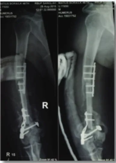
Figure 3 Right Humerus X-Ray Anteroposterior-Lateral View
DISCUSSION
Management of bone defects after severe open fractures of the distal humerus encompasses many technical difficulties. In these cases, appropriate osteosynthesis may not always be feasible, and bone grafting should be considered for the restoration of normal elbow anatomy. Autografts usually contain viable osteogenic cells, bone matrix proteins and support bone growth which are obtained from vascularized and non vascularized cortical and
autologous bone marrow grafts.11 Vascularized cortical grafts heal rapidly at the host-graft- interface, and their remodeling is similar to that of normal bone. On the contrary, non-vascularized grafts do not undergo resorption and revascu-larization and, therefore, they provide superior strength during the first six weeks.12 Vascularized bone transfers are more efficient than conventional corticocancellous inter-position grafting for the management of massive bone loss (>6 cm).9 Site of grafting is another important factor influencing osteogenesis. Resorption and replacement of a bone graft in a skeletal bed occurs more rapidly at the end of a long bone (cancellous) than at the center of shaft (cortical bone).13,14
The free osteocutaneous fibula flap is a compos-ite tissue transfer suitable to address combined defects in the 1-stage procedure. The fibula is a long and straight tubular bone, which is not difficult to harvest, while donor site morbidity is minimal up to a graft length of 20 cm.5,6
Clinical monitoring and early detection of anas-tomoses failure can be done within hours after the surgery by evaluating the skin paddle viability. In the herein case, the interposed vascularized graft completed the triangle formed by the lateral and medial bony columns and the trochlea, and also promoted fracture healing and patient recovery.4,5
In conclusion, the free osteocutaneous fibular graft should be further considered as a reconstruc-tive option for the treatment of metaphyseal or juxta-articular complex defects of the elbow joint.
CONCLUSION
The surgeon should decide elbow reconstruction in severe injuries that compromise several structures and create tissue defects by careful evaluation. The kind of lesion and mechanism of trauma are not necessarily the most important factors for planning treatment. The profile of the patient and general clinical conditions, surgical team experience and the level of multidisciplinary assistance have to be considered as well. The ideal procedure is an ‘‘all-in-one’’ reconstruction; however, this cannot always be achieved immediately. In these cases, the surgeon should choose reconstruction methods while taking into consideration the future steps for optimal treatment.
REFERENCES
1. Herman H. De Boer, Michael, B, Wood. Bone Changes
in the Vascularised Fibular Graft. From the Mayo Clinic, Rochester, USA. The Journal of Bone and Joint Surgery. 1989.
Figure 6 Angiography Study Post Operation
Figure 7 Clinical Post Operation
2. Weiland AJ, Baltimore, Kleinert HE, Kutz JE, Louisville K,
Daniel RK. Free Vascularized Bone Grafts in Surgery of
the Upper Extremity. American Society for Surgery of the Hand. The Journal of Hand Surgery. Montreal, Quebec, Canada. 1979.
3. Rose PS, Shin AY, Bishop AT, Moran SL, Sim FH. Vascularized Free Fibula Transfer for Oncologic Reconstruction of the Humerus. Lippincott Williams & Wilkins. 2005.
4. Canalle Terry S, Beaty James H. Campbell’s Operative
Orthopaedics 12th ed. Philadephia: Mosby Elsevier. 2013.
5. Kouvidis, George K, Byron E, Chalidis, Mark I,
Liddington FRCS, Peter V, Giannoudis EEC.
Reconstruction of a Severe Open Distal Humerus Fracture With Complete Loss of Medial Column by Using a Free Fibular Osteocutaneous Graft. 2008:225-230.
6. Battiston B, Vasario G, Ciclamini D, Rollero L, Tos P. Reconstruction of traumatic losses of substance at the elbow. Injury, Int. J. Care Injured. 2013;45:437–443.
7. Adani R, Tarallo L, Mugnai R. Reconstruction of
Post-Traumatic Bone Defect of the Upper-Limb with Vascularized Fibular Graft. Croatia: Intech. 2012.
8. Samartzis D, Shen FH, Goldberg EJ, An HS. Is autograft the gold standard in achieving radiographic fusion in one-level anterior cervical discectomy and fusion with rigid anterior plate fixation? Spine 2005; 30 : 1756-61.
9. Nandi SK, Roy S, Mukherjee P, Kundu B, De DK, Basu D. Orthopaedic applications of bone graft & graft substitutes: a review. Indian J Med Res. 2010;132:15-30.
10. Dell PC, Burchardt H, Glowczewskie FP Jr. A roentgeno-graphic, biomechanical, and histological evaluation of vas-cularized and non-vasvas-cularized segmental fibular canine autografts. J Bone Joint Surg Am. 1985;67:105-12. 11. Anderson KJ. The behavior of autogenous and
homoge-nous bone transplants in the anterior chamber of the rat’s eye. A histological study of the effect of the size of the implant. J Bone Joint Surg Am 1961; 43-A : 980-95. 12. Marx RE. Bone and bone graft healing. Oral Maxillofac
Surg Clin North Am. 2007;19:455-66.
13. Bauer TW, Muschler GF. Bone graft materials : an overview of the basic science. Clin Orthop Relat Res. 2000;371:1027. 14. Johan VDS. Bone Graft Substitutes: Developed for Trauma
and Orthopaedic Surgery. [thesis] Optima Grafische Communicatie. 2015.
15. Pape HC, Evans A, Kobbe P. Autologous bone graft: prop-erties and techniques. J Orthop Trauma. 2010;24 Suppl 1:S36-40.