www.fm.viamedica.pl
Address for correspondence: Mariusz Koda MD, Department of General Pathology, Medical University of Bialystok, ul. Waszyngtona 13, 15–269 Bialystok, Poland, tel: +48 85 748 59 45, tel/fax: +48 85 748 59 44, e-mail: kodamar@zeus.amb.edu.pl
Expression of the apoptotic markers
in normal breast epithelium, benign
mammary dysplasia and in breast cancer
Mariusz Koda
1, Luiza Kanczuga-Koda
2, Joanna Reszec
2, Mariola Sulkowska
1,
Waldemar Famulski
2, Marek Baltaziak
1, Wojciech Kisielewski
2, Stanislaw Sulkowski
11Department of General Pathology, Medical University, Białystok, Poland 2Department of Clinical Pathology, Medical University, Białystok, Poland
[Received 25 November 2003; Revised 9 March 2004; Accepted 9 March 2004]
Apoptosis and proliferation are processes associated with the development and progression of breast cancer. The sensitivity of tumour cells to the induction of apoptosis depends on the balance between pro- and anti-apoptotic proteins. The expression of Bak and Bcl-2 was examined using an immunohistochemical method in 71 primary breast cancers. Furthermore, Bcl-2 and Bak were assessed in the normal mammary gland as well as in benign mammary dysplasia adjacent to breast cancer. Positive immunostaining for Bcl-2 was observed in 77.8% of cases of normal breast epithelium (NBE), 93% of benign dysplasia without intra-ductal proliferation (BBD) as well as in 94% of intraintra-ductal proliferative lesions of the breast (BIPL). Expression of Bak was detected in 39% of cases of NBE, 45% of BBD and in 67% of BIPL. In breast cancer Bcl-2 and Bak expression was found in 83% and 70% of the cases studied, respectively. Increased Bcl-2 expression in primary tumours significantly correlated with favourable prognostic factors, name-ly pT1, G2 and lack of metastases to the regional name-lymph nodes (p < 0.01, p < 0.03, p < 0.02, respectively). There were no relationships between Bak and the clinicopathological features studied, but our results indicate changes in the expression of Bak during breast cancer development and progression. It would appear to be important to assess and compare pro- and anti-apoptotic proteins between normal mammary gland, benign mammary dysplasia and the primary tumours of breast cancer. This knowledge should be helpful in understanding breast cancer development and progression.
Key words: Bak, Bcl-2, normal mammary gland, benign mammary dysplasia, breast cancer
INTRODUCTION
INTRODUCTION
INTRODUCTION
INTRODUCTION
INTRODUCTION
The main group of genes controlling apoptosis is the Bcl-2 family, which comprises both inhibitors (Bcl-2, Bcl-Xl, Mcl-1) and promotors (Bax, Bak, Bad,
Bcl-Xs) [19]. Bcl-2 family members regulate mammary
gland development and homeostasis [6–8]. The
and maintaining cells in the G0 phase [3, 17]. The anti-apoptotic and anti-proliferative effects of Bcl-2 on the epithelial cells can diverge during breast can-cer development [4]. Moreover, changes in the ex-pression levels of anti- and pro-apoptotic proteins influence breast cancer development and progres-sion [14, 18]. Pro-apoptotic Bax and Bak heterodimer-ise with anti-apoptotic Bcl-2 and Bcl-Xl and through
this process Bcl-2 could inhibit apoptosis, increase the lifetime of invasive cells and induce clone selec-tion and the metastatic process of breast cancer [10]. The purpose of the study was to evaluate the ex-pression of the proteins involved in the regulation of apoptosis (Bcl-2 and Bak) in primary breast cancer as well as in the normal mammary gland and in benign mammary dysplasia adjacent to breast cancer.
MATERIAL AND METHODS
This study comprised 71 women, who underwent surgery for primary breast cancer in the years 2000– –2002. The age of the patients ranged from 30 to 80 years and the mean age was 54.6 years. The patients had not received any preoperative chemotherapy or hormonotherapy. Tumour samples were collected im-mediately after tumour removal, fixed in 10% buff-ered formaldehyde solution and embedded in paraf-fin blocks at 56oC. Histopathological examination,
ac-cording to the WHO classification of tumours [25], was performed using standard haematoxylin-eosin staining. Immunohistochemical studies were per-formed according to Koda et al. [12, 13], using the following antibodies: goat polyclonal Bak (Santa Cruz Biotechnology, USA) at a 1:200 dilution and mouse monoclonal Bcl-2 (Dako, Denmark) at a dilution 1:100. The reactions were performed by the Labelled Strepta-vidin Biotin (LSAB) technique (Dako). Appropriate im-munohistochemical controls were carried out. The evaluation of the immunostaining of Bak and Bcl-2 was analysed in 10 different tumour fields and the mean percentage of tumour cells with positive stain-ing was evaluated. The cut-off point for classifystain-ing the sections examined as positive was 25% of posi-tively stained cells for Bak and Bcl-2. Their expression was also assessed in 18 normal breast tissues (NBE) adjacent to the breast cancer, 29 cases of benign dys-plasia without intraductal proliferative lesions (BBD) and 18 cases of intraductal proliferative lesions (BIPL), mainly including usual ductal hyperplasia.
The differences between Bak and Bcl-2 status and the correlation with various clinicopathological fea-tures (stage, grade of tumour and lymph node
sta-tus) were evaluated using the Mann-Whitney U test. Statistical significance was assumed at p < 0.05. Correlation coefficients were used to assess associa-tions between the parameters studied.
RESULTS
In the study group of 71 women with primary breast cancer at the time of diagnosis 36 (50.7%) were without metastases to the regional lymph nodes [N(–)] and 35 (49.3%) involved the regional lymph nodes [N(+)]. Our studies included only inva-sive ductal carcinomas, representing grades G2 and G3 (49 and 22 cases, respectively) as well as the pT1 (43 cases) and pT2 (28 cases) stages.
Bcl-2 and Bak expression in the normal mammary gland and in benign breast lesions adjacent to breast cancer
Immunohistochemical analysis of NBE, BBD and BIPL revealed cytoplasmic localisation and microgran-ular staining for the Bcl-2 and Bak proteins. Strong immunostaining for Bcl-2 was observed in 77.8% of cases of NBE, 93% of BBD as well as in 94% of BIPL. Bcl-2 expression in NBE, BBD and BIPL positively cor-related with Bcl-2 expression in breast cancer. Weak positive immunostaining for the Bak protein was ob-served in 39% of NBE, 45% of BBD and 67% of BIPL.
Associations of Bcl-2 and Bak expression in breast cancer with selected
clinicopathological features
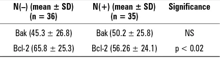
The expression of Bcl-2 and Bak was detected in 83% and 70% of the breast cancers, respectively. In the majority of cases Bcl-2 and Bak immunostaining was cytoplasmic, similar to that reported for NBE, BBD and BIPL, but in some tumours we also observed peri-nuclear staining. Increased Bcl-2 expression in prima-ry tumours significantly correlated with lack of me-tastases to the regional lymph nodes [N(–); p < 0.02; Table 1] as well as with the pT1 stage of the tumours
Table 1. Bcl-2 and Bak expression in breast cancer in
re-lation to lymph node status
N(–) (mean ± SD) N(+) (mean ± SD) Significance (n = 36) (n = 35)
Bak (45.3 ± 26.8) Bak (50.2 ± 25.8) NS
Bcl-2 (65.8 ± 25.3) Bcl-2 (56.26 ± 24.1) p < 0.02
(p < 0.01; Table 2) and grade G2 (p < 0.03; Table 3). There were no relationships between Bak and the clini-copathological features studied (Tables 1–3).
DISCUSSION
The expression of the Bcl-2 family of proteins changes during mammary gland development. Bcl-2 is expressed in the non-pregnant and early pregnan-cy female, but not in the lactating mammary gland [15]. An increase in the expression of pro-apoptotic Bak occurs during late pregnancy and lactation as well as during apoptotic involution [22]. Bcl-2 levels in normal breast epithelium undergo periodic changes during the menstrual cycle and this protein is regu-lated in a hormone-dependent manner within the premenopausal breast [21].
Dysregulation of the balance between prolifer-ation, differentiation and apoptosis in the normal mammary gland can lead to breast cancer devel-opment. The up-regulation of cell proliferation as well as the down-regulation of apoptosis contrib-ute to the accumulation of mutations, which lead to the subsequent development of breast cancer [15, 25]. It has been shown that a high apoptotic rate is associated with a high grade of tumour, large
tumour size and with a shortened disease-free sur-vival period [16].
There are many theories about the lack of activi-ty of anti-cancer drugs in breast cancer. The disrup-tion of the apoptotic pathways may be one of rea-sons. We therefore decided to assess the expression of those factors involved in apoptosis in the normal mammary gland, benign mammary dysplasia and primary cancer. The most significant findings of our study are: 1) a positive correlation in the expression of Bcl-2 between normal breast epithelium (NBE), benign dysplasia without intraductal proliferation (BBD) and intraductal proliferative lesions of the breast (BIPL) and breast cancer; 2) increased expres-sion of Bak in breast cancer compared to NBE and BBD (70%, 39%, 45%, respectively); 3) a lack of dif-ferences in Bak expression between breast cancer and BIPL (70% and 67%, respectively); 4) an associa-tion between expression of Bcl-2 and favourable prognostic factors [pT1, G2 and N(–)]; 5) increased, but not statistically significant, expression of Bak in poorly differentiated cancers.
In the study of Ioachim et al. [9] Bcl-2 protein was detected in 85.2% of benign hyperplastic lesions of the mammary gland and 40% of breast cancers.
Table 3. Bcl-2 and Bak expression in breast cancer in relation to histological grade
Lymph node status, number of patients G2 (mean ± SD) G3 (mean ± SD) Significance
N(–) and N(+), n = 49 (G2), n = 22 (G3) Bak (44.9 ± 26.7) Bak (54.1 ± 24.7) NS
Bcl-2 (66.1 ± 21.9) Bcl-2 (50.1 ± 28.2) p < 0.03
N(+), n = 22 (G2), n = 13 (G3) Bak (48.9 ± 26.6) Bak (52.5 ± 25.4) NS
Bcl-2 (60.8 ± 21.1) Bcl-2 (48.5 ± 27.5) NS
N(–), n = 27 (G2), n = 9 (G3) Bak (41.6 ± 26.8) Bak (56.4 ± 24.9) NS
Bcl-2 (70.3 ± 22.1) Bcl-2 (52.2 ± 30.7) NS
Mean ± SD — mean percentage of Bcl-2 or Bak-positive cells in 10 different tumour fields (as described in Material and methods)
Table 2. Bcl-2 and Bak expression in breast cancer in relation to tumour size
Lymph node status, number of patients pT1 (mean ± SD) pT2 (mean ± SD) Significance
N(–) and N(+), n = 43 (pT1), n = 28 (pT2) Bak (47.1 ± 26.7) Bak (48.8 ± 26.1) NS
Bcl-2 (67.1 ± 23.3) Bcl-2 (51.8 ± 25.1) p < 0.001
N(+), n =15 (pT1), n = 20 (pT2) Bak (49.9 ± 27.9) Bak (50.5 ± 24.9) NS
Bcl-2 (58.7 ± 24.8) Bcl-2 (54.4 ± 23.9) NS
N(–), n = 28 (pT1), n = 8 (pT2) Bak (45.5 ± 26.4) Bak (48.8 ± 26.1) NS
Bcl-2 (71.6 ± 21.6) Bcl-2 (45.4 ± 28.2) p < 0.01
On the other hand, Bargou et al. [1] observed no difference with regard to Bcl-2 (and Bcl-XL) expres-sion between normal breast epithelium and breast cancer tissue. Similarly to Bargou et al. [1], we found no significant differences in the percentage of Bcl-2--positive cases between breast cancers (83%) and NBE (77.8%), BBD (93%) and BIPL (94%). In the study by Gee et al. [5] Bcl-2 was detected in 70% of breast cancers. It has also been shown that Bcl-2-positive patients had a better prognosis than Bcl-2-nega-tive patients [11, 24, 26]. Rochaix et al. [20] sug-gested that Bcl-2 and Bak expression were associ-ated with a regulation of apoptosis in breast can-cer. They found that Bcl-2 expression in tumours was associated with a better differentiation of the cancers (G1 — 100% of Bcl-2-positive tumours, G2 — 81%, G3 — 60%), but there was no relationship between Bak and tumour grade [20]. In the present study an association was observed between tumour differentiation and Bcl-2 expression — the mean percentage of tumour cells with positive staining for Bcl-2 was increased in grade G2 breast cancers. In the study of Berardo et al. [2] high Bcl-2 expres-sion was associated with favourable prognostic fac-tors such as. ER positivity, low S phase fraction, a lower number of positive lymph nodes and over-all survival. Our findings confirm the results of Be-rardo et al. [2] with regard to the favourable prog-nostic significance of Bcl-2 expression in breast can-cer. Moreover, we observed a positive correlation between expression of Bcl-2 and ERa (unpublished data). On the other hand, our observations are not concordant with the results of Sierra et al. [23] which showed that the over-expression of Bcl-2 correlated with lymph node involvement.
In the present study we observed an increased expression of pro-apoptotic Bak in breast cancer com-pared with NBE and BBD. On the other hand, we did not find any differences in Bak expression between breast cancer and BIPL. Our results indicate that over-expression of pro-apoptotic proteins could contrib-ute to an increase in cell turnover and breast cancer development and progression, but we suggest that further studies should be carried out to fully assess Bak expression in breast cancer progression.
ACKNOWLEDGEMENTS
We are grateful to Edyta Jelska and Wojciech Mytnik for their expert technical assistance.
REFERENCES
1. Bargou RC, Daniel PT, Mapara MY, Bommert K, Wage-ner C, Kallinich B, Royer HD, Dorken B (1995) Expres-sion of the bcl-2 gene family in normal and malignant breast tissue: low bax-alpha expression in tumor cells correlates with resistance towards apoptosis. Int J Can-cer, 60: 854–859.
2. Berardo MD, Elledge RM, de Moor C, Clark GM, Os-borne CK, Allred DC (1998) Bcl-2 and apoptosis in lymph node positive breast carcinoma. Cancer, 82: 1296–1302.
3. Borner C (1996) Diminished cell proliferation associat-ed with the death-protective activity of Bcl-2. J Biol Chem, 271: 12695–12698.
4. Furth PA, Bar-Peled U, Li M, Lewis A, Laucirica R, Jager R, Weiher H, Russell RG (1999) Loss of anti-mitotic effects of Bcl-2 with retention of anti-apop-totic activity during tumor progression in a mouse model. Oncogene, 18: 6589–6596.
5. Gee JM, Robertson JF, Ellis IO, Willsher P, McClelland RA, Hoyle HB, Kyme SR, Finlay P, Blamey RW, Nicholson RI (1994) Immunocytochemical localization of Bcl-2 pro-tein in human breast cancers and its relationship to a series of prognostic markers and response to endo-crine therapy. Int J Cancer, 59: 619–628.
6. Heermeier K, Benedict M, Li M, Furth P, Nunez G, Hen-nighausen L (1996) Bax and Bcl-xs are induced at the onset of apoptosis in involuting mammary epithelial cells. Mech Dev, 56: 197–207.
7. Humphreys RC (1999) Programmed cell death in the terminal endbud. J Mammary Gland Biol Neoplasia, 4: 213–220.
8. Humphreys RC, Krajewska M, Krnacik S, Jaeger R, Weiher H, Krajewski S, Reed JC, Rosen JM (1996) Apo-ptosis in the terminal endbud of the murine mamma-ry gland: a mechanism of ductal morphogenesis. De-velopment, 122: 4013–4022.
9. Ioachim EE, Malamou-Mitsi V, Kamina SA, Goussia AC, Agnantis NJ (2000) Immunohistochemical expression of Bcl-2 protein in breast lesions: correlation with Bax, p53, Rb, C-erbB-2, EGFR and proliferation indices. An-ticancer Res, 20: 4221–4225.
10. Jacotot E, Costantini P, Laboureau E, Zamzami N, Susin SA, Kroemer G (1999) Mitochondrial membrane permeabilization during the apoptotic process. Ann NY Acad Sci, 887: 18–30.
11. Joensuu H, Pylkkanen L, Toikkanen S (1994) Bcl-2 pro-tein expression and long-term survival in breast can-cer. Am J Pathol, 145: 1191–1198.
12. Koda M, Sulkowski S, Garofalo C, Kanczuga-Koda L, Sulkowska M, Surmacz E (2003) Expression of the insu-lin-like growth factor-I receptor in primary breast cancer and lymph node metastases: correlations with estrogen receptors a and b. Horm Metab Res, 35: 794–801. 13. Koda M, Sulkowski S, Kanczuga-Koda L, Surmacz E,
14. Krajewski S, Blomqvist C, Franssila K, Krajewska M, Wasenius VM, Niskanen E, Nordling S, Reed JC (1995) Reduced expression of proapoptotic gene Bax is associ-ated with poor response rates to combination chemo-therapy and shorter survival in women with metastatic breast adenocarcinoma. Cancer Res, 55: 4471–4478. 15. Kumar R, Vadlamudi RK, Adam L (2000) Apoptosis in
mammary gland and cancer. Endocr Relat Cancer, 7: 257–269.
16. Liu S, Edgerton SM, Moore DH 2nd, Thor AD (2001) Measures of cell turnover (proliferation and apopto-sis) and their association with survival in breast can-cer. Clin Cancer Res, 7: 1716–1723.
17. O’Reilly LA, Huang DC, Strasser A (1996) The cell death inhibitor Bcl-2 and its homologues influence control of cell cycle entry. EMBO J, 15: 6979–6990.
18. Reed JC (1994) Bcl-2 and the regulation of programmed cell death. J Cell Biol, 124: 1–6.
19. Reed JC (1998) Bcl-2 family proteins. Oncogene, 17: 3225–3236.
20. Rochaix P, Krajewski S, Reed JC, Bonnet F, Voigt JJ, Brousset P (1999) In vivo patterns of Bcl-2 family pro-tein expression in breast carcinomas in relation to apoptosis. J Pathol, 187: 410–415.
21. Sabourin JC, Martin A, Baruch J, Truc JB, Gompel A, Poitout P (1994) Bcl-2 expression in normal breast tis-sue during the menstrual cycle. Int J Cancer, 59: 1–6. 22. Schorr K, Li M, Krajewski S, Reed JC, Furth PA (1999) Bcl-2 gene family and related proteins in mammary gland involution and breast cancer. J Mammary Gland Biol Neoplasia, 4: 153–164.
23. Sierra A, Castellsague X, Coll T, Manas S, Escobedo A, Moreno A, Fabra A (1998) Expression of death-related genes and their relationship to loss of apoptosis in T1 ductal breast carcinomas. Int J Cancer, 79: 103–110. 24. Silvestrini R, Benini E, Veneroni S, Daidone MG,
Toma-sic G, Squicciarini P, Salvadori B (1996) P53 and bcl-2 expression correlates with clinical outcome in a series of node-positive breast cancer patients. J Clin Oncol, 14: 1604–1610.
25. Tavassoli FA, Devilee P (2003) Pathology and genetics of tumours of the breast and female genital organs. IARC Press, Lyon.