ISSN 0015–5659 www.fm.viamedica.pl
Address for correspondence: S. Marinković, MD, PhD, Professor of Neuroanatomy and Gross Anatomy, Institute of Anatomy, Faculty of Medicine, University of Belgrade, Dr. Subotić 4/2, 11000 Belgrade, Serbia, e-mail: mocamarinkovic@med.bg.ac.rs
Craniovertebral anomalies associated
with pituitary gland duplication
I. Milić, M. Samardžić, I. Djorić, G. Tasić, V. Djulejić, S. Marinković
Neuroanatomy and Gross Anatomy, Institute of Anatomy, Faculty of Medicine, University of Belgrade, Serbia
[Received 27 January 2015; Accepted 10 September 2015]
Background: An extremely rare occurrence of the pituitary gland duplication inspired us to examine in detail the accompanying craniovertebral congenital anomalies in a patient involved.
Materials and methods: T1-wighted magnetic resonance imaging (MRI) was performed, as well as the multislice computerised tomography (MSCT) and MSCT angiography in our patient, as well as in a control group of 10 healthy subjects. Results: In a 20-year-old male a double pituitary gland was identified, as well as hypothalamic enlargement, tuberomamillary fusion and hamartoma. In addition, the patient also showed a duplicated hypophyseal fossa and posterior clinoid processes, notch of the upper sphenoid, prominent inner relief of the skull, inverse shape of the foramen magnum, third occipital condyle, partial aplasia of the anterior and posterior arches of the atlas with a left arcuate foramen, duplication of the odontoid process and the C2 body, and fusion of the C2–C4 and T12–L1 vertebrae. The MSCT angiography presented a segmental dilatation of both vertebral arteries and the A2 segment of the anterior cerebral artery, as well as a duplication of the basilar artery.
Conclusions: This patient is unique due to complex craniovertebral congenital anomalies associated with a duplication of the pituitary gland. (Folia Morphol 2015; 74, 4: 524–531)
Key words: atlas aplasia, craniovertebral anomalies, vertebrae fusion, pituitary duplication, third occipital condyle, odontoid duplication
INTRODUCTION
The craniovertebral junction in the narrow sense comprises the right and left occipital condyles, at-las and axis [16, 31, 47]. Most of the anomalies of these bony elements occur very infrequently [1–3, 6, 8–10, 12, 15, 17, 19, 24, 25, 27, 28, 35, 36, 38–40, 46, 48, 50, 51, 54, 55]. Duplication of the pituitary gland is an extremely rare event [4, 5, 7, 11, 20, 23, 26, 30, 34, 37, 42, 44, 45, 49, 52, 53]. Because of that, we decided to examine in detail a patient with those two types of the congenital anomalies. In order
to achieve this, we performed magnetic resonance imaging (MRI), multislice computerised tomography (MSCT) and MSCT angiography, as well as standard radiological morphometric methods. In addition, 10 volunteers were also examined by the MSCT ap-paratus in order to provide normal measurements of the relevant structures.
MATERIALS AND METHODS
per-formed in a 1.5 tesla Philips Intera apparatus. Two days later MSCT was performed in a Siemens Soma-tom Definition AS 128-slice scanner (rotation time 0.5 s, pitch 0.5, slice thickness 0.6 mm, 120–140 kV interval, manual 260 mA, noise index 3). This type of the scanner has the lowest level of the X-radiation. The MSCT cerebral angiography was performed as well, using Ultravist 370 as a contrast (bolus 100 mL, flow of contrast 4 mL/s). Multiplanar reconstruction and volume rendering were made of the skull, spine and blood vessels.
The control group consisted of 10 healthy sub-jects (6 males and 4 females) aged 19–39 (mean 31.8) years. The written consent of each person was obtained. The whole procedure was approved by the authorities of the Clinic of Neurosurgery and the Ethics Committee of the University Clinical Centre. All the volunteers underwent examination in the same MSCT apparatus. Linear measurements of certain parameters were performed in 3 planes (axial, coronal and sagittal) using standard software installed in the MSCT equipment.
RESULTS
Our patient will be presented first, and then the 10 subjects within the control group.
Case report
A 20-year-old man was admitted to the Clinic of Neurosurgery due to an intracranial tumour and pitu-itary gland duplication diagnosed in a local hospital by the MRI examination of the head and brain. The patient was previously examined because of the hypo-gonadism, an initial gynecomastia and delayed puber-ty. Two years previously, cryptorchism was diagnosed and a bilateral orchiopexy was performed. Because of a low level of testosterone (0.18 ng/mL), the patient started to receive a supplementary endocrine therapy. The other endocrinological and biochemical analyses were normal, as well as his karyotype (46XY). He was born vaginally at 39 week to a healthy 24-year-old mother. His family history is unremarkable.
The patient was tall (182.3 cm) and obese (body weight 105 kg) with a slight gynecomastia and moderate hypogonadism. He had a broad forehead, slightly prominent superciliary arches, progenia, a high palate, and short neck with some rotational difficulties. The examination showed no neurolog-ical symptoms or signs. The psychologneurolog-ical testing obtained a general IQ of 74.
The T1-weighted MRI of the brain showed 2 pi-tuitary glands, each of them with its own pipi-tuitary stalk. Of the other possible anomalies of the brain, only some alterations of the hypothalamus were no-ticed. Namely, the region of the median eminence, i.e. the posterior part of the tuber cinereum and infundibulum, was thicker than usual and was fused with the mammillary bodies. Just left to the fusion, a small suprasellar parasagittal mass was noticed, measuring 12 mm × 10 mm in size. The mass, which was radiologically described as a hamartoma, slightly displaced the left internal carotid artery.
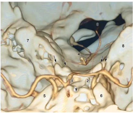
The MSCT examination presented 2 hypophyseal fossae in the sellar region, a bony notch at the level of the tuberculum sellae, and a broad dorsum sellae with a duplication of both right and left posterior clinoid processes (Fig. 1). The relief of the tabula interna of the skull interior was very expressed. The foramen magnum was oval in shape, and measured 35.6 mm in the sagittal diameter and 39.1 mm in the transverse diameter (Table 1).
The atlas (C1) showed, firstly, a partial aplasia of the posterior arch in its middle part (Fig. 2). The gap between the remnants of the right and left parts of that arch measured 26.1 mm. The right and left
lateral masses normally articulated with the occipital condyles above and the facets of the axis below. Both transverse foramina were incomplete in their anterior part. The longest transverse diameter of the atlas measured 91.9 mm (Table 1). Finally, a partial aplasia of the middle part of the anterior arch was present as well (Fig. 3). The gap between the remnants of the arch measured 22.7 mm.
The odontoid process (dens) of the axis (C2) was duplicated. The left process was a little bit higher (8.6 mm) than the right one (7.7 mm) (Table 1). The superior (cranial) transverse diameter, just below the apex, of the left process measured 8.1 mm, and of the right one 7.6 mm, whereas the inferior (caudal) transverse diameter, at the level of the dens base, was 14.5 mm of the left process and 14.7 mm of the right process (Table 1). The diameter of the cleft between the two processes (Figs. 2, 3) measured 3.7 mm in the upper part, and 3.0 mm in the basal part.
The cleft narrowed and continued through the body of the axis. This vertical cleft, from the level of the dens tip to the C3 body, measured 23.7 mm in length. Finally, the axis, C3 and C4 vertebrae were completely fused together (Figs. 2, 3). Such a fusion was also observed between the T12 and L1 vertebrae. A separate bone, located above and partially in front of the duplicated odontoid process, was merged with the middle part of the anterior rim of the fo-ramen magnum (Figs. 2, 3). This osseous junction measured 10.8 mm in a transverse direction. The maximum transverse diameter of the bone had a value of 12.9 mm, and its maximum sagittal diameter reached the value of 10.7 mm.
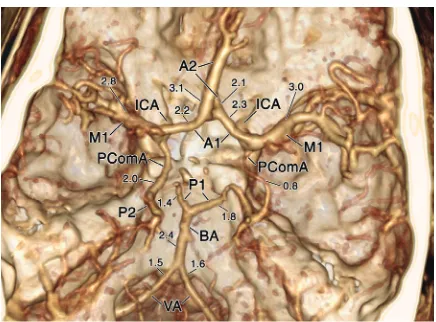
The MSCT angiography (Figs. 2, 4) showed some cerebral arteries of an uneven diameter, hypoplasia or “hyperplasia” of some other vessels, and an early bifurcation of the basilar artery. Thus, a dilatation
Table 1. Measurements [mm] of the bones and foramina: range (mean)
Subjects Foramen magnum Atlas (transverse
diameter) Height Odontoid processDiameter
Superior Inferior
Patient 35.6 × 39.1 91.9 8.6 8.1 14.5
7.7 7.6 14.7
Volunteers 35.3 × 28.0 75.3–90.4 (82.5) 14.4–19.4 (16.9) 7.8–12.1 (10.1) 11.4–16.1 (13.4) 40.1 × 32.8
(37.3 × 30.2)
Figure 2. Posterior view of the craniovertebral junction. Note the aplasia of the posterior arch (between the two asterisks) of the atlas, a duplication (1 and 2) of the odontoid process, and a separate bone (3) merged with the anterior rim (4) of the foramen magnum; 5 — dilatation of the vertebral artery between the atlas and the occipital bone; 6 — transverse process of the atlas; 7 — petrous part of the left temporal bone; 8 — basilar artery on the clivus; 9 — dorsum sellae; 10 — left internal carotid artery; 11 — left lamina of the axis, close to its spinous process (cut). Note the fused laminae of the C2–C4 vertebrae.
of the extradural vertebral artery on both sides was ob-served (Fig. 2), which measured 3.4 mm and 3.3 mm in diameter, respectively, whereas the intradural (med-ullary) segment of both arteries was much thinner (Fig. 4). These 2 arteries formed a short basilar artery (only 20.6 mm in length), which presented an early bifurcation, i.e. a duplication. Mild hypoplasia of the left P1 segment was present, as well as “hyperplasia” of the left posterior communicating artery, which was continuous with the ipsilateral P2 segment. A local dilatation of the A2 segment of the left anterior cer-ebral artery was observed, which measured 3.1 mm in size (Fig. 4). The vascular diameters are presented in Figure 4.
Control group
In order to compare the obtained radiological data and measurements in our patient with normal parameters, 10 healthy volunteers underwent the MSCT examination. We measured in these subjects the sagittal and transverse diameters of the foramen magnum (mean, 37.3 mm × 30.2 mm), the transverse diameter of the atlas (82.5 mm on average), the height of the odontoid process (mean 16.9 mm), and its superior (mean 10.1 mm) and inferior transverse diameters (mean 13.4 mm) (Table 1).
In addition, a 29-year-old male showed an incom-plete left transverse foramen of the atlas, whereas a 19-year-old female presented an asymptomatic aplasia of the middle part of the posterior arch of the atlas.
DISCUSSION
We distinguished in this section a presentation of the craniovertebral anomalies, and a description of a double pituitary gland with the accompanying brain alterations.
Craniovertebral anomalies
The infrequent congenital anomalies may affect any of the main parts of the craniovertebral junction. They are manifested in several ways.
Firstly, they can be expressed as hypoplasia of the clivus or the occipital condyles with atlanto-axial subluxation [25, 46]. In some other patients a narrow foramen magnum was observed, as well as a clivus-odontoid articulation [32, 33]. A specific bone, the so-called median or third condyle, occurs very rarely [17]. The foramen magnum, which was not measured in other reports, was oval in our pa-tient. Its diameters are inversed when compared to the measurements within the volunteers’ group, i.e. the foramen had a shorter sagittal than a transverse diameter.
As regards the atlas, its agenesis, partial aplasia, hypoplasia or a cleft of the C1 posterior arch can appear, or the agenesis or aplasia of the anterior arch
[1, 2, 6, 8, 9, 24, 27, 28, 35, 36, 38, 51]. Occurrence of the supernumerary facets of the atlas is also pos-sible, as well as its partial fusion with the odontoid or with the anterior rim of the foramen magnum, or an incomplete transverse foramen [18, 25, 39, 46].
The latter event is called the arcuate foramen [50]. If there is a combination of the partial aplasia of both arches, such a C1 vertebra is known as a bipartite atlas
[24], which mainly consists of the 2 lateral masses and transverse processes. In the case of an arch absence or its partial aplasia, a bony defect is bridged by dense fibrous tissue [27].
The anomalies affecting the C2 vertebra can be manifested as an absence of its laminae [8] or the presence of supernumerary facets, as well as a cleft or a double body [30, 39]. Certain changes in the region of the odontoid process may also occur: its aplasia [1, 19], hypoplasia [10, 12, 48, 54, 55], fusion with the C1 anterior arch or with the clivus [18], as
well as cleft, bifurcation [43] or duplication [15, 30].
Occasionally, basilar invagination occurs, or os odon-toideum or os terminale close to the dens [1, 3, 12, 13, 17, 28, 46, 55].
The adjacent vertebrae can be partially or com-pletely fused [33, 55]. These congenital fusions of the cervical vertebrae are consistent with the Klippel-Feil anomaly, Rubinstein-Taybi syndrome and some other
disorders [16, 21, 28, 30, 40, 55].
Most of the mentioned craniovertebral anomalies are so uncommon, that they were often published separately as case reports in renowned international journals. Two of them were accidentally revealed in our group of healthy volunteers, i.e. partial aplasia of the posterior arch of the atlas, as well as an arcuate foramen of the same vertebra.
In 55% of the patients with a double pituitary gland, at least one of the above mentioned cranio-vertebral anomalies could be found, e.g. hypoplasia, cleft or duplication of the odontoid process, the C2 body duplication or widened C1 arch [30, 43, 45, 52].
In some cases, a cleft of another cervical vertebra was seen, as well as a duplication of the cervical vertebral bodies. In other patients, partial or complete fusion affected various cervical vertebrae: C2–C3, C3–C5 or C2–C5 [28, 40, 45]. In 1 patient thoracolumbo-sacral rachischisis was reported [36]. Our patient presented a complete fusion of the C2–C4 vertebrae. As a con-sequence, he had a shortness of the neck with some limitation of the lateral rotation. In addition, the patient showed fusion of the T12 and L1 vertebrae. In our patient with a double pituitary, we did not find a single craniovertebral anomaly, but a combina-tion of several of them: partial aplasia of the C1 pos-terior and anpos-terior arches, that is a bipartite atlas, and a left arcuate foramen of the atlas, then duplication of the odontoid and body of the C2, the third occipital condyle, and fusion of the C2–C4 and T12 and L1 vertebrae. As for the bipartite atlas, it is produced by a disorder of the embryonic ossification centres, one of which forms the anterior arch and the other two create the posterior arch and the lateral masses [38].
The bipartite atlas in our patient, whose right and left halves were completely separated (Figs. 2, 3), had a transverse diameter longer than in the volunteers’ group (91.1 mm in the patient, compared with a maximum 90.4 mm in healthy subjects). The latter is most likely the reason for the mentioned inverse oval shape of the foramen magnum in our patient. Finally, his broad sella showed a double hypophyseal
fossa, a superior sphenoid notch and duplication of the posterior clinoid processes (Fig. 1).
A bone, which was located superior and anterior to the dens, was merged with the middle part of the anterior rim of the foramen magnum, i.e. with the basion region (Figs. 2, 3). This bone, which was named as the median or third condyle, is actually a remnant of the occipital vertebra [17]. Embrylogi-cally, the hypocentrumof the 4th occipital sclerotome (proatlas) normally develops into the anterior tubercle of the clivus, whereas its centrum forms the apical cap of the dens [3, 17]. The rostral ventral neural arch takes part in the formation of the anterior rim of the foramen magnum, the occipital condyles and third condyle, whilst the dorsocaudal part of the neural arch partially forms the arches of the atlas and its lateral masses. The first cervical (C1) sclerotome gives rise to the rest of the dens and atlas, whereas the C2 sclerotome forms the largest part of the axis body and its arch. It is obvious that disorders of all the mentioned sclerotomes were expressed in our patient. The odontoid process in our patient was duplicat-ed. The upper and lower diameters of the two were smaller than those in the group of volunteers (Table 1). Their height was shorter as well, i.e. 8.1 mm on aver-age, compared to 16.1 mm in volunteers. According to some authors [3, 15], the odontoid process has 2 foetal ossification centres on each side of the midline, which fuse in the postnatal period, i.e. by 1 year in age. A delay in fusion results in a cleft which separates the two halves of dens. This cleft also passed virtually through the whole thickness of the axis body in our patient.
As for the skull, brachycephaly or microcephaly was sometimes present in patients with a double pituitary [45, 53]. Within the skull, duplication of the sella was noticed, or a broad sella with 2 hypophyseal fossae (like in our patient), or cleft of the body of the sphenoid or basi-sphenoid [5, 41, 42, 45]. We also noticed in our patient a very expressed inner relief of the skull bones, which is like “gyral impressions” in shape.
Double pituitary gland
Pituitary duplication is an extremely rare event. According to some reports, from 1880 until 2011 only 38 such patients were reported [30]. The pi-tuitary duplication can be associated with certain face abnormalities: a bifid or double tongue, cleft lips or palate, macrostomia, supernumerary teeth, micrognathia or retrognathia, palatine dermoid, epig-nathus, pharyngeal teratoma, hypertelorism, broad forehead, and an altered shape of the auricle [5, 20, 23, 26, 42, 45, 49, 52].
The described craniofacial anomalies or duplica-tions, i.e. median cleft face syndrome, can also be seen in persons without a double pituitary gland
[22, 43]. In some of them, even a double nose can be present, as well as a third orbit [22]. Our patient presented only mild face anomalies, i.e. a broad fo-rehead, somewhat prominent superciliary arches, low positioned auricles, and progenia.
In addition to the face anomalies in patients with a double pituitary, certain changes were sometimes noticed in various organs or body parts: a short neck, torticollis, an absent thyroid isthmus, cardiac anoma-lies including ventricular septal defect and transposi-tion of great vessel, agenesis of the hemidiaphragm, congenital diaphragmatic hernia, 11 pairs of ribs, urinary abnormalities, renal agenesis or a kidney hy-pertrophy, colorectal atresia, etc. [30, 45, 49].
The pituitary duplication was usually associated with a double pituitary stalk and infundibulum, and rarely with a duplication of the anterior 3rd ventricle. In 1 case even a triplication of the pituitary gland was revealed [29]. Some patients with duplication present precocious puberty [11, 52, 53]. On the contrary, some other individuals, like our patient, show hypogonadism and delayed sexual development [45].
As regards the brain structures, hypothalamic enlargement was noticed occasionally, as well as tuberomamillary fusion [7, 29, 34, 45, 53]. In some patients, hypoplasia, or a partial or total agenesis of
the corpus callosum were revealed, then agenesis of the anterior commissure or the septum pellucidum, absence of the olfactory bulb and/or tract, broad optic nerves or chiasm, hydrocephalus or ventricular enlar-gement, then duplication of the anterior 3rd ventricle, Sylvian aqueduct or mammillary bodies, as well as third cerebral peduncle, hypoplastic or broad pons, hypoplastic cerebellum, clival encephalocoele, Dandy--Walker syndrome, and cleft or partial duplication of the spinal cord [4, 5, 7, 20, 23, 30, 34, 37, 45, 49].
Of all the mentioned brain anomalies and neopla-stic changes, our patient showed hypothalamic enlar-gement and its fusion with the mammillary bodies, as well as a suprasellar hamartoma. The latter pathologic alteration, with a similar parahypothalamic location, was found in several of the reported patients with pituitary duplication [30].
Some neurological symptoms and signs can be manifested in those patients, but in many of them, including our patient, the neurological deficits were absent [30, 45]. The psychological status of the pa-tients is either normal or in the domain of mental retardation. Our patient had a general IQ of 74.
The cerebral vasculature in our patient comprised several hypoplastic or “hyperplastic” arterial seg-ments, but also an early bifurcation, i.e. duplication, of the basilar artery, as was noticed in some other patients [7, 23, 44, 49]. The latter anomaly seems to be a characteristic features in patients with the pituitary duplication [44]. Like us, some authors also found a segmental dilatation of the distal anterior cerebral artery, i.e. the pericallosal vessel [7], but we also noticed a dilatation of the extradural segment of both vertebral arteries (Fig. 2). As regards the con-genital cerebrovascular malformations, the arterial aneurysms were not found, but only an arteriovenous malformation was present in 1 patient [7, 45].
The cause and mechanism of the pituitary du-plication, facial and craniovertebral anomalies are still being debated. Partial twinning is supposed, as well as prenatal teratogen exposure, a variant of the median cleft face syndrome, and splitting of the notochord during embryonic development [30, 45].
CONCLUSIONS
to our knowledge, has never been described in these patients. On the other hand, most of the associated face and cerebral anomalies were absent in our patient.
ACKNOWLEDGEMENTS
This work was supported by grant No. 175061 from the Ministry of Science, Serbia. We are very grateful to Mrs. Elza Holt for reviewing the English text of our manuscript
REFERENCES
1. Ahmed R, Traynelis VC, Menezes AH (2008) Fusions at the craniovertebral junction. Child Nerv Syst, 24: 1209–1224. 2. Alvarez Caro F, Pumarada Prieto PM, Alvarez Berciano F (2008) Congenital defect of the atlas and axis. A cause of misdiagnose when evaluating an acute neck trauma. Am J Emerg Med, 26: 840.e1–e2.
3. Arvin B, Fournier-Gosselin MP, Fehlings MG (2010) Os odontoideum: etiology and surgical management. Neu-rosurgery, 66 (3 suppl.): A22–A31.
4. Bagherian V, Graham M, Gerson LP, Armstrong DL (1984) Double pituitary glands with partial duplication of facial and forebrain structures with hydrocephalus. Comput Radiol, 8: 203–210.
5. Bale PM, Reye RD (1976) Epignathus, double pituitary and agenesis of corpus callosum. J Pathol, 120: 161–164. 6. Bliemel C, Kuehl H, Ruchholtz S, Kühne CA (2009) Partial aplasia of the atlas in a child. Unfallchirurg, 112: 513–516. 7. Burke M, Zinkovsky S, Abrantes MA, Riley W (2000)
Dupli-cation of the hypophysis. Pediatr Neurosurg, 33: 95–99. 8. Chau AM, Wong JH, Mobbs RJ (2009) Cervical myelopathy
associated with congenital C2/3 canal stenosis and defi-ciencies of the posterior arch of the atlas and laminae of the axis: case report and review of the literature. Spine, 34: E886–E891.
9. Currarino G, Rollins N, Diehl JT (1994) Congenital defects of the posterior arch of the atlas: a report of seven cases including an affected mother and son. Am J Neuroradiol, 15: 249–254.
10. Davis D, Gutierrez FA (1977) Congenital anomaly of the odontoid in children. A report of four cases. Child Brain, 3: 219–229.
11. de Penna GC, Pimenta MP, Drummond JB, Sarquis M, Martins JC, de Campos RC, Dias EP (2005) Duplication of the hypophysis associated with precocious puberty: presentation of two cases and review of pituitary emb-ryogenesis. Arq Bras Endocrinol Metabol, 49: 323–327. 12. El Asri AC, Akhaddar A, Gazzaz M, Okacha N, Boulhroud O,
Baallal H, Belfquih H, Belhachmi A, Mandour C, El Mo-starchid B, Boucetta M (2010). Dynamic CT scan of the craniovertebral junction: a role in the management of os odontoideum. Neurol Neurochir Pol, 44: 603–608. 13. Fielding W, Hensinger RN, Arbor A, Hawkins RJ (1980) Os
odontoideum. J Bone Joint Surg Am, 62: 376–383. 14. Ganau M, Spinelli R, Tacconi L (2013) Complex
deve-lopmental abnormality of the atlas mimicking a Jefferson fracture: diagnostic tips and tricks. J Emerg Trauma Shock, 6: 47–49.
15. Garant M, Oudjhane K, Sinsky A, O’Gorman AM (1997) Duplicated odontoid process: plain radiographic and CT appearance of a rare congenital anomaly of the cervical spine. Am J Neuroradiol, 18: 1719–1720.
16. Garrett M, Consiglieri G, Kakarla UK, Cang SW, Dickman CA (2010) Occipitoatlantal dislocation. Neurosurgery, 66 (3 suppl.): A48–A55.
17. Goel A, Shah A (2010) Unusual bone formation in the anterior rim of foramen magnum: cause, effect and tre-atment. Eur Spine J, 19 (suppl. 2): S162–S164.
18. Gupta S, Phadke RV, Jain VK (1993) C1–C2 block vertebra with fusion of anterior arch of atlas and the odontoid. Australas Radiol, 37: 95–96.
19. Gurney JW (1986) Absent dens on submentovertex view of the skull: new sign of an abnormal odontoid. Can Assoc Radiol J, 37: 38–39.
20. Hamon-Kérautret M, Ares GS, Demondion X, Rouland V, Francke JP, Pruvo JP (1998) Duplication of the pituitary gland in a newborn with median cleft face syndrome and nasal teratoma. Pediatr Radiol, 28: 290–292.
21. Hankinson TC, Anderson RCE (2010) Craniovertebral jun-ction abnormalities in Down syndrome. Neurosurgery, 66 (3 suppl.): A32–A38.
22. Hähnel S, Schramm P, Hassfeld S, Steiner HH, Seitz A (2003) Craniofacial duplication (diprosopus): CT MR imaging, and MR angiography indings. Case report. Radiology, 226: 210–213.
23. Hori A (1983) A brain with two hypophyses in median cleft face syndrome. Acta Neuropathol, 59: 150–154. 24. Hu Y, Ma W, Xu R (2009) Transoral osteosynthesis C1 as
a function-preserving option in the treatment of bipartite: atlas deformity: a case report. Spine, 34: E418–E421. 25. Iikko E, Tikkakoski T, Pyhtinen J (1998) The helical
three--dimensional CT in the diagnosis of torticollis with occipi-tocondylar hypoplasia. Eur J Radiol, 29: 55–60.
26. Ilina EG, Laziuk GI (1989) A new case of the “double hypophysis-multiple congenital developmental defect” complex. Tsitol Genet, 23: 45–46.
27. Klimo P JR, Blumenthal DT, Couldwell WT (2003) Congeni-tal partial aplasia of the posterior arch of the atlas causing myelopathy: case report and review of the literature. Spine, 28: E224–228.
28. Lampropoulou-Adamidou K, Athanassacopoulos M, Ka-rampinas PK, Vlamis J, Korres DS, Pneumaticos SG (2013). Congenital variations of the upper cervical spine and their importance in preoperative diagnosis. A case report and a review of the literature. Eur J Orthop Surg Traumatol, 23 (suppl. 1): S101–S105.
29. Manara R, Citton V, Rossetto M, Padoan A, D’Avella D (2009) Hypophyseal triplication: case report and embryologic considerations. Am J Neuroradiol, 30: 1328-1329.
30. Manjila S, Miller EA, Vadera S, Goel RK, Khan FR, Crowe C, Geertman RT (2012) Duplication of the pituitary gland as-sociated with multiple blastogenesis defects: Duplication of the pituitary gland (DPG)-plus syndrome. Case report and review of literature. Surg Neurol Int, 3: 23–35. 31. Martin MD, Bruner HJ, Majman DJ (2010) Anatomic and
32. Menezes AH, VanGilder JC, Graf CJ, McDonnell DE (1980) Craniocervical abnormalities. A comprehensive surgical approach. J Neurosurg, 53: 444–455.
33. Michie I, Clark M (1968) Neurological syndromes asso-ciated with cervical and craniocervical anomalies. Arch Neurol, 18: 241–247.
34. Mutlu H, Paker B, Bunes N, Emektar A, Keceli M, Kantarci M (2004) Pituitary duplication associated with oral der-moid and corpus callosum hypogenesis. Neuroradiology, 46: 1036–1038.
35. Phan N, Marras C, Midha R, Rowed D (1998) Cervical myelopathy caused by hypoplasia of the atlas: two case reports and review of the literature. Neurosurgery, 43: 629–633.
36. Quinteiro Antolin T, Castellano Romero I, Yáñze Calvo J (2012) Aplasia of the posterior arches of the atlas: a presentation of 2 cases. Rev Esp Cir Ortop Traumatol, 56: 381–384.
37. Roessmann U (1984) Duplication of the pituitary gland and spinal cord. Arch Pathol Lab Med, 109: 518–520. 38. Sabuncuoglu H, Ozdogan S, Karadag D, Kaynak ET (2011)
Congenital hypoplasia of the posterior arch of the atlas: case report and extensive review of the literature. Turk Neurosurg, 21: 97–103.
39. Salunke P, Futane S, Sharma M, Sahoo S, Kovilapu U, Khandelwal NK (2015) “Pseudofacets” or “supernumerary facets” in congenital atlanto-axial dislocation: boon or bane? Eur Spine J, 24: 80-87.
40. Samartzis D, Kalluri P, Herman J, Lubicky JP, Shen FH (2008) 2008 young investigator award: The role of congenitally fused cervical segments upon the space available for the cord and associated symptoms in Klippel-Feil patients. Spine, 33: 1442–1450.
41. Shah P, Likeman M, Munro-Davies L (2007) Bradycardia in minor trauma: don’t be slow on the uptake! Emerg Med J, 24: e13.
42. Shah S, Pereira JK, Becker CJ, Roubal SE (1997) Duplication of pituitary gland. J Comput Assist Tomogr, 21: 459–461. 43. Shen W, Cui J, Chen J, Ji Y, Zou J, Chen H, Xiongzheng M (2013) Partial midfacial duplication. J Craniofac Surg, 24: 934–936.
44. Shroff M, Blaser S, Jay V, Chitayat D, Armstrong D (2003) Basilar artery duplication associated with pituitary duplication: a new finding. Am J Neuroradiol, 24: 956–961.
45. Slavotinek A, Parisi M, Heike C, Hing A, Huang E (2005) Craniofacial defects of blastogenesis: Duplication of pi-tuitary with cleft palate and oropharyngeal tumors. Am J Med Genet A, 35: 13–20.
46. Smith JS, Shaffrey CI, Abel MF, Menezes AH (2010) Basilar invagination. Neurosurgery, 66 (3 suppl.): A39–A47. 47. Steinmetz MP, Mroz TE, Benzel EC (2010) Craniovertebral
junction: biomechanical considerations. Neurosurgery, 66 (3 suppl.): A7–A12.
48. Stevens CA, Pearce RG, Burton EM (2009) Familial odon-toid hypoplasia. Am J Med Genet A, 49A: 1290–1292. 49. Tagliavini F, Pilleri G (1986) Mammillo-hypophyseal
dupli-cation (diplo-mammillo-hypophysis). Acta Neuropathol, 69: 38–44.
50. Travan L, Saccheri P, Sabbadini G, Crivellato E (2011) Bi-lateral arcuate foramen associated with partial defect of the posterior arch of the atlas in a medieval skeleton: case report and review of the literature. Looking backward to go forward. Surg Radiol Anat, 33: 495–500.
51. Tsuang FY, Chen JY, Wang YH, Lai DM (2011). Neuro-logical picture. Occipitocervical malformation with atlas duplication. J Neurol Neurosurg Psychiatry, 82: 1101–1102.
52. Vieira TC, Chinen RN, Ribeiro MR, Nogueira RG, Abucham J (2007) Central precocious puberty associated with pitui-tary duplication and midline defects. J Pediatr Endocrinol Metab, 20: 1141–1144.
53. Vittore CP, Murray RA, Martin LS (2005) Case 79: pituitary duplication. Radiology, 234: 411–414.
54. Westermeyer RR (2003) Odontoid hypoplasia presenting as torticollis: a discussion of its significance. J Emerg Med, 24: 15–18.

![Table 1. Measurements [mm] of the bones and foramina: range (mean)](https://thumb-us.123doks.com/thumbv2/123dok_us/9915339.1979003/3.581.63.282.508.670/table-measurements-mm-bones-foramina-range-mean.webp)
