R E S E A R C H A R T I C L E
Open Access
Intestinal barrier function of Atlantic salmon
(Salmo salar
L.) post smolts is reduced by
common sea cage environments and suggested
as a possible physiological welfare indicator
Henrik Sundh
1*, Bjørn Olav Kvamme
2, Frode Fridell
2,5, Rolf Erik Olsen
3, Tim Ellis
4, Geir Lasse Taranger
2,
Kristina Sundell
1Abstract
Background:Fish farmed under high intensity aquaculture conditions are subjected to unnatural environments that may cause stress. Therefore awareness of how to maintain good health and welfare of farmed fish is important. For Atlantic salmon held in sea cages, water flow, dissolved oxygen (DO) levels and temperature will fluctuate over time and the fish can at times be exposed to detrimentally low DO levels and high temperatures. This experimental study investigates primary and secondary stress responses of Atlantic salmon post smolts to long-term exposure to reduced and fluctuating DO levels and high water temperatures, mimicking situations in the sea cages. Plasma cortisol levels and cortisol release to the water were assessed as indicators of the primary stress response and intestinal barrier integrity and physiological functions as indicators of secondary responses to changes in environmental conditions.
Results:Plasma cortisol levels were elevated in fish exposed to low (50% and 60% saturation) DO levels and low temperature (9°C), at days 9, 29 and 48. The intestinal barrier function, measured as electrical resistance (TER) and permeability of mannitol at the end of the experiment, were reduced at 50% DO, in both proximal and distal intestine. When low DO levels were combined with high temperature (16°C), plasma cortisol levels were elevated in the cyclic 1:5 h at 85%:50% DO group and fixed 50% DO group compared to the control (85% DO) group at day 10 but not at later time points. The intestinal barrier function was clearly disturbed in the 50% DO group; TER was reduced in both intestinal regions concomitant with increased paracellular permeability in the distal region. Conclusions:This study reveals that adverse environmental conditions (low water flow, low DO levels at low and high temperature), that can occur in sea cages, elicits primary and secondary stress responses in Atlantic salmon post smolts. The intestinal barrier function was significantly affected by prolonged hypoxic stress even when no primary stress response was observed. This suggests that intestinal barrier function is a good experimental marker for evaluation of chronic stress and that it can be a valuable tool to study the impact of various husbandry conditions on health and welfare of farmed Atlantic salmon.
Background
The global increase in production of fish under high intensity aquaculture conditions has increased the awareness of husbandry conditions to maintain good health and welfare of farmed fish [1-4]. The concept of
welfare for fish is under discussion but has been defined as the absence of suffering by some authors [5]. Fish farming results in a range of unnatural environments that may constitute a threat to the homeostasis of the fish and can be defined as stressors [6]. Thus, it is important to include stress as a central topic in the dis-cussions on welfare and health of all intensively farmed animals including fish [7,8] as long term stress can result in pathological conditions [6,9,10]. In aquaculture,
* Correspondence: [email protected]
1
Fish Endocrinology Laboratory, Department of Zoology/Zoophysiology, University of Gothenburg, PO Box 463, S-405 30, Gothenburg, Sweden Full list of author information is available at the end of the article
stress can be caused by a suboptimal or rapidly chan-ging environment or by various husbandry procedures. The initial stress response includes alterations in beha-vioral and physiological mechanisms to cope with the new and challenging situations and is generally regarded as adaptive [11,12]. However, prolonged or repeated per-iods of stress as well as severe stress can result in adverse effects on various physiological systems which may have consequences for welfare and lead to compro-mised health and performance [6,9].
Water oxygen levels have been suggested as a key lim-iting factor in intensive farming of salmonids and are considered one of the most important factors affecting health and welfare [1]. In salmon aquaculture, dissolved oxygen (DO) levels in a sea cage can be highly variable both temporally and spatially and is affected by factors like stocking density, degree of stratification, water cur-rents, position of the sea cages and daily and seasonal variations [13,14]. Tidal cycles have a strong influence on farms situated in fjords sheltered from other causes of water movement like wind, waves and prevailing cur-rents. In these environments cyclic drops in DO level occur when the tidal current changes direction (slack water). Further, the combination of slack water and high water temperature during late summer causes the lowest and most critical DO levels within the cages [13]. Thus, during unfavourable environmental conditions these fac-tors create a sub-optimal, stressful environment which can be detrimental for the fish health and welfare.
Similar to other vertebrates, a stress response in fish starts with the recognition of a potential threat to homeostasis by the central nervous system, which in turn activates primary stress response pathways. A key pathway is the hypothalamic-pituitary-interrenal (HPI) axis which results in elevated levels of the glucocorticoid hormone cortisol in the circulation [15]. Cortisol elicits secondary stress responses by affecting metabolism, osmoregulation, immune and barrier functions [15]. If sustained, these secondary responses will, in turn, affect whole animal performance such as growth, swimming capacity, disease resistance and reproduction, referred to as the tertiary stress responses [16].
Cortisol is a widely used indicator of stress in fish [17]. A vast amount of data show that plasma cortisol levels increase in response to a wide variety of physical, chemical, and biological stressors often found in aqua-culture [18,19]. However, depending on the severity and duration of the stressor, plasma cortisol can either stay slightly elevated or return to basal levels even though the stressor is still present [9,15,19,20]. Cortisol may therefore serve as a reliable indicator for acute stress, but may be less useful as an indicator for chronic stress. Furthermore, due to its rapid release into the circulation after initiation of stress, blood sampling for cortisol
measurements needs to be standardized in order to exclude possible effects of the sampling itself, which is a major obstacle when sampling large scale farming condi-tions. In order to avoid possible effects of sampling, the development of non-invasive methods is ongoing. In sal-monids, free (unconjugated) cortisol is released through the gills, its release rate is correlated to plasma levels of cortisol which can therefore be used as a non-invasive indicator of cortisol status [21].
Stress in mammals has been demonstrated to affect gastrointestinal (GI) functions and cause for example impaired transport and disturbed barrier function which may, in turn, lead to increased permeability for macro-molecules, microbial products, antigens and bacterial translocation [22-26]. These effects have mainly been attributed to endocrine stress responses through gluco-corticoids [22,23]. Stress has similarly been shown to affect GI barrier integrity in fish. Acute stress in Atlantic salmon caused immediate damage to the intercellular junctional complexes and occasionally total loss of cell-cell contact [27]. Also, cell-cellular damage was evidenced by loosening of microvilli and loss of cell content [27]. These damages were present up to 12 h after stress, whereas at later time points post stress, no or very few histological alterations could be observed [27]. Similar histopathological results were evident in acutely stressed rainbow trout (Oncorhynchus mykiss Walbaum) in an experiment where also an increased intestinal perme-ability 4 and 48 h after stress was demonstarted [28]. The permeability of the GI barrier is an important phy-siological feature as it will affect the ability to transport nutrients, water and salts as well as the translocation of harmful agents like pathogens. Thus, GI secondary stress responses may directly contribute to tertiary stress responses like decreases in growth and increases in sus-ceptibility to pathogens and disease. Intestinal barrier function is therefore potentially valuable measures to assess detrimental effects of prolonged stress and wel-fare in aquaculture.
Methods
Experimental setup Fish
The experiments were carried out at the Matre research station, Institute of Marine Research, Norway (61° N). Atlantic salmon post smolts (Aquagen strain) with a start weight at day 0 of 390.5 ± 3.3 g, (N = 900, n = 75 per tank) in Experiment 1 and 359.5 ± 18.9 g, (mean ± SEM., N = 27, n = 9 from three random tanks) in Experiment 2 (see below) were stocked in indoor tanks. Four treatments were run in triplicate, giving a total of 12 tanks. The fish were fed twice daily with 15-25% sur-plus feed. Waste feed was collected 0.5 h after each meal in order to measure feed intake and appetite at tank level, and to calculate feed rations. Fish were deprived of food for 48 h before sampling. The fish were treated according to the Norwegian national legis-lation for laboratory animals. All fish had been vacci-nated during the freshwater stage using an oil based vaccine containing formalin inactivated bacteria and virus. Morphometric and food intake data (e.g. size, growth, feed intake rates, food conversion ration) were gathered during the experiments and are reported else-where (Kvamme et al., in preparation); the current manuscript is restricted to physiological measures with focus on the GI tract.
Experiment 1: Fixed oxygen saturations at 4 levels This experiment aimed at mimicking the overall situa-tion often found in sea cages when water flow decreases, DO levels are reduced to around 50%, and the low water exchange rate may cause a build up of toxic metabolites. On 23 of February 2006, 3516 individuals were netted from a holding tank, transported for 5 minutes, netted again, bulk weighed and distributed to the experimental, octagonal tanks (water depth 76 cm, 6.5 m3). An equal density of 16.1 kg m-3 and 293 individuals in each treat-ment tank was obtained and all tanks were supplied with 100 L min-1 of SW. Mean ± SD temperature was 8.6 ± 0.1°C and salinity was 34.2 ± 0.1 throughout the experiment. The oxygen saturation of the inlet water was measured by an Oxyguard 420 probe with a Com-mander logger unit (Oxyguard international A/S, Den-mark, http://www.oxyguard.dk), logged every hour, and showed a value of 99.7 ± 1.8% (mean ± SD). Conductiv-ity of the inlet water was measured using an Indumax P CLS50 sensor (Endress + Hauser) and logged every 15 or 30 minute. Following an acclimation period of 13 days (day 0 = March 8th), four treatment oxygen regimes in triplicate tanks were set with fixed DO levels at 50%, 60%, 70% and 80% saturation. These oxygen levels were automatically regulated by adjusting inflow (range 41-137 L min-1) based on the outlet oxygen read-ings for each individual tank. The levels of carbon diox-ide (CO2) were well below recommended safe levels
(Oxyguard 420 probe, Oxyguard International, Den-mark, http://www.oxyguard.dk). Target oxygen levels and oxygen cyclic variations were achieved by supplying additional oxygen through a separate inlet with hyperox-ygenated water (300-400% O2). Throughout the acclima-tion period of 27 days the DO levels were kept above 85%. After the acclimation period, at day 0 (Feb 19th), four different oxygen treatment regimes were initiated: fixed 50% or 85% DO levels (equating to DO concentra-tions of 4.01 and 6.82 mg L-1 respectively), or 50% or 85% DO levels in two different 6 hour cycles (4:2 h at 85:50%; 1:5 h at 85:50%). These fluctuations were selected to mimic the variations in DO levels associated with periods of slack water at tidal reversals [13,14]. The oxygen levels were automatically computer regulated by adjusting inflow of hyperoxygenated water based on the outlet oxygen readings. Salinity and temperature was logged at several positions throughout the facilities using TST 487-1A2B temperature probe and Liquisys M CLM223/253 salinity probe (Endress + Hauser). Water flow, temperature, conductivity and oxygen data were monitored and processed by computer software (SD Matre, Normatic, Nordfjordeid, Norway). The mean ± SD temperature throughout the experiment was 16.0 ± 0.1°C. The experimental period lasted for 38 days, end-ing on March 29th.
Blood sampling
Sequential blood samplings for plasma cortisol analysis were performed in both Experiment 1 and 2 (9 fish tank-1). In Experiment 1, blood was sampled from all treatment groups on days 9, 29 and 48, and in Experi-ment 2, on days 10 and 29. In ExperiExperi-ment 1, blood was collected from additional fish (4 fish tank-1) sampled between days 41-43 from the 80% and 50% DO groups and in Experiment 2 between days 36-38 from the fixed 85% and 50% DO groups. In order to minimize the use of experimental animals, these blood samples were obtained from fish sampled for intestines (see below). Further, an experimental design did not include a time 0-sampling point. Previous studies on the Aquagen-strain have shown low and stable plasma cortisol levels in unstressed, control fish suggesting a very low or zero risk for inherent differences between the randomly assigned fish groups at the start of the present experi-ment. This made the exclusion of a 0-sampling point, in order to reduce experimental animals, valid. Blood sam-pling was performed in two steps: 1) the fish were sampled from the tank by one quick dip netting of an excess number of fish. From these, the appropriate number of fish (4 or 9) were randomly selected and anesthetised in SW containing metomidate (12.5 mg L -1
). 2) The fish were quickly killed with a sharp blow to the head and the blood withdrawn from the caudal vein
using heparinised syringe and needle. The blood was centrifuged at 13 000 g for 3 min. The plasma was transferred to new tubes and snap frozen in liquid nitro-gen and stored at -80°C until further analysis.
Water sampling
In addition to plasma cortisol measurements, water samples were collected from both experiments for mea-surement of water cortisol concentrations. Assessment of the release rate of unconjugated cortisol into the water is a non-invasive approach to explore the cortisol status of fish [21]. In Experiment 1, water samples were collected at 00:00 on days 0, 3, 6, 14, 29 and 48; in Experiment 2, water cortisol samples were collected at 08:00 (prior to first feeding) on days 0, 1, 3, 7, 10, 15 and 28. The water samples (1 L) were collected in poly-ethylene bottles from the outflow of each tank (without disturbing the fish) and also from the main inlet water and stored at -20°C until extraction.
Plasma and water cortisol analysis
Plasma cortisol levels were measured in unextracted plasma based on a radio immunoassay (RIA) procedure described elsewhere [31] using a cortisol antibody pre-viously validate for this method [32]. Briefly, a sheep-anti cortisol sheep-antibody (Code: S020; Lot: 1014-180182) from Guildhay Ltd. (Guildford, Surrey, U. K.) was used. Hydrocortisone-[1,2,6,7-3H(N)] (NET 396, NEN Life Sciences Products, USA) was used as tracer and cortisol standards were prepared from hydrocortisone (Sigma, St. Louis, USA). For determination of radioac-tivity in the samples a b-counter (Wallac 1409 Liquid Scintillation Counter, Turku, Finland) was used. Intra-and inter-assay coefficient of variation (CV) for cortisol was 3.9% and 5.4% respectively. The detection limit was 0.8 ng mL-1. Samples below the limit of detection were assigned the value of the assay detection limit. Water cortisol samples were extracted and concentra-tions (ng L-1) were determined by RIA as described elsewhere [33] and then converted to cortisol release rates (ng g-1h-1) using flow rate (L min-1) and interpo-lated biomass data, as previously described [21].
Intestinal barrier integrity and permeability
were carefully removed. The intestine, from the last pyloric caeca to the anus, was removed and opened longitudinally, divided into a proximal and a distal part and thereafter washed and placed in ice-cold salmon Ringer solution [34]. The serosa was peeled off the intestinal segments to minimize diffusion distance and ensure sufficient oxygenation of the tissue, before mounted into Ussing chambers as previously described [34]. Four mL of Ringer solution was added to each side of the intestinal epithelium and the preparations were allowed a period of 60 minutes for stabilization of the electrical parameters before the start of the experiment. The intestinal area of exposure was 0.75 cm2. Oxygenation and stirring were ensured by an air-lift (synthetic air containing 0.3% CO2) on both sides of the intestinal segments. The temperature in the Ussing chambers was maintained at the tank water temperature using water-filled cooling mantles. The electrical parameters; transepithelial resistance (TER), short-circuit current (SCC) and transepithelial poten-tial (TEP) were measured every 5 min throughout the experimental period (150 min) as a continuous moni-toring of preparation viability, transporting functions and integrity. The three electrical parameters obtained give information on the physiological and barrier func-tions of the intestinal epithelium. In short, TER is mainly a measure of the paracellular permeability, and SCC describes the sum of active transports taking place in the apical and basolateral membranes of the enterocytes. This, together with the passive leakage of charged molecules across the epithelium is reflected in the TEP. Results for the electrical parameters were cal-culated as the mean TER, TEP or SCC between 130-150 min ie. the last 5 measuring points of the experi-ments. The paracellular permeability of the intestinal epithelium was also assessed as the apparent perme-ability (Papp) of the hydrophilic marker14C-mannitol, a well documented paracellular permeability marker [35]. After the stabilizing period of 60 minutes, the experi-ment started (t = 0) by renewing the Ringer solution on the serosal side. On the mucosal side, the Ringer solu-tion was replaced with fresh Ringer solusolu-tion containing 14
C-mannitol (spec. act. 0.04 MBq mL-1). The electrical parameters were continuously monitored from t = 0 and for assessment of Papp, 50μl of the serosal Ringer was sampled at time points 10, 15, 20, 50, 80, 85 and 90 min after start of the experiment. Radioactivity was deter-mined in a liquid scintillation counter (Beckman LS 1801; Beckman, Fullerton, CA, USA) after adding 5 ml of Optiphase High Safe II (Wallac, Turku, Finland). Papp was calculated using Equation (1):
Papp=dQ/dt×1/ACo (1)
where dQ/dtis the appearance rate of mannitol in the serosal compartment of the Ussing chamber, Ais the area of intestinal surface exposed in the chamber andCo
is the initial concentration on the mucosal side.
Histology
Intestinal segments were analyzed for histological damage to the epithelium in the extreme groups in each experiment (ie. 80% and 50% DO in Experiment 1 and constant 85% and 50% DO in Experiment 2). On day 48 in Experiment 1 and day 29 in Experiment 2, respectively, 10 fish from each group were sampled and the intestinal regions were removed as described above, but the intes-tines were not opened longitudinally. From Experiment 1 the proximal region was sampled, whereas from experi-ment 2, both proximal and distal regions were sampled. The sections were fixed in McDowell’s fixative [36], stored in closed containers at 6°C in darkness until further processing. For histological preparation, the seg-ments were washed in phosphate buffer (300-320 mOsm, pH 7.4), post-fixed for 1.5 h in osmium tetroxide, rewashed in phosphate buffer and staineden blocin 2% aqueous uranyl acetate. After dehydration in a graded series of ethanol concentrations, the samples were embedded in Epon/Araldite via propylene oxide. Semi-thin (1.0 μm) sections were stained with 2% toluidine blue and examined under a Leica DMLB light micro-scope (LM) at 400 × and 630 × magnification. Images were acquired by a Leica DC 300 digital camera and further processed using Olympus Cell a-d™software.
Statistics
and DO treatment levels as fixed factors and with tank nested within treatment. For these analyses, SPSS 18.0 (SPSS Inc., Chicago, Illinois) was used. Cortisol release rate data were analysed using a repeated measures ANOVA (Stata 9.0) to assess the effects of treatment, tank (nested within treatment) and sampling occasion. All data are expressed as means ± SEM and p < 0.05 was regarded as significant and indicated as *, p < 0.01 as ** and p < 0.001 indicated as ***.
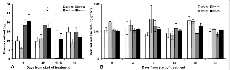
Results Plasma cortisol Experiment 1
DO levels had an effect on plasma cortisol levels (p < 0.001) but were not affected by time (p = 0.607) and no interaction could be observed (p = 0.128) (Figure 1A). Further post hoc (SNK) analysis of DO treatment levels revealed two subsets of the four oxygen treatments, with 80% and 70% DO in one subset showing lower plasma cortisol values compared to the other subset, containing 60% and 50% DO (p < 0.05). Plasma cortisol levels were also analysed in blood collected from the fish sampled for Ussing chamber experiments, but only in the two extreme treatments at day 41-43, and revealed no differ-ences between these groups.
Experiment 2
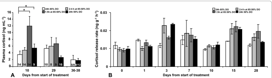
In this experiment plasma cortisol samples below limit of detection were removed in order not to violate the statis-tical analysis. At day 10, 14 fish from the 3 least extreme
treatments (85% DO, 2:4 h at 85:50% DO, 1:5 h at 85:50% DO) and 9 fish from the 50% DO group were excluded (Figure 2A). At day 29, 10 samples were excluded from the 85% DO group, 8 samples from the 2:4 h at 85:50% DO group, 9 samples from the 1:5 h at 85:50% DO group and 16 from the 50% DO group (Figure 2A). Plasma cor-tisol levels were not affected by sampling occasion (p = 0.572) but slightly influenced by DO treatment levels (p = 0.088). An interaction was observed (p = 0.029) and Bonferroni corrected pair-wise comparison of plasma cortisol levels revealed elevated cortisol levels in the 1:5 h at 85:50% DO group and 50% DO group compared to the 85% DO group (p = 0.002 and p = 0.012 respectively) at the first sampling after 10 days (Figure 2A). Thereafter no differences could be observed. Moreover, all treatment groups showed the same pattern in time,ie. no difference between sampling occasions (p = 0.572). Also in this experiment, plasma cortisol levels were analysed in sam-ples from the fish used for Ussing chamber experiments, but only in the the two extreme groups at day 36-38 but no differences could be revealed.
Water cortisol Experiment 1
No significant differences were observed between the dif-ferent DO levels (p = 0.18) indicating that DO levels did not affected the release rate for cortisol at any of the sam-pling occasions (Figure 1B). Cortisol release rate was not affected by tank (p = 0.35) nor sampling day (p = 0.28).
e (ng
g
-1h -1)
g
mL
-1)
20 25 30
80% DO 70% DO 60% DO 50% DO
0.02
80% DO 70% DO 60% DO 50% DO
◊◊
sol release
rat
e
sm
a cortisol
(n
g
10
15 0.01
Corti
Pla
s
0 5
48 A 9 Days from start of treatment29 41-43
0
0 3 6 14 29 48
B Days from start of treatment
Experiment 2
The cortisol release rate was not affected by tank (p = 0.97), but was affected by sampling day (p < 0.001; Figure 2B). Although the effect the different DO level regimes did not significantly affect the cortisol release rate there was a tendency (p = 0.10) towards lower release rate in the fixed 85% DO group compared to all other groups (Figure 2B).
Intestinal barrier function Experiment 1
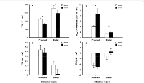
Both the proximal and distal intestines from the 50% DO treatment showed increased paracellular permeabil-ity as revealed by decreased TER (p = 0.011) (Figure 3A). In agreement, the other permeability marker Papp, describing the diffusion rate of mannitol, was elevated in both proximal and distal intestine in the 50% DO group (p = 0.020) compared to 85% DO group (Figure 3B). No major differences were observed in TEP (Figure 3C) or SCC (Figure 3D) in the either intestinal region.
Experiment 2
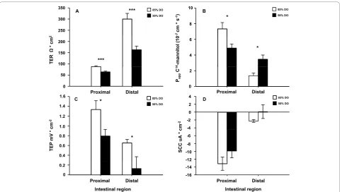
Also in this experiment both intestinal regions showed disturbed paracellular integrity as shown by decreased TER (p < 0.001) (Figure 4A) and affected Papp (Figure 4B) where an interaction between intestinal region and treatment was found (p < 0.001), revealing a decrease in Pappof the proximal intestine whereas there was an ele-vation in the distal region in the 50% DO group
compared to the 85% DO group (Figure 4B). Further, both intestinal regions from the 50% DO group had a reduced capability to maintain an electrochemical gradi-ent between mucosa and serosa as shown by the decrease in TEP (p = 0.025) (Figure 4C). No significant differences were observed in SCC (Figure 4D).
Histology
The proximal intestine was characterized by an outer layer of tightly packed granulocytes, the stratum granu-losum, located on the peritoneal side of the stratum compactum. Some individual granulocytes were also located at the luminal side of this layer but the density was low. The columnar enterocytes were nicely arranged and non-vacuolarised. On occasion, small lipid droplets, stained by osmium, could be seen in the apical part of cells of the fish from Experiment 1. Mucus producing goblet cells were scattered throughout the enterocyte layer. Subjecting the fish to hypoxia tended to shorten villi height, noted in 50% of the fish examined in Experi-ment 1 and in 60% of the fish from ExperiExperi-ment 2, while the same measures for control fish were 20 and 30% respectively. No gross morphological damage to the proximal intestine could be observed in either Experi-ment 1 or 2. The distal intestine had a more complex structure in that it contained complex folds with sub-stantial connective tissue and simple folds with less con-nective tissue. Although a distinct stratum granulosum 10
Plasm
a cortisol
(ng
mL
-1)
A 29 36-38
0 2 4 6 8 10 12 14 16
14 * *
9 10 8 9 16 5/88/12 14 14
2:4 h at 85:50% DO 24h 50% DO 24h 85% DO
1:5h at 85:50% DO
0 0.01 0.02 0.03
0 1 3 7 10 15 28
Days from start of treatment
Cortisol release
rate (ng
g
-1h -1)
B
2:4 h at 85:50% DO 24h 50% DO 24h 85% DO
1:5h at 85:50% DO
Days from start of treatment
is found in the distal intestine as well, there is a clear impression of higher number of granulocytes on the luminal side in this region compared to the proximal intestine. In the distal intestine the supranuclear cyto-plasm of the enterocytes were heavily vacuolarised, ran-ging from very small to large vacuoles. Fish from the 50% DO group in Experiment 2 appeared to display altered appearance of the intestinal segments. When compared, reduced villi height and increased size of sub-mucosa of the enterocyte layer was observed in 70% of the fish compared to only 30% for control fish. Although no gross morphological damage could be observed in the distal intestine, a disturbed morphology was still noticeable.
Discussion
In this study, two experiments on Atlantic salmon post smolts were conducted in order to mimic different water
currents and low oxygen conditions as well as diurnal cyclic variations in oxygen levels in combination with high temperatures, as observed during sea cage condi-tions [13,14]. The imitation of the complex sea cage water conditions were successfully created and main-tained in controlled tank situations over several weeks. This renders, to our knowledge, the first study to achieve this and thus giving the opportunity to study physiologi-cal responses to this type of husbandry conditions.
Two main approaches were used to mimic water qual-ity conditions that commonly occur in the sea cage environment during salmon farming [13,14]. In Experi-ment 1, oxygen levels were maintained at fixed levels by regulating water inflow according to the oxygen con-sumption of the fish, which resulted in very stable oxy-gen tensions over time. This reflects the sea-cage situation (oxygen levels being dependent upon the rates of consumption and replacement by water flow). Figure 3Intestinal barrier function after long term hypoxia (Experiment 1). This experiment aimed at mimicking an overall situation often found in sea cages when water flow decreases and DO levels are reduced to levels as low as around 50%. Decreased water exchange rate may also cause increased concentrations of toxic metabolites. Four fish from each tank in triplicate was sampled between days 41-43, from 80% and 50% DO levels groups created by adjusting inflow (range 41-137 L min-1) in response to oxygen consumption of the fish. The intestine was removed and opened longitudinally, divided into a proximal and a distal part, washed in ice-cold salmon Ringer solution and mounted in Ussing chambers. The electrical parameters; transepithelial resistance (TER), short-circuit current (SCC) and transepithelial potential (TEP) were measured. TER is mainly a measure of the paracellular permeability, and SCC describes the sum of active transports. This, together with the passive leakage of charged molecules across the epithelium is reflected in the TEP. The paracellular permeability of the intestinal epithelium was also assessed as the apparent permeability (Papp) of a well documented paracellular marker14C-mannitol. Ussing chamber data was analysed
using a genral linear model with intestinal region and treatment (with tank nested within treatment) as factors. Paracellular permeability was higher in both intestinal regions in the 50% DO group as indicated by decreased TER (p < 0.05) (A) and the increased Pappfor mannitol which
Biomass and food supply were consistent across treat-ments and low but increases in CO2 and ammonium was observed whereas no changes in pH were present. The highest CO2 levels observed never exceeded 10 mg L-1. This was thus well below levels where detrimental effects of CO2(> 20 mg L-1) for salmonids are observed [29]. Thus, most likely the results observed in Experi-ment 1 mainly are effects of the different experiExperi-mentally set DO levels. However, it cannot be excluded that even small differences in CO2 and ammonium could have an impact on the physiology of the fish together with low DO levels. Therefore, although the results of Experiment 1 are discussed in relation to oxygen treatment and hypoxia in order to simplify nomenclature, other aspects of compromised water should be included in the inter-pretation. In contrast, in Experiment 2 the effect of
oxygen was isolated from other water quality para-meters: water flow was consistent between treatment groups and oxygen level was regulated by balancing uptake with addition of hyperoxic water. Treatments effects in this experiment can therefore be expected to be due solely to hypoxia. Furthermore, in Experiment 2, the temperature was set to 16°C (compared to 8.6°C in Experiment 1) as field observations indicate that the lowest DO levels in sea cages are most often observed during late summer/autumn when oxygen production from algae is low and when temperature usually is ele-vated as well. Thus, the two experiments have been designed to create two different types of scenarios in an attempt to mimic different environmental situations in a sea cage and are therefore expected to affect the phy-siology of the fish in different ways.
Figure 4Intestinal barrier function after long term hypoxia at high temperature (Experiment 2). This experiment aimed at mimicking DO levels measured in sea cages in fjords sheltered from waves, wind and strong currents. In these situations, cyclic drops in DO levels are frequently observed during slack water at tidal reverse and further decreased DO levels are observed during high temperatures. Four oxygen treatment regimes were initiated: fixed 50% or 85% DO levels, or 50% or 85% DO levels in two different 6 hour cycles (4:2 h at 85:50%; 1:5 h at 85:50%) at 16°C. Four fish per tank from fixed 85% and 50% DO groups were sampled. The intestine was removed and opened longitudinally, divided into a proximal and a distal part, washed in ice-cold salmon Ringer solution and mounted in Ussing chambers. The electrical parameters; transepithelial resistance (TER), short-circuit current (SCC) and transepithelial potential (TEP) were measured. TER is mainly a measure of the paracellular permeability, and SCC describes the sum of active transports. This, together with the passive leakage of charged molecules across the epithelium is reflected in the TEP. Ussing chamber data was analysed in a genral linear model with intestinal region and treatment (with tank nested within treatment) as factors. The paracellular permeability of the intestinal epithelium was also assessed as the apparent permeability (Papp) of
14
C-mannitol. TER was lower in the 50% DO group in both intestinal regions (p < 0.001) (A). An interaction between intestinal region and treatment was found for Papp(p < 0.001), revealing a decrease in Pappof the proximal intestine whereas there was an increase in the distal
Environmental conditions creating a threat to homeos-tasis results in alloshomeos-tasis. This means a change in beha-vioural and physiological parameters to meet the environmental changes and maintain homeostasis. In allostasis, cortisol is an important regulator [9,37,38]. Salmonids are regarded as relatively intolerant to hypoxic conditions [39]. In fish, the minimum oxygen tension to provide fully saturated haemoglobin has been suggested to be 60% [29]. It can therefore be assumed that the lowest DO treatment in both experiments (50%) were stressful to the fish and thus a threat to the homeostasis. Previous work has shown that exposure to a hypoxic environment activates a cortisol response, but that there can be a rapid acclimation (habituation). In rainbow trout subjected to fixed 55% DO levels, plasma cortisol level doubled after 2 h (from ~20 ng mL-1 to ~40 ng mL-1) but returned to basal levels after 24 h [40]. The magnitude of this plasma cortisol response to hypoxia is similar to that observed in Experiment 1, although the duration of the response differs as the ele-vation in plasma cortisol levels were sustained several weeks after start of treatment. Hypoxia itself can induce a primary stress response but toxic metabolites (ammo-nia and CO2) can also induce this stress response in sal-monids [18,41] suggesting that a combination of these factors reveals the cortisol response in the present experiments. Even though the lack of interaction between DO levels and sampling occasion in Experi-ment 1 indicates that there are still a difference in plasma cortisol levels between the 50 and 60% DO level groups compared to the 70 and 80% DO level groups, the plasma cortisol levels in the lower oxygen environ-ment have clearly decreased at the end of the experi-ment (day 41-43 and 48). In Experiexperi-ment 2, no differences were seen between the treatment groups after day 10 and the plasma cortisol levels were low and close to basal levels [15] in all groups, as also indicated by the numerous samples excluded for being below the limit of detection for the RIA. The data on plasma corti-sol levels from the two experiments taken together indi-cates that the fish have acclimated (habituated) to the new environments. However, it may not necessarily reflect a decreased activity of the corticosteroid system as plasma cortisol concentrations reflect the balance of the production and plasma clearance rates of the hor-mone from circulation. During chronic stress, increased activity including increased receptor levels and clearance rate may even render decreased plasma cortisol levels [17]. Thus, activation of the HPI axis may not be reflected by maintained increases in plasma cortisol levels during chronic stress and may therefore not be a reliable marker for chronic stress. Stocking density has been shown to reduce somatic growth although no
changes, or even a decrease in plasma levels of cortisol was observed (reviewed in [1]).
The long term effect of physiological responses to stress can result in an allostatic load which is a“wear and tear”on the body and thus the cost for acclimation to sub optimal environments [42]. The allostatic load exhibited by the mimicked husbandry conditions in the present study resulted in decreased intestinal barrier functions as evident by increased intestinal permeability. TER was significantly reduced in the fixed 50% DO groups in both intestinal regions in both experiments. Pappfor mannitol increased concomitant with decreased TER in both intestinal regions in Experiment 1 and in the distal intestine in Experiment 2. Moreover, by com-paring TER between the two experiments, TER appeared more reduced in Experiment 2 compared to Experiment 1 suggesting that the allostatic load from low DO levels is more severe at an elevated temperature and that hypoxia and high temperature per semay act as additional stressors. Increases in temperature raise the metabolism of the fish and thus oxygen demand. Concomitantly, increased temperature decreases the car-rying capacity of the water to oxygen. Thus, the effects of the increased temperature, apart from being a prob-able stressor in itself, further ads to the hypoxic situa-tion already created in the experimental design. Studies in mammals show that stress reduces the barrier func-tion of the intestinal epithelium and causes increased paracellular permeability, increased uptake of macromo-lecules, bacterial products and antigens [24-26,43-45]. These effects are probably mediated by glucocorticoids as administration of dexametasone directly induces increased intestinal permeability in rats, an effect that was blocked by the glucocorticoid receptor antagonist RU-486 [22,23]. There is evidence that stress and corti-costeroids have similar effects on intestinal integrity and permeability also in fish. Slow release cortisol implant increases the paracellular permeability of rainbow trout intestine for mannitol (K. Sundell, personal obs). Pro-longed stress (hyperoxygenation and reduced water flow) in Atlantic salmon elevated plasma cortisol levels and increased paracellular permeability concomitant with increased translocation rate of pathogenic bacteria
Aeromonas salmonicida [46]. Further, subjecting rain-bow trout to an acute exhaustive stress elevated intest-inal permeability up to 48 h in both the proximal and distal region [28]. Although the exact mechanism whereby hypoxia causes the detrimental changes to the intestinal barrier is unknown, it can be argued that cor-tisol may be an important mediator of the permeability increase.
a rather sensitive and sustainable physiologic indicator of prolonged effects of stress when other measures can be hard to interpret. Several different stressors: hyperox-ygenation and reduced water flow [46], hypoxia (present study), hypoxia at high temperature (present study), IPNV infection [46], chronic feed stress [47] and high stocking density and low DO levels (Sundh et. al. in pre-paration) have all been shown to increase the intestinal permeability,ie. decrease the intestinal barrier functions after long term exposure. In agreement, although both increased cortisol levels, ultrastructural damage to inter-cellular junctions as well as gross morphological damage to epithelium coincided with increased permeability in acutely stressed rainbow trout, the increase in intestinal permeability was maintained and still significant at time points long after stress when ultrastructural damage and differences in plasma cortisol levels had returned to basal [28]. A similar pattern has been documented in rats where increased permeability was observed in the absence of morphological damage to intestinal tissues after stress [48]. This further indicates that the perme-ability increase in response to prolonged stress may not always be a result of damages to the epithelium but can also be the result of a physiological and/or immunologi-cal regulation [49-51].
In mammals, stress is discussed in relation to intest-inal diseases like the inflammatory bowels diseases (IBD) [51]. These diseases are thought to result from a contin-uous and inappropriate activation of the mucosal immune system, driven by the enteric microflora [52]. Increased paracellular permeability, macromolecular uptake and bacterial translocation are important media-tors behind the development of IBD which have major impact on the health and welfare of the diseased indivi-duals [52,53]. The results from the current experiments suggest that prolonged stress in Atlantic salmon results in reduced barrier function similar to the observations in patients with IBD or with increased risk of developing IBD. In support, stress in Atlantic salmon has previously been shown to increases the adherence of both enteric and pathogenic bacteria to the intestinal epithelium and also cause increased translocation of liveAeromonas sal-monicida[27,46]. The reduced barrier function will not only increase the risk for pathogenic infection, but also the delivery of antigens to the mucosal immune system with an increased immune response as a result. This may, in turn, lead to development of an intestinal inflammatory response. Intestinal inflammation, observed as increased infiltration of neutrophils, could be measured in Atlantic salmon after prolonged stress (Sundh H et. al. in preparation). All together, increased intestinal permeability in Atlantic salmon may increase the disease susceptibility and generate an IBD like phy-siological status with increased permeability, bacterial
translocation and intestinal inflammation as common features. This should be considered as a major threat to the health and welfare of the farmed fish and thus, the intestinal permeability could be suggested to function as a physiological indicator of stress as well as welfare of Atlantic salmon.
Other aspects of the intestinal function were also affected by the experimental treatments. TEP but not SCC, was affected by the hypoxic treatment, as observed as a tendency in Experiment 1 and as a significant decrease in Experiment 2, in both proximal and distal region of the intestine. While SCC is a reflection of the overall active transports taking place in the intestinal epithelium, TEP reflects the active transport functions as well as the intestinal barriers ability to maintain an electrochemical gradient across the epithelium. Thus, the drop in TEP observed in Experiment 2 is most prob-ably caused by a decrease in TER rather than by decreased active transport activities. The physiological consequence of a reduced TER and TEP could be impairment of nutrient uptake. The absorption of water soluble nutrients (ie. amino acids and glucose) are per-formed against their gradient mainly by use of the elec-trochemical gradient for Na+ into the cell as created by basolaterally located Na+/K+- ATPases [54-56]. How-ever, when TER is reduced, the paracellular leakage of Na+ is increased which in turn will reduce the potential difference between the lumen and the enterocyte and reduce the gradient for Na+into the cell. This could be compensated for by increasing the activity of the Na+/K +
- ATPases. However, this does not appear to occur in the present studies and therefore a reduced nutrient uptake could be an additional consequence of the stress-ful husbandry conditions studied. Thus, apart from increasing disease susceptibility and risk of inflamma-tion, intestinal permeability alterations can also be a contributing factor to reduced somatic growth.
non-treatment factor not present at the times the water sam-pling were collected 2) a lack of sensitivity in the water samples as they represent an integration over the whole tank population and over the residence time of the water within the tank, or 3) a loss of sensitivity due to normalisation of the release rate assuming a constant water flow, while in reality flow could change very rapidly as it was regulated in response to oxygen con-sumption of the fish. The latter reason illustrate that the non-invasive method, although having benefits [21,33], is not suitable when water flow fluctuates which is the case also in the sea cage situation. This problem in application is being addressed by searching for normalis-ing compounds released by fish at a constant rate [21].
Conclusions
This study demonstrates that environmental conditions with low water flow and low levels of DO, which often occurs in sea cages, can affect Atlantic salmon physiol-ogy. The environment is recognized by the fish as a threat to which they respond through activation of the HPI axis and the fish appear to be able to acclimate in terms of plasma cortisol status. However, despite accli-mation in plasma cortisol, the intestinal barrier function was significantly reduced in the longer term and the dis-turbance was more severe at high temperatures. A reduced intestinal barrier may increases the disease sus-ceptibility, decrease nutrient up take rendering decreased somatic growth and increases the risk of developing a chronic intestinal inflammation. Cortisol is a widely used indicator of stress in fish [17] and a vast body of evidence show that plasma cortisol levels increase in response to a wide variety of physical, che-mical, and biological stressors often encountered in to aquaculture [18,19]. Although cortisol may serve as a reliable marker for acute stress, it is recognized as less suitable as an indicator of chronic stress. This, and pre-vious studies [27,28] demonstrate that GI integrity may be compromised although plasma cortisol has returned to basal levels. Meaningful secondary responses such as the intestinal barrier function may therefore be prefer-able as indicators of the impact of chronic stress condi-tions on fish performance and can be used when evaluating the impact of husbandry conditions on the health and welfare of farmed fish.
Acknowledgements
This work was financed by the WEALTH project (Welfare and health in sustainable aquaculture, EU FP6 project no: No 501984). Financial support was also given by the Swedish Research Council for Environment, Agricultural Sciences and Spatial Planning to KS and the Norwegian Ministry of Fisheries and Coastal Affairs to GLT. Further financial support was given by the Helge Ax:son Johnsons Foundation, Wilhelm & Martina Lundgrens Foundation and Adlerbertska Scientific Foundation to HS. We would also like to acknowledge Lars Niklasson and Dean Basic for excellent technical
assistance and Frode Oppedal for synthesising all information on the experimental design. We would also like to acknowledge the technical staff at IMR both at Matre Research Station. Without your good spirit and outstanding knowledge these experiments would never have been manageable.
Author details
1Fish Endocrinology Laboratory, Department of Zoology/Zoophysiology,
University of Gothenburg, PO Box 463, S-405 30, Gothenburg, Sweden.
2Institute of Marine Research, P.O. Box 1870 Nordnes, 5817 Bergen, Norway. 3Institute of Marine Research, N-5984 Matredal, Norway.4Cefas Weymouth
Laboratory, Barrack Road, The Nothe, Weymouth, Dorset, DT4 8UB, UK.
5PHARMAQ AS, P.O. Box 267 Skøyen, Oslo, Norway.
Authors’contributions
HS conducted the intestinal and blood samplings, all physiological experiments and analysis as well as plasma cortisol RIA analyses, statistical analysis, interpretation of results and are responsible for and corresponding author of the manuscript. BOK participated in the planning and performing of the experimental trials, blood samplings, interpretation of results and editing of the manuscript. FF participated in the planning and performing of the experimental trials, blood samplings, interpretation of results and editing of the manuscript. REO performed the histology samplings and analysis and interpretation of results and editing of the manuscript. TE performed the water samplings, analysis of water cortisol, interpretation of results and editing of the manuscript. GLT contributed with scientific ideas, in developing the experimental design, interpretation of results and editing of the manuscript. KS conceived the study, contributed with scientific conception and development of the experimental design, interpretation of results and writing of the manuscript. All authors read and approved the final manuscript.
Received: 22 July 2009 Accepted: 9 November 2010 Published: 9 November 2010
References
1. Ellis T, North B, Scott AP, Bromage NR, Porter M, Gadd D:The relationships between stocking density and welfare in farmed rainbow trout.J Fish Biol2002,61(3):493-531.
2. Hastein T, Scarfe AD, Lund VL:Science-based assessment of welfare: aquatic animals.Rev Sci Tech Off Int Epiz2005,24(2):529-547. 3. Turnbull J, Bell A, Adams C, Bron J, Huntingford F:Stocking density and
welfare of cage farmed Atlantic salmon: application of a multivariate analysis.Aquaculture2005,243(1-4):121-132.
4. Ashley PJ:Fish welfare: Current issues in aquaculture.Appl Anim Behav Sci 2007,104(3-4):199-235.
5. Huntingford FA, Adams C, Braithwaite VA, Kadri S, Pottinger TG, Sandoe P, Turnbull JF:Current issues in fish welfare.J Fish Biol2006,68(2):332-372. 6. Moberg GP:Biological response to stress: implications for animal welfare.
InThe Biology of Animal Stress: Basic Principles and Implications for Animal Welfare.Edited by: Moberg B and Mench JA. New York: CABI Publishing; 2000:1-21.
7. Pottinger TG:The stress response in fish - mechanisms, effects and measurement.InFish welfare.Edited by: Branson EJ. Oxford: Blackwell Publishing Ltd; 2008:32-48.
8. Conte FS:Stress and the welfare of cultured fish.Appl Anim Behav Sci 2004,86(3-4):205-223.
9. Schreck CB:Accumulation and long-term effects of stress in fish.InThe Biology of Animal Stress: Basic Principles and Implications for Animal Welfare. Edited by: Moberg B and Mench JA. New York: CABI Publishing; 2000:147-158.
10. Pickering AD:Endocrine-induced pathology in stressed salmonid fish.
Fisheries Research1993,17(1-2):35-50.
11. Pickering AD:Rainbow trout husbandry: management of the stress response.Aquaculture1992,100(1-3):125-139.
12. Korte SM, Koolhaas JM, Wingfield JC, McEwen BS:The Darwinian concept of stress: benefits of allostasis and costs of allostatic load and the trade-offs in health and disease.Neurosci Biobehav Rev2005,29(1):3-38. 13. Johansson D, Juell JE, Oppedal F, Stiansen JE, Ruohonen K:The influence
swimming behaviour of Atlantic salmon (Salmo salar L.) in production cages.Aquaculture2007,265(1-4):271-287.
14. Johansson D, Ruohonen K, Kiessling A, Oppedal F, Stiansen JE, Kelly M, Juell JE:Effect of environmental factors on swimming depth preferences of Atlantic salmon (Salmo salar L.) and temporal and spatial variations in oxygen levels in sea cages at a fjord site.Aquaculture2006, 254(1-4):594-605.
15. Barton BA:Stress in fishes: A diversity of responses with particular reference to changes in circulating corticosteroids.Integ Comp Biol2002,
42(3):517-525.
16. Wendelaar Bonga SE:The stress response in fish.Physiol Rev1997,
77(3):591-625.
17. Mommsen TP, Vijayan MM, Moon TW:Cortisol in teleosts: dynamics, mechanisms of action, and metabolic regulation.Rev Fish Biol Fisheries 1999,9:211-268.
18. Donaldson EM:The pituitary-interrenal axis as an indicator of stress in fish.InStress and fish.Edited by: Pickering AD. London: Academic press Inc; 1981:11-47.
19. Barton BA, Iwama GK:Physiological changes in fish from stress in aquaculture with emphasis on the response and effects of corticosteroids.Ann Rev Fish Dis1991,1:3-26.
20. Fridell F, Gadan K, Sundh H, Taranger GL, Glette J, Olsen RE, Sundell K, Evensen Ø:Effect of hyperoxygenation and low water flow on the primary stress response and susceptibility of Atlantic salmon Salmo salar L. to experimental challenge with IPN virus.Aquaculture2007, 270(1-4):23-35.
21. Ellis T, James JD, Sundh H, Fridell F, Sundell K, Scott AP:Non-invasive measurement of cortisol and melatonin in tanks stocked with seawater Atlantic salmon.Aquaculture2007,272(1-4):698-706.
22. Meddings JB, Swain MG:Environmental stress-induced gastrointestinal permeability is mediated by endogenous glucocorticoids in the rat.
Gastroenterology2000,119(4):1019-1028.
23. Spitz JC, Ghandi S, Taveras M, Aoys E, Alverdy JC:Characteristics of the intestinal epithelial barrier during dietary manipulation and glucocorticoid stress.Crit Care Med1996,24(4):635-641.
24. Groot J, Bijlsma P, Van Kalkeren A, Kiliaan A, Saunders P, Perdue M: Stress-induced decrease of the intestinal barrier function. The role of muscarinic receptor activation.Ann N Y Acad Sci2000,915:237-246. 25. Santos J, Benjamin M, Yang PC, Prior T, Perdue MH:Chronic stress impairs
rat growth and jejunal epithelial barrier function: role of mast cells.Am J Physiol Gastrointest Liver Physiol2000,278(6):G847-854.
26. Velin AK, Ericson AC, Braaf Y, Wallon C, Söderholm JD:Increased antigen and bacterial uptake in follicle associated epithelium induced by chronic psychological stress in rats.Gut2004,53(4):494-500.
27. Olsen RE, Sundell K, Hansen T, Hemre GI, Myklebust R, Mayhew TM, Ringø E:Acute stress alters the intestinal lining of Atlantic salmon,Salmo salarL.: An electron microscopical study.Fish Physiol Biochem2002,
26:211-221.
28. Olsen RE, Sundell K, Mayhew TM, Myklebust R, Ringø E:Acute stress alters intestinal function of rainbow trout,Oncorhynchus mykiss(Walbaum).
Aquaculture2005,250(1-2):480-495.
29. Wedemeyer GA:Physiology of Fish in Intensive Culture SystemsNew York: Chapman & Hall; 1996.
30. Hitchman ML:Measurement of dissolved oxygenNew York: Wiley; 1978. 31. Young G:Cortisol secretion in vitro by the interrenal of coho salmon
(Oncorhynchus kisutch) during smoltification relationship with plasma thyroxine and plasma cortisol.Gen Com Endocrinol1986,63(2):191-200. 32. Sundh H, Calabrese S, Jutfelt F, Niklasson L, Olsen RE, Sundell K:
Translocation of infectious pancreatic necrosis virus across the intestinal epithelium of Atlantic salmon (Salmo salarL.).Aquaculture.
33. Ellis T, James JD, Stewart C, Scott AP:A non-invasive stress assay based upon measurement of free cortisol released into the water by rainbow trout.J Fish Biol2004,65(5):1233-1252.
34. Sundell K, Jutfelt F, Agustsson T, Olsen RE, Sandblom E, Hansen T, Björnsson BT:Intestinal transport mechanisms and plasma cortisol levels during normal and out-of-season parr-smolt transformation of Atlantic salmon, Salmo salar.Aquaculture2003,222(1-4):265-285.
35. Blikslager AT, Moeser AJ, Gookin JL, Jones SL, Odle J:Restoration of barrier function in injured intestinal mucosa.Physiol Rev2007,87(2):545-564. 36. Olsen RE, Myklebust R, Kaino T, Ringø E:Lipid digestibility and
ultrastructural changes in the enterocytes of Arctic char (Salvelinus
alpinusL.) fed linseed oil and soybean lecithin.Fish Physiol Biochem1999,
21(1):35-44.
37. McEwen BS, Wingfield JC:The concept of allostasis in biology and biomedicine.Horm Behav2003,43(1):2-15.
38. Landys MM, Ramenofsky M, Wingfield JC:Actions of glucocorticoids at a seasonal baseline as compared to stress-related levels in the regulation of periodic life processes.Gen Comp Endocrinol2006,148(2):132-149. 39. Bickler PE, Buck LT:Hypoxia tolerance in reptiles, amphibians, and fishes:
life with variable oxygen availability.Annu Rev Physiol2007,69:145-170. 40. Lai JCC, Kakuta I, Mok HOL, Rummer JL, Randall D:Effects of moderate
and substantial hypoxia on erythropoietin levels in rainbow trout kidney and spleen.J Exp Biol2006,209(14):2734-2738.
41. Fivelstad S, Olsen AB, Kloften H, Ski H, Stefansson S:Effects of carbon dioxide on Atlantic salmon (Salmo salarL.) smolts at constant pH in bicarbonate rich freshwater.Aquaculture1999,178(1-2):171-187. 42. McEwen BS:Protective and damaging effects of stress mediators.N Engl
J Med1998,338(3):171-179.
43. Kiliaan AJ, Saunders PR, Bijlsma PB, Berin MC, Taminiau JA, Groot JA, Perdue MH:Stress stimulates transepithelial macromolecular uptake in rat jejunum.Am J Physiol Gastrointest Liver Physiol1998,275:G1037-G1044. 44. Santos J, Yang PC, Söderholm JD, Benjamin M, Perdue MH:Role of mast
cells in chronic stress induced colonic epithelial barrier dysfunction in the rat.Gut2001,48(5):630-636.
45. Söderholm JD, Yang PC, Ceponis P, Vohra A, Riddell R, Sherman PM, Perdue MH:Chronic stress induces mast cell-dependent bacterial adherence and initiates mucosal inflammation in rat intestine.
Gastroenterology2002,123(4):1099-1108.
46. Sundh H, Olsen RE, Fridell F, Gadan K, Evensen Ø, Glette J, Taranger GL, Myklebust R, Sundell K:The effect of hyperoxygenation and reduced flow in fresh water and subsequent infectious pancreatic necrosis virus challenge in sea water, on the intestinal barrier integrity in Atlantic salmon,Salmo salarL.J Fish Dis2009,32(8):687-689.
47. Knudsen D, Jutfelt F, Sundh H, Sundell K, Koppe W, Frøkiær H:Dietary soya saponins increase gut permeability and play a key role in the onset of soyabean-induced enteritis in Atlantic salmon (Salmo salarL.).Brit J Nutr 2008,100:120-129.
48. Saunders PR, Kosecka U, McKay DM, Perdue MH:Acute stressors stimulate ion secretion and increase epithelial permeability in rat intestine.Am J Physiol Gastrointest Liver Physiol1994,267(5):G794-799.
49. Gareau MG, Silva MA, Perdue MH:Pathophysiological Mechanisms of Stress-Induced Intestinal Damage.Curr Molec Med2008,8:274-281. 50. Capaldo CT, Nusrat A:Cytokine regulation of tight junctions.Biochim
Biophys Acta - Biomembranes2009,1788(4):864-871.
51. Söderholm JD, Perdue MH:Stress and the gastrointestinal tract II. Stress and intestinal barrier function.Am J Physiol Gastrointest Liver Physiol2001,
280(1):G7-G13.
52. Podolsky DK:Inflammatory bowel disease.N Engl J Med2002,
347(6):417-429.
53. Caso JR, Leza JC, Menchen L:The Effects of Physical and Psychological Stress on the Gastrointestinal Tract: Lessons from Animal Models.Curr Molec Med2008,8:299-312.
54. Buddington RK, Krogdahl A, Bakke-McKellep AM:The intestines of carnivorous fish: structure and functions and the relations with diet.
Acta Physiol Scand Suppl1997,638:67-80.
55. Loretz CA:Electrophysiology of ion transport in teleost intestinal cells.In Fish physiology. Cellular and molecular approaches to fish ionic regulation. Volume 14.Edited by: Wood CM, Shuttleworth TJ. New York; Academic press; 1995:25-56.
56. Collie NL:Hormonal Regulation of Intestinal Nutrient Absorption in Vertebrates.Amer Zool1995,35(6):474-482.
doi:10.1186/1472-6793-10-22