13
Research Paper:
Effects of Aloe Vera Gel Extract on
Aging-Associated Histological Changes of the Sciatic Nerve
Babak Barmaki1, Hamidreza Ghaffari2* , Soghra Gholami3, Malihe Alipoor Tabrizi41. Department of Physiology, School of Medicine, Zabol University of Medical Sciences, Zabol, Iran. 2. Department of Anatomical Sciences, School of Medicine, Zabol University of Medical Sciences, Zabol, Iran. 3. Department of Anatomy, School of Medicine, Shiraz University of Medical Sciences, Shiraz, Iran. 4. Department of Heart, Emam Reza Hospital, Mashhad University of Medical Sciences, Mashhad, Iran.
* Corresponding Author: Hamidreza Ghaffari, PhD
Address: Department of Anatomical Sciences, School of Medicine, Zabol University of Medical Sciences, Zabol, Iran. Tel: +98 (54) 32230764
E-mail: [email protected]
A B S T R A C T
Article info:
Received: 13 Feb 2018
Accepted: 05 Jul 2018
Available Online: 01 Jan 2019
Keywords:
Aging, Aloe vera, Neuropathy, Sciatic nerve, Rats
Hamidreza Ghaffari is the assistant professor of anatomy in the Department of Physiology, School of Medicine, Zabol University of Medical Sciences, Zabol, Iran. His research interests are anatomy, histology and embryology.
Introduction: Prior qualitative and quantitative studies have reported morphological changes that occur in nerve fibers and non-neuronal cells of peripheral nerve during the lifetime of the rats. Previous studies suggest that Aloe Vera has some beneficial and therapeutic effects such as anti-oxidant effects. In the current study, we have evaluated histological and histomorphometric changes of Sciatic nerve in aged rats treated with Aloe Vera gel extract.
Methods: Sixty healthy male albino Sprague-Dawley rats weighing 200-250 g were randomly divided into two groups: control (normal diet), and experimental group (normal diet+Aloe Vera extract). Rats of the experimental group received 400 mg/kg Aloe Vera extract daily. Within 16, 20 and 24 months, animals were anesthetized with sodium thiopental (40 mg/kg) via IP injection. After removal of Sciatic nerve on the left side, nerve dissected and fixed in 4% glutaraldehyde, post fixed in osmium tetroxide 1%, dehydrated and then embedded in TAAB resin. Thin sections (1 μm) were stained with toluidine blue stain and ten slides were obtained from each animal and got examined on light microscope. Masson’s trichrome stain was used to evaluate the development of fibrosis. Histomorphometric and histologic criteria on Sciatic nerve were determined and data were recorded. Data were analyzed using SPSS software. Results: Resultsof the current study confirmed accumulation of collagen in the perineurium by aging. Our data suggested that aging caused a decrease in diameter of nerve trunk and nerve fibers, thickness of myelin and nerve fiber area. The nerve fascicle diameter decreased in the control group, only. The numbers of myelinated fibers with enfolding into the axoplasm and out folding, irregularity of nerve fibers, myelin sheath with unclear boundaries and alteration in myelin compaction were also increased. Treatment with Aloe Vera gel prevented nerve trunk, as well as fiber and myelin thickness changes.
Conclusion: In the current study, we observed some alterations in sciatic nerve caused by aging, such as accumulation of collagen in perineurieum and endoneurieum. Also, a significant decrease was seen in nerve fibers,nerve trunks diameter and myelin thickness which were highly evident in rats of the 24 months age group. Treatment with Aloe vera gel extract improved histological changes and retarded neuropathy signs, significantly.
Citation: Barmaki B, Ghaffari H, Gholami S, Alipoor Tabrizi M. Effects of Aloe Vera Gel Extract on Aging-Associated Histological Changes of the Sciatic Nerve. Anatomical Sciences. 2019; 16(1):13-22.
14
1. Introduction
eurologic diseases are common among the elderly and account for approximately fifty percent of disabilities after age 65 [1]. Peripheral neural function abnormalities are commonly seen during physical examina-tion of elderly patients. Aging accompanies with changes in structure and function of peripheral nerves that may be results of the aging process; however in absence of dis-eases, relatively minor changes happen [2].Peipheral neu -ropathy in aging may occur due to trauma, regenerative dis-turbance, poor perfusion and local nerve entrapment[3].
Previous studies indicated that functional deficits may be the consequence of nerve fiber loss [4], myelin abnor-malities [1,5,6] and/or alterations in connective tissue and vascularization [7] as well as changes in neuronal or glial expression of membrane channels, trophic fac-tors and cell adhesion molecules, slow axonal transport or membrane turn over. Aging also deteriorates the capa-bility of peripheral nerve regeneration and reinnervation of effectors, different in motor and sensory nerves [8]. However, in some studies regarding aging, differences between adult and old animals have often been due to comparison of only two experimental groups, whereas the life span and the duration of growth periods should be carefully taken into account to ensure that adult and old animals are compared specifically. Thus the results of these studies performed on rats aged 20±4 months [5] or 30 months [7] are not strictly comparable.
The need for multiple time points in aging studies has been pointed out in earlier research [9]. On the other hand, methodological aspects including the type of ex-plored nerve fibers, the proximity to the neuronal soma, and selected inclusion criteria are of significance in evaluating the aging changes of the peripheral nerves. Severity of morphological alterations is the most pro-nounced distally [10]. A number of quantitative studies have been demonstrated on the morphological changes that appear in peripheral nerve trunks over time, and have mainly focused on Myelinated nerve Fibers (MF). This is whilst comprehensive, detailed investigations concerned with Unmyelinated Axons (UA), Schwann Cells (SC) and other non-neuronal cells are so limited. Many rare appearances have been described in aged or pathological nerves but an exhaustive ultrastructural study in normal adult and aged nerves is still required.
Aloe vera is a succulent plant with medical benefits and has been used for many years. The plant has a vis-cous clear gel core. Clinical evaluations have revealed
that some pharmacologically active ingredients are con-centrated in both the gel and rind of Aloe vera leaves
[11-13]. Aloe vera contains a lot of potentially active constituents, such as: vitamins, enzymes, minerals, sug-ars, lignin, Saponins, salicylic acids, and amino acids. The pharmacological actions of Aloe vera, are studied in vitro or arthritic activity, and in terms of antibacterial and hypoglycemic effects [13, 14]. The aim of the pres-ent work was to evaluate the effects of hydro alcoholic extract of Aloe vera gel on histomorphometric and histo-logic parameters of Sciatic nerve tissue among aged rats at different time points of the experimental period.
2. Materials and Methods
Animal intervention
Ninety albino Sprague-Dawley adult male rats weigh-ing 200-250 g were received from the Razi Vaccine and Serum Research Institute and housed in a standard con-dition (25°C temperature and 12 h/12 h light-dark cycle). The rats had been fed a standard rat diet along with the tap water ad libitum. The animals were divided into two groups of control and intervention. We have studied the animals of each group at 16, 20 and 24 months (n=15 at any time point). The animals from the control group received normal diet (subgroup I:16 months , subgroup III: 20 months, subgroup V: 24 months), whilst 300 mg/ kg Aloe vera extract was added to daily diet daily of the experimental group (subgroup II: 16 months , subgroup IV: 20 months, subgroup VI: 24 months).
Tissue processing
At the end of the experimental period, all animals were euthanized by an anesthetic overdose. For dissection purpose, the left Sciatic nerve exposed before dividing into distal branches. Eventually, the animals were eutha-nized by injecting sodium thiopental overdose.
Histologic and histomorphometric evaluation of sciatic nerve
The separated Sciatic nerves were immersed in glutar-aldehyde 4% for 3 to 4 hours, rinsed with buffer, post fixed in osmium tetroxide 1% and dehydrated through a graded ethanol series. The tissue were then placed in a mixture of propylene oxide and TAAB resin (1:1) (TAAB 812, DDSA, MNA, DMP 30) and got transferred into pure resin, afterwards. Semi thin transverse section 2-3 µm thick were cut using a ultramicrotome (Micro-tome: C. reichert, Austria om U3) and stained with 1%
N
15 Barmaki B, et al. Effects of Aloe Vera Gel Extract on Aging-Associated Histological Changes of the Sciatic Nerve. ASJ. 2019; 16(1):13-22.
toluidine blue (fluka) on a 80ºC hot plate for 30-45 sec-onds and 10 slides were obtained from each rat [15].
Connective tissue evaluation of sciatic nerves
Samples were fixed for 24 hours in a 10% formalin so-lution and phosphate-buffered saline (pH 7.4) at room temperature. The samples were then washed in distilled water, dehydrated in graded ethanol, embedded in par-affin (Merck, Darmstadt, Germany) and cut into 5-mm sections, accordingly. The sections were stained apply-ing Masson’s trichrome stain (Sigma-Aldrich, St. Louis, MO, USA) [16]. The following histomorphometric vari-ables were analyzed by Dino software (version 5) for all groups of our study: A. Thickness of myelin sheath; B. Diameter of myelinated nerve fibers; C. Thickness of whole Sciatic nerve; D. Diameter of sciatic nerve Fas-cicles; E. Nerve fiber area.
Preparation of Aloe vera gel extract
According to the results of previous studies [12] Fresh Aloe vera leaves with approximately 75 to 90 cm length -with slight modifications- were prepared. After cleans-ing, the leaves were cut transversely into pieces. The vis-cous gel in the center of the leaves were homogenized. Lyophilization procedure was demonstrated on the ho-mogenate. in the following step, the lyophilized samples were extracted using 95% ethanol and water. The filtrate was vaporized in a rotary evaporator. The intervention group received 400 mg Aloe vera extract per kilogram body weight once daily, to gavage [11].
Statistical analysis
Statistical analysis of the obtained data was performed by SPSS software Version 18. Histomorphometric data were analyzed by ANOVA test at different time points (months 16, 20 and 24) in each group, followed by Post-Hoc Duncan test. Comparison of values between groups
at each time point was demonstrated by student’s t-test. P≤0.05 was considered as significant.
3. Results
Body weight
Changes in body weight among different experimental groups and at different time points (months 16, 20, 24) are shown in Tables 1 and 2. Body weight was signifi-cantly decreased at corresponding time points in the ex-perimental groups (subgroups II, IV and VI), compared to the control groups (P<0.001, Table 1).
Morphological aspects
All nerves included in this study showed acceptable structures preservation. One, two or more fascicles were present in the sections, at with equal percentages of pre-viously described [17]. Morphological differences were observed between the groups, not only on the myelinated fibers, but also on the fibers enclosed by endoneurium and perineurium (Figures 1and 2). Morphologic alterations of the endoneurium of fibers were present in subgroups I and III of the rats in control groups, whilst the same were observed in the most severe condition, in the nerves of the oldest rats (Figure 2, subgroup V). There was an obvious excess of connective tissue correlated with aging with an increase in the connective tissue irregularity.
Also there was morphologic alterations of the my-elinated fibers in the control group with the most se-vere rates in the nerves of the oldest rats (subgroup V) (P<0.05). Main changes consisted of presence of con-torted and enfolded myelin sheaths, as well as myelin loops and splitting (Figure 3a). Our results indicated an increased irregularity in myelinated fibers which af-fected nerve circularity by aging. In addition, myelin thickness decreased and was more evident in large fi-bers, also some grossly swollen demyelinated fibers
Table 1. The effects of treatment with Aloe vera hydro alcoholic extract on body weight
Group
Age Control Experimental
16 months 303.4±5a, b 248±5.3a
20 months 341.1±3.9b 262.8±4.3a
24 months 346.9±4.5b 272.8±4
a: Significant difference were observed between group I, III & V and the animals of the control groups, as well as II and IV subgroups and VI in animals treated with Aloe vera (P<0.001).
16
were present. According to our data, large myelinated fibers were affected more (P<0.001).
On the light microscopy, semi-thin sections of Sciatic nerve in the rats of the intervention group were compared with the control group rats and consisted of a large num-ber of anomalies; endonurial swelling along with rupture of nerves, destruction and degeneration of nerve myelin sheath, irregularities in shape and about uncertain re -sults (Figure 3 a, band 4 a, b). Our findings suggested that, the number of myelinated nerve fibers with myelin folding into axoplasm (in folding) and folds to the
my-elin sheath of Schwann cell cytoplasm (out folding) in the intervention group and a Treatment prolonged for 16 and 24 months with Aloe vera, could reduce and balance all abnormalities in the above-mentioned areas among the control group, to a great extent of progress that prevents the nerve myelinated, abnormal forms increased (Figure 3 aand 4 a) slightly similar to the control group (Figure 3 a and 4 a). As a result, the number of myelinated nerve fibers with myelin abnormalities decreased significantly after re-ceiving treatment with Aloe vera(Figure 3 aand 4 a). Table 2. Morphometric parameters of sciatic nerves. The diameter of the whole sciatic nerve and its fascicles, nerve fibers
and axon, myelin thickness, internodal distance (μm) and nerve fiber area (μm2) in male rats at the 16th, 20th and 24th month
of the experiment
Groups
Variables
Control Intervention
16 Months
Subgroup I Subgroup III 20 Months Subgroup V24 Months Subgroup II16 Months Subgroup IV20 Months Subgroup VI24 Months
SN 2008.31±99.8 1823.25±83.9 1604.8±46.8** 2283.72±41a 2116±29.4**b 1881.2±17.9***c
F 338.78±46.27 360.74±62 404.4±48.1 665.2±101.4c 512.5±22.6** 340.5±40.03***
NF 8.68±.26 7.69±.13** 6.1±.16*** 9.9±.25b 9.3±.14c 8.7±.2***c
MT 3.4±0.07 3.13±.17 2.2±.5*** 3.3±.05c 3±.13 2.7±.13**c
NFA 76.2±0.8 67.4±1.2*** 53.8±1.2*** 93.5±2.8c 79.5±0.7***c 74±1.2***c
AD 3.67±0.18 3.9±.14 2.9±.16*** 3.9±.10 3.9±.17 3.3±.12*
IND 46.06±1.6 64.64±3.6 63.7±3.6 64.3±10.4 63.6±1.4 63.8±1.7
a: Significant differences between subgroups III and V of the of animals in the control group and between subgroups II and IV and VI in animals of the intervention group treated with Aloe Vera; b: Significant differences between groups at same age; ***: Significant differences between subgroups at months 20 and 24 and month 16 subgroup in the control and experimental groups (Mean±SE); SN: Whole sciatic nerve diameter; F: Sciatic Fascicle diameter; NF: Nerve Fiber diameter; MT: Myelin Thickness; NFA: Nerve Fiber Area; AD: Axon Diameter; IND: Inter Nodal Distance
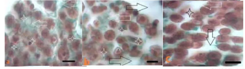
Figure 3. Light photomicrographs of semi thin transverse sections of the Sciatic nerve in the intervention group of 24 (a), 20 (b) and 16 (c) months rats showing stained with Masson's trichrome (a= 24 months), (b= 20 months) and (c =16 months) rats. The arrow indicates an increasing amount of connective tissue of the endoneurium and perineurium. * Indicates the abnormal fibers (Magnification 400X).
Figure 4. Light photomicrographs of semi thin transverse sections of the Sciatic nerve in the control groups stained with Masson's trichrome (a= 24 months, b=20 months, and c= 16 months). (1) Irregularity in shape of myelin (2) increased small myelinated fibers (3) increased connective tissue on the endoneurium and perineurium were observed in the sections (magnification 400X).
4. Discussion
Current study investigated the effects of aging on histologic and histomorphometric changes in rat’s sciatic nerve . We also examined possible therapeutic effects of Aloe Vera gel extract
Figure 3. Light photomicrographs of semi thin transverse sections of the Sciatic nerve in the intervention group of 24 (a), 20 (b) and 16 (c) months rats showing stained with Masson's trichrome (a= 24 months), (b= 20 months) and (c =16 months) rats. The arrow indicates an increasing amount of connective tissue of the endoneurium and perineurium. * Indicates the abnormal fibers (Magnification 400X).
Figure 4. Light photomicrographs of semi thin transverse sections of the Sciatic nerve in the control groups stained with Masson's trichrome (a= 24 months, b=20 months, and c= 16 months). (1) Irregularity in shape of myelin (2) increased small myelinated fibers (3) increased connective tissue on the endoneurium and perineurium were observed in the sections (magnification 400X).
4. Discussion
Current study investigated the effects of aging on histologic and histomorphometric changes in rat’s sciatic nerve . We also examined possible therapeutic effects of Aloe Vera gel extract Figure 1. Light photomicrographs of semi thin transverse sections of the Sciatic nerve in the intervention group of 24 (a), 20
(b) and 16 (c) months rats showing stained with Masson's trichrome (a=24 months), (b=20 months), and (c=16 months) rats. The arrow indicates an increasing amount of connective tissue of the endoneurium and perineurium. * Indicates the abnormal fibers (Magnification 400X).
17 Morphometric aspects
The diameter of whole nerve fiber, sciatic fascicle, my-elin thickness, nerve fiber diameter and area, internodal width and axon diameter are all presented in Table 2 in two groups evaluated at months 16, 20 and 24. There was a significant progressive reduction of the whole sci-atic nerve diameter , nerve fiber, myelin thickness and nerve fiber area in two groups in respect of aging. The re-duction of whole nerve diameter was lower in the group treated by Aloe vera (P<0.01 at month 20; P<0.001 at month 24). Treatment with Aloe vera caused a signifi-cant increase in nerve fiber diameter ,compared to the control group (P<0.01 at month 16; P<0.001 at months 20 and 24). Myelin thickness was decreased by aging, but treatment has led to improvements in this variable (P<0.001 at months 16 and 24). Aging caused a reduc-tion of axon diameter in the control (P<0.001 during
months 16 and 24) and the experimental (P<0.05 during months 16 and 24) groups; but no significant difference was observed between the studied groups at any time points. Also, our data declared a significant decrease in the nerve fiber area at months 20 and 24 (P<0.001) in both groups, which could be concluded that the treat-ment with Aloe vera caused significant improvetreat-ment (P<0.001 at all time points).
Nerve fascicle measurement showed a significant dif-ference between the control and experimental groups of animals at month 16 (P<0.001). In summary, Aging caused a significant increase of this variable, in the inter-vention group, only. There was no significant changes of internodal distance between both groups during the ex-perimental period. In addition, by aging, rats developed rare age-related pathologies, such as glomerulonephritis and pneumonia (two rats from the control groups).
SN 2008.31
±99.8 1823.25±83.9 1604.8±46.8** 2283.72±41 a 2116±29.4**b 1881.2±17.9*** c
F 338.78±
46.27 360.74±62 404.4±48.1 665.2±101.4 c 512.5±22.6** 340.5±40.03***
NF 8.68±.26 7.69±.13** 6.1±.16 *** 9.9±.25b 9.3±.14c 8.7±.2***c
MT 3.4±0.07 3.13±.17 2.2±.5 *** 3.3±.05 c 3±.13 2.7±.13 ** c
NFA 76.2±0.8 67.4±1.2*** 53.8±1.2*** 93.5±2.8c 79.5±0.7**
*c 74±1.2***c
AD 3.67±0.1
8 3.9±.14 2.9±.16*** 3.9±.10 3.9±.17 3.3±.12*
IND 46.06±1.
6 64.64±3.6 63.7±3.6 64.3±10.4 63.6±1.4 63.8±1.7
a Significant differences between subgroups III and V of the of animals in the control group
and between subgroups II and IV and VI in animals of the intervention group treated with Aloe Vera . b Significant differences between groups at same age. *** Significant differences
between subgroups at months 20 and 24 and month16 subgroup in the control and experimental groups (Mean±SE). SN: Whole Sciatic nerve diameter, F: Sciatic Fascicle diameter, NF: Nerve fiber diameter, MT: myelin thickness, NFA: Nerve fiber area, AD: axon diameter, IND: inter nodal distance
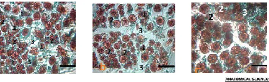
Figure 1. Light photomicrographs of semi thin transverse sections of the sciatic nerve of Sprague-Dawley male rats in control group at 24th (a), 20th (b) and 16th (c) months showing: (1) Irregularity in shape of myelin (2) disruption of myelin density (3) enfolding and out folding of myelin (4) Wallerian degeneration (5) large and small myelinated fiber with very thin myelin sheath. Note that, there are an increase in the number of myelinated fibers with deformed and folded myelin sheaths (enfolding and out folding) and myelin splitting from C (16 months old) to A (24 months old) and B (20 months old) photomicrographs. The arrow indicates the increasing number of nerve fibers with small diameter. * Indicates the normal fibers. Toluidine blue stained. (Magnification 400X)
Commented [y15]: هبوناسکیروط هبرادومنودرهتاعلاطا تروص SE ای SD دوشهتشون.
Commented [d16]: دش حلاصا
Figure 3. Light photomicrographs of semi thin transverse sections of the sciatic nerve of Sprague-Dawley male rats in control
group at 24th (a), 20th (b) and 16th (c) months showing: 1. Irregularity in shape of myelin; 2. Disruption of myelin density; 3.
Enfolding and out folding of myelin; 4. Wallerian degeneration; and 5. Large and small myelinated fiber with very thin myelin sheath. Note that, there are an increase in the number of myelinated fibers with deformed and folded myelin sheaths (enfolding and out folding) and myelin splitting from C (16 months old) to A (24 months old) and B (20 months old) photomicrographs. The arrow indicates the increasing number of nerve fibers with small diameter.
* Indicates the normal fibers. Toluidine blue stained. (Magnification 400X)
Figure 2. Light photomicrographs of semi thin transverse sections of the Sciatic nerve in control groups stained with Masson's
trichrome (a=24 month, b=20 month, and c= 16 month). 1. Irregularity in shape of myelin; 2. Increased small myelinated fibers; and 3. Increased connective tissue on the endoneurium and perineurium were observed in the sections(magnification 400X).
18
Winter & Spring 2019, Volume 16, Number 1
Figure 2 light photomicrographs of semi thin transverse sections of the Sciatic nerve in the control groups stained with Masson’s trichrome (a=24 months, b=20 months, and c=16 months). 1. Irregularity in shape of myelin; 2. Increased small myelinated fibers; and 3. Increased con-nective tissue on the endoneurium and perineurium were observed in the sections (magnification 400X).
4. Discussion
Current study investigated the effects of aging on his-tologic and histomorphometric changes in rat’s sciatic nerve. We also examined possible therapeutic effects of Aloe vera gel extract on histologic and histomorphomet-ric changes. Our results have demonstrated that aging was statistically associated with decreased diameter of whole sciatic nerve and nerve fiber, myelin thickness and nerve fiber area. Also, nerve fascicle diameter decreased in the intervention group, only. Our results showed that treatment with Aloe vera has led to an improvements in the Sciatic nerve, nerve diameter and myelin thickness. It is well known that aging phenomenon affects periph-eral nerve, both in humans and animal models; and these changes are not linearly correlated with age [1].
Accumulation of collagen in perineurium and myelin deformities were seen in peripheral nerves by aging. The main morphological alterations found in the present study were enfolding and out folding of myelin sheath that increased irregularity of nerve fibers. Some myelin deformities exist in normal nerves such as myelin loops and splitting, relevant with alterations of fiber size [18]. However, the increased frequency of such alterations by aging has been referred to an early response of large fi-ber myelin sheaths to axonal atrophy [19] as shown by the Morphometric Approach in the present study. These
alterations may reduce conduction velocity of the large myelinated fibers, described for other species [20]. Al -teration in myelinated fibers may be a consequence of perfusion deficit of endoneurium that affects Schwann cells function.
Schwann cells numbers are important in the regenera-tion process, due to their augmented division following stressful conditions such as a trauma [21]. Our results suggested that subgroups II, IV and VI nerves might have been under regeneration, although this process was not sustained in the animals of control subgroups I, III and V nerves. Regeneration process could be impaired by a poor blood supply of the endoneural spaceince, since the blood vessels of control animals were severely damaged(Figure 3 aand 4 a).
The number of fibers in a peripheral nerve is known to be constant during the adulthood lifespan [17, 22,23], and not correlated with age. On the other hand, it is well known that the myelinated fiber density decreases by de-velopment and aging due to the increased amount of the endoneural connective tissue[17,18,22]. In the present study, aging increased density of small myelinated fibers and decreased that of large myelinated fibers in the ani-mals of the control groups(Figure 3). Such tendency to -wards decreasing amount of nerve fibers in subgroups I, III and V was associated with an increase in the amount of the endoneural connective tissue (Figures 1 and 2).
This observation is in line with those regarding the tibial nerves of mice [18] and the sural nerve of female Fisher rats. They did not observe any significant change in the number of myelinated nerve fibers by aging, de-spite reporting remarkable regeneration in their samples. An important difference between Knox study and ours Figure 2. Light photomicrographs of semi thin transverse sections of the sciatic
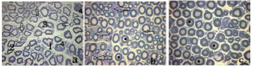
nerve of Sprague-Dawley male rats in the intervention group aged 24 (a), 20 (b) and 16 (c) months showing: (1) Irregularity in shape and out folding of myelin (2) disruption of myelin density (3) folding of myelin into Axoplasm. Note that, photomicrographs there are an increase in the number of myelinated fibers with deformed and folded myelin sheaths (enfolding and out folding) and myelin splitting from C (16 months old) to A (24 months old) and B (20 months old). In ‘a’ section , the arrow indicates large and small myelinated fiber with very thin myelin sheath. Number 4 indicates Wallerian degeneration. The arrow in b section, indicates the increasing number of nerve fibers with small diameter. * Indicates the normal fibers (Toluidine blue stained, Magnification 400X).
Figure 2. Light photomicrographs of semi thin transverse sections of the sciatic nerve of Sprague-Dawley male rats in the intervention group aged 24 (a), 20 (b) and 16 (c) months showing: (1) Irregularity in shape and out folding of myelin (2) disruption of myelin density (3) folding of myelin into Axoplasm. Note that, photomicrographs there are an increase in the number of myelinated fibers with deformed and folded myelin sheaths (enfolding and out folding) and myelin splitting from C (16 months old) to A (24 months old) and B (20 months old). In ‘a’ section , the arrow indicates large and small myelinated fiber with very thin myelin sheath. Number 4 indicates Wallerian degeneration. The arrow in b section, indicates the increasing number of nerve fibers with small diameter. * Indicates the normal fibers (Toluidine blue stained, Magnification 400X).
Figure 4. Light photomicrographs of semi thin transverse sections of the sciatic nerve of Sprague-Dawley male rats in the
inter-vention group aged 24 (a), 20 (b) and 16 (c) months showing: 1. Irregularity in shape and out folding of myelin; 2. Disruption of myelin density; and 3. Folding of myelin into Axoplasm. Note that, photomicrographs there are an increase in the number of myelinated fibers with deformed and folded myelin sheaths (enfolding and out folding) and myelin splitting from C (16 months old) to A (24 months old) and B (20 months old). In ‘a’ section , the arrow indicates large and small myelinated fiber with very thin myelin sheath. Number 4 indicates Wallerian degeneration. The arrow in b section, indicates the increasing number of nerve fibers with small diameter. * Indicates the normal fibers (Toluidine blue stained), (Magnification 400X).
19 is that they have evaluated nerve fibers only ,according
to their selected method of sampling, whereas whole nerve was investigated in our study. It is well known that nerves with a large population of different size fi-bers may have a heterogeneous distribution, which could cause a bias in assay [24].
Coleman et al. surveyed aging morphological and functional elements involved in tactile detection, encod-ing and transmission changes, as well as the skin itself. A combination of several changes contributed to the ef-fect of aging on touch sensation have led to tactile de-fect with advancing age [9]. Evaluation at different age points, demonstrated that most age-related changes are not necessarily linear, throughout life [1,18, 25].
Our results revealed an axonal atrophy in the myelin-ated fibers of the sciatic nerves by aging. This observa-tion .ties well with previous studies of the alteraobserva-tions of sural nerve of 60 years old human with dispropor-tionately thick myelin sheaths in mall and medium size nerve fibers [26]. Sharma et al. showed reduced aver-age diameter of the myelinated fibers in the tibial and plantar nerves of aged rats.
We have discovered that, morphological alterations were more evident in the large myelinated fibers. These conclusions suggested the presence of a neuropathy due to demyelination in aged rats. As mentioned ear-lier, decreased number of large myelinated nerve fibers were associated with increased endoneurial connec-tive tissue. Esquisatto et al. investigated structure and composition of sciatic extracellular matrix in rats by aging and observed the most changes in perineurium. Biochemical analyses showed highest rates of non-col-lagenous proteins accumulations in aged rats (730 days old) and levels of MMP2 and MMP9 enzyme were also high in those rats [27].
Romanovsky et al. declared that density of medium and large sized nerve fibers of dorsal root ganglia that are a3NaK positive decreased significantly by aging. This phenomenon could lead to tendon reflex impairment as an important factor in falling and postural disability in the elderly population. Failure of muscle spindles afferent by aging may contribute to such reflex disturbance. [28].
Our results provided evidence for preventive effects on some morphometric parameters of Sciatic nerve tissue in aged rats receiving hydro alcoholic extract Aloe vera gel .Aloe vera is among few medical plants with a long-term popularity. Clinical evaluations have revealed that the
pharmacologically active ingredients are concentrated in both the gel and rind of Aloe vera leaves [12].
Nagamatsu showed that an increased ROS generation in nerve ischemia, like hydrogen peroxide is accompa-nied with damages to nerve fibers [29]. Aloe vera ex-tracts have antioxidants compounds like vitamins E and C with the potential to reduce blood lipids in people who suffer from diabetes [11]. Ghaffari et al. have dosu-mented that Aloe vera can reduce blood sugar, and may exert a protective effect on the vulnerable tissues, as an antioxidant [30]. Also, prior research identified benefi-cial effects of Aloe vera on carbohydrate metabolism and optic nerve structure in diabetic rats [31].
Guven et al. investigated neuroprotective effects of Aloe vera in ischemia reperfusion mediated injury of sciatic nerve and indicated that it caused significant de-crease in pathologic changes of the nerve via antioxidant and anti-inflammatory effect as compared with meth-ylprednisolone [32]. Lopez jornet et al. found that ap-plication of Aloe vera gel topically decreased burns and pain sensation in patients suffered from burning tongue syndrome [33].
The present study confirmed that consumption of Aloe vera could ameliorate some degenerative process, as per mentioned above, in the sciatic nerve of aged rats. Nev-ertheless, the morphometric approach used in the pres-ent study revealed the existence of an axonal neuropa-thy, mainly of large myelinated fibers. In conclusion, the present study indicated that consuming Aloe vera could delay the process of neuropathy aging in the Sciatic nerve of male rats.
In the current study, we observed some alterations in sciatic nerve caused by aging, such as accumulation of collagen in perineurieum and endoneurieum. Also, a sig-nificant decrease was seen in nerve fibers,nerve trunks diameter and myelin thickness which were highly evi-dent in rats of the 24 months age group. Treatment with Aloe vera gel extract improved histological changes and retarded neuropathy signs, significantly.
Ethical Considerations
Compliance with ethical guidelines
The study was performed in accordance with the guide-lines of National Institute of Health for the care and use of laboratory animals.
20
Funding
This research did not receive any specific grant from funding agencies in the public, commercial, or not-for-profit sectors.
Authors contributions
All authors have read and approved the manuscript.
Conflict of interest
The authors declare no conflict of interest.
Acknowledgement
We would like to sincerely thank Dr. khodakaram Tafti for pathological examination of specimens.
References
[1] Verdú E, Ceballos D, Vilches JJ, Navarro X. Influence of ag-ing on peripheral nerve function and regeneration. Jour-nal of the Peripheral Nervous System. 2000; 5(4):191-208.
[DOI:10.1046/j.1529-8027.2000.00026.x] [PMID]
[2] Mold JW, Vesely SK, Keyl BA, Schenk JB, Roberts M. The prevalence, predictors, and consequences of peripheral sensory neuropathy in older patients. The Journal of the American Board of Family Practice. 2004; 17(5):309-18.
[DOI:10.3122/jabfm.17.5.309] [PMID]
[3] Verghese J, Bieri P, Gellido C, Schaumburg H, Herskovitz S. Peripheral neuropathy in young-old and old-old pa-tients. Muscle & Nerve. 2001; 24(11):1476-81. [DOI:10.1002/ mus.1171] [PMID]
[4] Hashizume K, Kanda K. Differential effects of aging on mo-toneurons and peripheral nerves innervating the hindlimb and forelimb muscles of rats. Neuroscience Research. 1995; 22(2):189-96. [DOI:10.1016/0168-0102(95)00889-3]
[5] Majeed S. Survey on spontaneous peripheral neuropathy in ag-ing rats. Arzneimittel-Forschung. 1992; 42(7):986-90. [PMID]
[6] Melcangi R, Magnaghi V, Cavarretta I, Martini L, Piva F. Age-induced decrease of glycoprotein Po and myelin ba-sic protein gene expression in the rat sciatic nerve: Repair by steroid derivatives. Neuroscience. 1998; 85(2):569-78.
[DOI:10.1016/S0306-4522(97)00628-3]
[7] Knox CA, Kokmen E, Dyck PJ. Morphometric alteration of rat myelinated fibers with aging. Journal of Neuropa-thology & Experimental Neurology. 1989; 48(2):119-39.
[DOI:10.1097/00005072-198903000-00001]
[8] Verdú E, Butí M, Navarro X. Functional changes of the pe-ripheral nervous system with aging in the mouse. Neu-robiology of Aging. 1996; 17(1):73-7. [DOI:10.1016/0197-4580(95)02010-1]
[9] Coleman P, Finch C, Joseph J. The need for multiple time points in aging studies. Neurobiology of Aging. 1990; 11(1):1-2. [DOI:10.1016/j.neurobiolaging.2003.10.002]
[10] Thomas P, King R, Sharma A. Changes with age in the peripheral nerves of the rat. Acta Neuropathologica. 1980; 52(1):1-6. [DOI:10.1007/BF00687222] [PMID]
[11] Rajasekaran S, Ravi K, Sivagnanam K, Subramanian S. Ben-eficial effects of Aloe vera leaf gel extract on lipid profile status in rats with streptozotocin diabetes. Clinical and Ex-perimental Pharmacology and Physiology. 2006; 33(3):232-7. [DOI:10.1111/j.1440-1681.2006.04351.x] [PMID]
[12] Rajasekaran S, Sivagnanam K, Subramanian S. Antioxidant ef-fect of Aloe vera gel extract in streptozotocin-induced diabetes in rats. Pharmacological Reports. 2005; 57(1):90-6. [PMID]
[13] Vogler B, Ernst E. Aloe vera: A systematic review of its clin-ical effectiveness. British Journal of General Practice. 1999; 49(447):823-8. [PMID] [PMCID]
[14] Sahu PK, Giri DD, Singh R, Pandey P, Gupta S, Shrivastava AK, et al. Therapeutic and medicinal uses of Aloe vera: A review. Pharmacology & Pharmacy. 2013; 4(08):599.
[DOI:10.4236/pp.2013.48086]
[15] Bozzola JJ, Russell LD. Electron microscopy: Principles and techniques for biologists. Burlington, Massachusetts: Jones & Bartlett Learning; 1999.
[16] Tseng H, Lin SE, Chang YL, Chen MH, Hung SH. Deter-mining the critical effective temperature and heat dispersal pattern in monopolar radiofrequency ablation using tem-perature-time integration. Experimental and Therapeutic Medicine. 2016; 11(3):763-8. [DOI:10.3892/etm.2015.2956] [PMID] [PMCID]
[17] Jeronimo A, Jeronimo CAD, Sanada LS, Fazan VPS. Mi-croscopic anatomy of the sural nerve in the postnatal de-veloping rat: A longitudinal and lateral symmetry study. Journal of Anatomy. 2005; 206(1):93-9. [DOI:10.1111/j.0021-8782.2005.00368.x] [PMID] [PMCID]
[18] Ceballos D, Cuadras J, Verdú E, Navarro X. Morphomet-ric and ultrastructural changes with ageing in mouse pe-ripheral nerve. Journal of Anatomy. 1999; 195(4):563-76.
[DOI:10.1046/j.1469-7580.1999.19540563.x] [PMID] [PMCID]
[19] Krinke G, Froehlich E, Herrmann M, Schnider K, Da Suva F, Suter J, et al. Adjustment of the myelin sheath to axonal atrophy in the rat spinal root by the formation of infolded myelin loops. Cells Tissues Organs. 1988; 131(3):182-7.
[DOI:10.1159/000146510]
[20] Peters A. The effects of normal aging on myelin and nerve fibers: A review. Journal of Neurocytology. 2002; 31(8-9):581-93. [DOI:10.1023/A:1025731309829] [PMID]
[21] Jeronimo A, Jeronimo CAD, Rodrigues Filho OA, Sanada LS, Fazan VPS. A morphometric study on the longitu-dinal and lateral symmetry of the sural nerve in mature and aging female rats. Brain Research. 2008; 1222:51-60.
[DOI:10.1016/j.brainres.2008.05.055] [PMID]
[22] Schellens RL, van Veen BK, Gabreëls-Festen AA, Noter-mans SL, van’t Hof MA, Stegeman DF. A statistical ap-proach to fiber diameter distribution in human sural nerve. Muscle & Nerve. 1993; 16(12):1342-50. [DOI:10.1002/ mus.880161212] [PMID]
21 [23] Berthold C, Carlstedt T, Corneliuson O. Anatomy of the
nerve root at the central-peripheral transitional region. Pe-ripheral Neuropathy. 1984; 1:156-70.
[24] Romero E, Cuisenaire O, Denef J-F, Delbeke J, Macq B, Ve-raart C. Automatic morphometry of nerve histological sec-tions. Journal of Neuroscience Methods. 2000; 97(2):111-22.
[DOI:10.1016/S0165-0270(00)00167-9]
[25] Bouche P, Cattelin F, Saint-Jean O, Leger J, Queslati S, Guez D, et al. Clinical and electrophysiological study of the pe-ripheral nervous system in the elderly. Journal of Neurol-ogy. 1993; 240(5):263-8. [DOI:10.1007/BF00838158] [PMID]
[26] Jacobs J, Love S. Qualitative and quantitative morphol-ogy of human sural nerve at different ages. Brain. 1985; 108(4):897-924. [DOI:10.1093/brain/108.4.897] [PMID]
[27] Esquisatto MAM, de Aro AA, Fêo HB, Gomes L. Changes in the connective tissue sheath of Wistar rat nerve with aging. Annals of Anatomy-Anatomischer Anzeiger. 2014; 196(6):441-8. [DOI:10.1016/j.aanat.2014.08.005] [PMID]
[28] Romanovsky D, Mrak RE, Dobretsov M. Age-dependent decline in density of human nerve and spinal ganglia neurons expressing the α3 isoform of Na/K-ATPase. Neuroscience. 2015; 310:342-53. [DOI:10.1016/j.neurosci-ence.2015.09.034] [PMID] [PMCID]
[29] Nagamatsu M, Schmelzer JD, Zollman PJ, Smithson IL, Nickander KK, Low PA. Ischemic reperfusion causes li-pid peroxidation and fiber degeneration. Muscle & Nerve. 1996; 19(1):37-47. [DOI:10.1002/mus.880190103] [PMID]
[30] Ghaffari H, Gholami S, Naghdi M. Histologic and histo-morphometric study of tibial nerve treated by hydro-alco-holic Aloe vera gel extract in diabetic male rats. Anatomical Sciences Journal. 2013; 10(2):63-72.
[31] Saberi M, Gholami S. An investigation on the effects of the Aloe vera extract on the thickness of the retina in male diabetic rats. Iranian Journal of Veterinary Research. 2012; 13(4):296-302.
[32] Guven M, Gölge UH, Aslan E, Sehitoglu MH, Aras AB, Akman T, et al. The effect of Aloe vera on ischemia— Reperfusion injury of sciatic nerve in rats. Biomedicine & Pharmacotherapy. 2016; 79:201-7. [DOI:10.1016/j.bi-opha.2016.02.023] [PMID]
[33] López Jornet P, Camacho Alonso F, Molino Pagan D. Pro-spective, randomized, double-blind, clinical evaluation of Aloe vera Barbadensis, applied in combination with a tongue protector to treat burning mouth syndrome. Jour-nal of Oral Pathology & Medicine. 2013; 42(4):295-301.
[DOI:10.1111/jop.12002] [PMID]