Eukaryon
Eukaryon
Volume 5
Celebrating Darwin's 100th Anniversary
Article 27
3-26-2009
Cystic Fibrosis: Channeling the Discovery of CFTR Mutations
Cystic Fibrosis: Channeling the Discovery of CFTR Mutations
Jennifer Brown
Lake Forest College
Jessica Disch
Lake Forest College
Jaymie Honold
Lake Forest College
Melissa Schramm
Lake Forest College
Nengding Wang
Lake Forest College
Follow this and additional works at: https://publications.lakeforest.edu/eukaryon
Disclaimer:
Eukaryon is published by students at Lake Forest College, who are solely responsible for its
content. The views expressed in Eukaryon do not necessarily reflect those of the College.
Articles published within Eukaryon should not be cited in bibliographies. Material contained
herein should be treated as personal communication and should be cited as such only with the
consent of the author.
Cystic Fibrosis: Channeling the Discovery of CFTR Mutations
Cystic Fibrosis: Channeling the Discovery of CFTR Mutations
Cover Page Footnote
Cover Page Footnote
We would like to thank Dr. Shubhik DebBurman for his help throughout this project. We would also like to
express our gratitude to our peer tutors Lokesh Krukeja, Mithaq Vahedi, and Lital Silverman for their
valuable input and feedback.
Eukaryon, Vol. 5, March 2009, Lake Forest College
Review Article
52
Cystic Fibrosis: Channeling the Discovery of CFTR Mutations
Jennifer Brown, Jessica Disch, Jaymie Honold, Melissa Schramm, and Nengding Wang*
Department of Biology Lake Forest College Lake Forest, Illinois 60045
Summary
Cystic fibrosis (CF) is an autosomal recessive disease caused by mutation of the cystic fibrosis transmembrane conductance regulator (CFTR). CFTR is a protein forming chloride channels in the membrane of epithelial cells. It consists of two transmembrane domains (TMD), two nucleotide binding domains (NBD), and an R domain. The channel is first activated by the phosphorylation of its R domain. ATP then binds to CFTR’s NBDs for the channel to conduct Cl- out of the
cell. Mutations in CFTR can cause misfolding and prevent adequate transportation of Cl-. The most
common cause of the disease is the deletion of phenylalanine 508 of CFTR. When F508 reaches the ER, it is recognized as irreparable and thus targeted for degradation, which causes the symptoms of CF. The third most common mutation, G551D, does not function properly at the membrane. Molecular chaperones such as Hdj-2/Hsc70, Hsp90, and HspBP1 may rescue CFTR misfolding. This has therapeutic value because once
F508 is sent to the membrane, it can function normally. There are many treatments for CF, including airway clearing techniques, antibiotics, and lung transplantation. Recent research has suggested that oligonucleotide insertion, curcumin treatment, and digotoxin treatment can reverse the F508 phenotype.
Introduction
Cystic Fibrosis (CF) is a disease marked by an ineffective chloride channel that afflicts thousands of individuals every year, yet there is still no known cure. It was first diagnosed in the 1700s by kissing a child on the forehead and if their sweat tasted salty, the parents knew their child had the disease (Littlewood, 2002). Currently, there are many more definitive ways of diagnosing this fatal disease. In 1938, Dorothy Anderson described the symptoms of a child with cystic fibrosis in detail, such that they could be applied in a clinical setting. By examining family inheritance, in 1946, Anderson discovered that cystic fibrosis was a recessive genetic disease. In 1953, it was revealed that children with cystic fibrosis had increased sweat electrolytes and therefore, sweat tests became an accurate means for diagnosis. Neonatal screening began in the 1970s, allowing for earlier diagnosis of this disease. In 1988, the first and most common mutation associated with cystic fibrosis,
F508, was found on chromosome 7 (Littlewood, 2002). Cystic fibrosis is an autosomal recessive disease caused by the mutation of the CFTR gene. In 1985, the CFTR gene was located at 7q21-34, on the long arm of chromosome 7, which was soon followed by the identification of the gene sequence (Welsh, et al., 2001). The CFTR gene codes for a protein which contains 1480 amino acids and has been named the cystic fibrosis transmembrane conductance regulator, also known as ________________________________________________
*This author wrote the paper for Biology 221: Cell and Molecular Biology taught by Dr. Shubhik DebBurman.
CFTR (Southern, 1997). A mutation in CFTR often leads to CF. CFTR is a chloride channel expressed on the surface of epithelial cells in the intestine, respiratory system, pancreas, and sweat glands. Currently, there are over 1000 mutations comprising four classes that result in CF; however, one mutation, the deletion of phenylalanine at position 508, accounts for over 70% of CF patients. The other mutations are very rare, and only 4 others have frequencies above 1% (Littlewood, 2002).
There are over 1,000 new cases of cystic fibrosis diagnosed each year, and about 90% are diagnosed before the child is 3-years-old. Cystic fibrosis is most common among people of European background, with carrier numbers of 1 in 25. Mortality rates have been dramatically altered since the 1960s. In 1969, the mean life expectancy was 14 years. In 2005, however, it was 36.8 years (McCutchen, 2007).
CF is characterized by thick mucus secretions in the pancreas, lungs, and reproductive organs. These thick mucus secretions block the airways and cause chronic bacterial infection and degeneration of the lung tissue. Patient death is often due to recurrent infections. Other symptoms of CF are the destruction of the pancreas and sterility in males (Quekett, 2007).
A common biological model for CF is a transgenic mice model (Clarke, et al., 1992). Recently, efforts have been made to map the entire genetic structure of the most common cause of CF infection: the Pseudomonas aeruginosa bacterium. This way researchers can identify specific genes that cause the infection and turn off the target genes when needed (CFF.org, 2007)
CF does not have a cure; the only available therapies currently address the symptoms of the disease. Patients that suffer from severe lung disease caused by CF have the option of lung transplantation (CFF.org, 2007). Another treatment option for CF is the use of airway-clearing techniques to loosen the thick mucus in the lungs, making it easier to expel by coughing. Antibiotics used to treat CF are administered orally, intravenously, or inhaled. Inhaled drugs are commonly used after airway-clearing techniques and have the unique ability to reach the airways quickly and easily. Gene therapy is not a currently available treatment for CF, but offers considerable promise if a proper vector can be found. These treatments present patients only temporary relief from their symptoms, and there is ongoing research to find a cure by specifically targeting mutations such as
F508.
Cystic fibrosis is a very well studied disease; however, there is still much to be learned. Over the past few years, tremendous advances have been made in this field. To understand the cellular impact of CF, we must first recognize how CFTR functions normally. Mutations causing CFTR to misfold and function abnormally, or not at all, must also be identified. To correct the adverse effects of these mutations, regulation of chaperones is a useful strategy. Addressing all of these factors lead to improvements in therapy, though there is still no cure for the disease.
Normal Function of CFTR
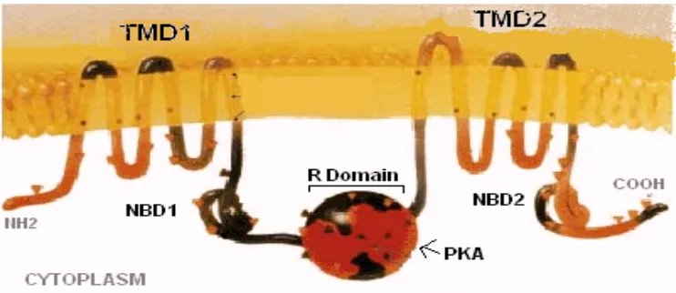
Figure 1. CFTR chloride channel structure. (Journal of NIH Research http://history.nih.gov/exhibits/genetics/10b-sm.gif).
a process called RVD response, the chloride channels are activated allowing chloride to flow passively through the channels with water to restore homeostasis. CFTR is found in cells comprising many organs including the lung, pancreas, liver, or any other systems that require fluid mucus for their function. The water that is transported along with the negatively charged ions hydrates the cellular secretions in the aforementioned organs, aiding in fluidity (Anderson, 1992). In normal lungs the stabilization of NaCl concentration is aided by the passive transport of water into and out of the cells, producing a low salt content in the surface liquid coating airway epithelial cells (Dorin, 2004). This low salt environment facilitates the growth of naturally occurring antimicrobial peptides that fight off bacterial infections.
Consisting of two transmembrane domains (TMD1 and TMD2) along with two nucleotide binding domains (NBD1 and NBD2) (Riordan et al., 1989), CFTR is a member of the ABC transporter family due to its utilization of ATP hydrolysis in function (see Fig 1).
CFTR is activated when ATP binds to both its nucleotide binding domains causing NBD1-NBD2 dimerization (Vergani, et al., 2005). However, before ATP binds, protein kinase A (PKA) must first phosphorylate the R Domain of the protein, bridging the two homologous halves (the TMDs), and giving the protein configuration as follows: TMD1-NBD1-R-TMD2-NBD2 (Gadsby, et al., 2006). Negatively charged ions, such as chloride, are thought to accumulate near the positively charged regions of the pore until phosphorylation and ATP binding activates its opening, allowing the ions to flow through, down their concentration gradient.
Upon the intramolecular dimer interaction initiated by ATP binding to NBD1 followed by ATP binding to NBD2, a signal is transmitted through cytoplasmic linking domains to open the gate in the transmembrane domain. The activated channel is characterized by two open conductance states: O1 and O2 (Gunderson and Kopito, 1995). The O2 confirmation results in a 15% larger opening than O1, but the open CFTR confirmation only exists in this state 20% of the time.
The channel closes in two different ways, ATP dissociation or ATP hydrolysis, both of which result in a decrease of hydrogen bonding between the NBD domains (Ramjeesingh, et al., 2003). Both cause a destabilization of the NBD dimer and consequently a lack of signal transmission through the cytoplasmic linking domains, causing the channel to close (Gadsby, et al., 2006).
The activity of the channel is characterized by the following directionality: CO1O2C. Because the pathway is strictly directional, this gives further evidence that an outside source of energy (i.e. ATP) is needed to drive the gating asymmetry (Gunderson and Kopito, 1995).
Mutations in CFTR directly affect chloride conductance across the cell membrane, and without efficient anion flow, water movement slows and dehydrated mucus clogs and accumulates in organ ducts, fostering bacterial growth (Gadsby, et al., 2006). In addition to the decreased fluidity and increase in bacterial growth, because antimicrobial activity requires a low salt concentration and because CF surface fluid has a high NaCl concentration, Cystic Fibrosis epithelia fail to kill the thriving bacteria (Smith, at al., 1996). Failure of CFTR to function properly is often the effect of a number of mutations.
Class II mutation F508: the Most Common Mutation in CF
There are four class mutations associated with CF, the most common form being the codon deletion of pheylalanine residue at position 508 from the NBD1 of the CFTR (Southern, 1997). The mutation of F508 is a class II mutation and affects the maturation of CFTR by inducing misfolding (Welsh, 2001). When the mutated CFTR protein reaches the ER, the quality-control mechanism of its cellular component recognizes that the protein is folded incorrectly and marks the defective protein for degradation. As a result, F508 does not reach the cell membrane (Cui, 2006). Unlike CFTR, once wild type (WT) CFTR is synthesized it is transported to the ER and proceeds to the plasma membrane through ER export. Interestingly, the F508 mutation does not alter the chloride channel ability of CFTR but rather corrupts the normal intracellular processing of the protein (Southern, 1997). Recently, scientists have concentrated on defining the class II mutation F508 and, in the process, are discovering its functions, leading to better understanding of this fatal defect.
Protein Kinase CK2, CFTR, F508: F508 Deletion Disrupts a Kinase-Binding Site
WT-54
CFTR but not F508 CFTR. Thus, CK2 appears to aid in the regulation of CFTR activity.
Knowing that F508 is dependent on CK2-CFTR interaction, Treharne, et al. (2007) investigated whether the deletion of F508 disrupts binding of signaling molecules. It was confirmed that CK2 phosphorylates WT but not F508. To test whether Ser-511 is phosphorylated by CK2 in WT-CFTR, Ser-511 was mutated in recombinant NBD1 (Treharne, et al., 2007). The results indicated that CK2 failed to phosphorylate the S-511 mutant (Treharne, et al., 2007). Thus, neither F508-NBD1 nor mutated Ser-511-NBD1 interacts with CK2.
It remains unclear how the loss of F508 leads to defective channel gating. Treharne, et al. (2007) examined the role of the Ser-511 mutation in CFTR function and found that both F508 and F508-Ser-511 disrupt CFTR channel gating—Ser-511 more so than F508. From this data, it may be interpreted that Ser-511 may be involved in trafficking (Treharne, et al., 2007).
CK2 can use either ATP or GTP as a phosphate donor for kinase activity, making it an attractive candidate for future research of CF. F508-CFTR interactions have shown that explanations for the multi-system nature of CF may reside in differences between proteins bound to WT and
F508 CFTR. These findings suggest that the normal function of CFTR is to either maintain the CK2 content of the apical membrane or to provide a F508-dependent anchoring protein for CK2 (Treharne, et al., 2007). CK2 regulated
F508 CFTR dependently, and Ser-511 is a critical residue in CK2-dependent gating of CFTR. Thus, F508 is critical for CK2 to bind CFTR and phosphorylate NBD1 at Ser-511.
The Role of CFTR Phenylalanine 508 Side Chain Ion Channel Gating
CFTR functions as a chloride channel and the deletion of Phe508 in the NBD1 impairs both maturation and function of the protein. Although F508 is retained in the ER, it is still able to function as an ion channel (Welsh, 2001). Through the substitution of Phe508 by several other residues rather than being deleted, Cui, et al. (2006) found that CFTR was able to mature and be transported to the cell surface. Cui, et al. (2006) investigated this proposition further by researching the role of the Phe508 side chain in the CFTR channel gating. Their results indicated that the interaction of the Phe508 side chain did play a role in channel gating and is required to keep the channel open (Cui, et al., 2006).
This is the first direct evidence that a specific interaction of the Phe508 side chain plays a role in determining residency time in the closed state. Although the side chain is not essential for CFTR folding, it is important in ion channel function.
Impact of F508 Mutation in NBD1 of CFTR on Domain Folding and Structure
It has been proposed by many that the primary effect of the
F508 mutation is to cause misfolding of the NBD1, which leads to the degradation of CFTR. Through structural and biophysical studies on human NBD1 domains, Lewis, et al. (2005) reported that they had failed to demonstrate significant changes of in vitro stability or folding kinetics in the presence or absence of F508 mutation. This raises the possibility that instead of causing the folding of NBD1, the primary effect of F508 is a disruption of proper interdomain interactions at the Phe508 site in CFTR (Lewis, et al., 2005).
Lewis, et al. (2005) examined the crystal structures of the Phe508 region in mNBD1 (mouse NBD1, gold), hNBD1-7a- F508 (human NBD1, 7 base mutations, deletion of Phe508, cyan), and hNBD1-2b- F508A (human NBD1, 2 base mutations, no yield of F508, blue). The three models were super positioned to each other and
Lewis, et al. (2005) found that the F508 hNBD1 shows minimal conformational changes compared to mNBD1.
Looking at the worm diagrams of hNBD1-7a-
F508 and hNBD1-2b- F508A Lewis, et al. (2005) were able to compare the structures of hNBD1 and mNBD1. They observed a difference between the conformations of the two hNBD1. The structure of hNBD1-7a- F508 indicated a deletion of Phe508 through the shortening of a loop present in hNBD1-2b- F508A.
Additionally, through surface topography, Lewis, et al. (2005) examined the worm diagrams of hNBD1-7a-
F508 and hNBD1-2b- F508A and found them to be dramatically different. The surface topography of hNBD1-7a-
F508 is altered at the site of the mutation, representing the presumed region of binding to the MSD1 of CFTR. When Phe508 is deleted, Val-510 (a side chain) is removeed from its normal position (Lewis, et al., 2005).
Thus, the domains of hNDB1 with and without the
F508 mutation cause slight conformational changes. Although there are no significant differences in the folding properties of the two version of hNBD1, the slight structural change of hNBD1-7a- F508 suggests that interdomain interactions are likely to be considerably altered by the
F508 mutation. These results demonstrate new insights into the molecular pathology of the predominance of the class II mutation, which may be of particular significance to efforts in discovery of treatment.
G551D: A Class III Mutation
A class III mutation affects CFTR function after the protein leaves the ER and is inserted in the plasma membrane. However, once it reaches the membrane, the chloride channel is defectively regulated. In the past few years, researchers have been focusing on the different ways the most common mutations act in a cystic fibrosis cell. G551D is the most common class III mutation and the third most common overall (Welsh, 2001); therefore, it is a good candidate for study. G551D is a missense mutation, which results in an exchange of asparagine for glycine at the 551 codon (McMorran, et al., 2001). This mutation occurs in 2-5% of CF patients. G551D is located on the apical membrane; however, it binds nucleotides less frequently and has reduced NBD1 ATPase activity (Derand, 2002). Therefore, the regulation of the chloride channel is defective.
The G551D mutation was originally suspected to be located at or near the ATP binding domain. However, Howell, et al. (2000) found that the binding site was actually not near the binding domain. They realized that the substitution of the two amino acids caused the helix to loosen, therefore changing the shape of active ATP binding site (Howell, et al. 2000).
G551D causes a defect in the immunological response to inflammatory stimuli. McMorran, et al. (2001) infected mice with the G551D missense mutation using Pseudomonas aeruginosa. This is a bacterium that causes a lung infection to which CF patients are extremely susceptible. The research showed the decreased immunological response by comparing body weight, number of bacteria in the lungs, and the amount of pro-inflammatory mediators (TNF-, KC and MIP-2) between G551D CF mice and mice with WT CFTR. The CF mice showed twice as much body weight decrease and a greatly increased amount of bacteria when compared to the WT mice. The CF mice also had a greater pro-inflammatory response with the
It was found that different mutations of CF would possibly require different types of treatment. The G551D mutation responded differently to three different agents that strongly influence chloride channels compared to the response of another CFTR mutation, G1349D,. The three agents used were phloxine B, pyrophosphate, and 2’-deoxy ATP. The phloxine B and 2’-deoxy ATP had an effect on the G551D patients, whereas only the 2’-deoxy ATP had an effect on the G1349D patients. The hypothesis given for 2’-deoxy ATP working on both was that the binding sites for ATP are impeded by both of these mutations. 2’-deoxy ATP was able to act as normal ATP and help both mutations function properly. The phloxine B increased the amount of chloride exiting the cell in the patients with the G551D mutation and in the patients with the G1349D mutation; however, it was not enough to save the cell in the latter (Cai, et al., 2006).
Another form of treatment for the G551D mutation would be the addition of genistein, a molecule that improves chloride channel activity by increasing the amount of time the channel is open (Suaud, et al., 2002). In WT CFTR cells, the normal chloride channel activity inhibits the epithelial Na+ channel, ENaC. However, when the chloride channel activity is low, ENaC works to stimulate it. In G551D CFTR cells, ENaC and CFTR do not interact normally. Genistein was able to restore normal interactions between the two channels (Suaud, et al., 2002).
Molecular Chaperones and Their Therapeutic Potentials
In most cases, CF is caused by the mutation F508 in the CFTR gene. The mutated protein is synthesized, but it is incapable of being sent to the plasma membrane through ER export due to misfolding. In this process, many molecular chaperones and co-chaperones are involved through either aiding the protein’s entrance into the secretory pathway or targeting the protein for degradation. Such chaperones include Hdj-2/Hsc70, Hsp90, and HspBP1. Depending on their functions, different chaperones play distinct roles in different stages of CFTR folding. Regulation of chaperone expression can be of great therapeutic value because once F508 is sent to the plasma membrane it will be at least partially functional (Pasyk and Foskett, 1995).
Hdj-2/Hsc70 Chaperone Pair Facilitates CFTR Biogenesis CFTR consists of TMD 1 and 2, NBD 1 and 2, and an R domain. The F508 mutation was found to be localized in NBD1 and causes CFTR to misfold (Lewis, et al., 2005). Thus chaperones that specifically help NBD1 to fold properly are of vital importance to the correct folding of CFTR. Human DnaJ2 (Hdj-2) is a co-chaperone of heat shock cognate 70 (Hsc70) (Meacham, et al., 1999). Both have been isolated in complexes with CFTR and F508 CFTR. In fact, the complexes that the chaperones formed with F508 CFTR were ~2 fold more abundant than those with CFTR. These data indicate that not only do the chaperones interact with the protein, they also interact more with F508 than CFTR.
To locate where the Hdj-2/Hsc70 chaperone pair works, the ability of these chaperones to form complexes with CFTR fragments was investigated. It was found that with the expression of NBD1, there was an increase in the binding abilities of Hdj-2 and Hsc70. When other domains were expressed, there was a decrease in their binding abilities. To further analyze how the Hdj-2/Hsc70 complex interacts with NBD1, studies were done to find out whether its aggregation can be prevented. The results show that Hdj-2/Hsc70 could suppress >85% of NBD1 aggregation. These data demonstrate that the Hdj-2/Hsc70 chaperone pair facilitates the early steps of CFTR folding, specifically of
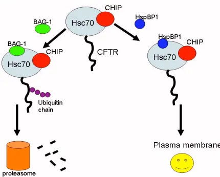
Figure 2. This figure depicts how HspBP1 and BAG-1 compete with
binding to Hsc70 and CHIP to either inhibit or stimulate the degradation of CFTR.
the NBD1 region. A theory for how CFTR folding is carried out was then proposed. The first step is the insertion and orientation of the TMD1 into the ER membrane. Then NBD1 is synthesized and bound by Hdj-2 and Hsc70. In the time before the R domain is synthesized, Hdj-2 and Hsc70 stabilize NBD1 or promote its folding. When the R domain and MSD2 are folded, Hdj-2 is released. NBD2 is the last to be synthesized and CFTR is then in its final mature form and ready to be sent to the plasma membrane. Thus, regulating the levels of Hdj-2/Hsc70 may help NBD1 folding and, in turn, facilitate the folding of CFTR. Current investigation of the Hdj-2/Hsc70 chaperone pair includes investigating whether it is a common component of the machinery that helps membrane protein biogenesis (Meacham, et al., 1999).
Hsp90 Contributes to CFTR Maturation
Hsp90 is an abundant chaperone in cells that plays an important role in preventing protein aggregation and helping proteins fold or degrade (Smith, et al., 1995). Thus, studies were done to find out whether Hsp90 can facilitate CFTR and F508 CFTR folding. Hsp90 was found to bind to both CFTR and F508 CFTR (Loo, et al., 1998). Ansamysin is a drug that inhibits that association between Hsp90 and its substrate. Then, whether ansamysin would also destroy the binding between Hsp90 and CFTR was investigated. In fact, no CFTR was found to be associated with Hsp90, indicating that the CFTR-Hsp90 complex was immediately perturbed when the drug was used. Once this was found, the effects of ansamysin on the synthesis of CFTR and F508 CFTR was studied. Ansamysin inhibited the maturation of both forms of CFTR. Connecting these results, it can be concluded that if the association between Hsp90 and CFTR is disrupted, the maturation of CFTR cannot be achieved. Another hypothesis was that the perturbation of the Hsp90 and CFTR association would also accelerate the degradation of immature CFTR by proteasomes. This was investigated by looking at the effects of ansamysin alone compared to the effects of ansamysin and a proteasome inhibitor, lactacystin, on the synthesis of CFTR. As predicted, with just ansamysin there was a decrease in the amount of mature CFTR and lactacystin corrected its negative effects.
56
being investigated to see if they play a role in facilitating Hsp90 in its function (Loo, et al., 1998).HspBP1 Inhibits CFTR Degradation and Stimulates CFTR Maturation
There are two pathways in which CFTR can be directed while trafficking through the ER depending on whether it is folded properly or misfolded. When it is correctly folded, the CFTR protein will be sent to the plasma membrane and function normally. When it is misfolded, it will be recognized by specific chaperones and targeted for degradation. One of these chaperones is Hsc70 associated with CHIP (Connell, et al., 2001). By binding to Hsc70, CHIP induces the ubiquitination of the target protein, thus targeting it for degradation by the proteasome (Meacham, et al., 2001). There are other co-chaperones that form complexes with Hsc70/CHIP, which either enhance or inhibit its degradation activity. BAG-1 is a co-chaperone that promotes degradation when it binds to the complex. HspBP1 is another important co-chaperone and studies were done to find out whether it regulates the function of the Hsc70/CHIP complex. First of all, HspBP1 and CHIP were found to bind to Hsc70 at the same time but at different positions. Thus they do not inhibit each other’s actions; instead they work in cooperation. When bound to the Hsc70/CHIP complex, HspBP1 was found to inhibit the ubiquitin ligase activity of CHIP, thus slowing the rate of degradation of CFTR. Thus, HspBP1 is another chaperone that, if it can be regulated, it will help the process of sending CFTR to the membrane (Meacham, et al., 2001).
Innovative Treatment Possibilities
Aside from common therapies such as lung transplantation, airway-clearing techniques, inhaled drugs, and antibiotics, recent research shows promise for other techniques addressing CF symptoms (CFF.org, 2007).
Curcuminsends F508 CFTR to the plasma membrane
Curcumin is a major component of the Indian curry spice turmeric, which can act as a low-affinity sarcoplasmic/ endoplasmic reticulum calcium pump inhibitor (Egan, et al., 2004). It was recently found to have possible therapeutic effects on F508 CFTR. When BHK cell lines containing
F508 CFTR were treated with curcumin, the surface density of the CFTR channels were found to significantly increase (Egan, et al., 2004). Using an iodide efflux assay with cAMP activation of the chloride channels, Egan, et al. (2004) were able to demonstrate that through its increased density at the plasma membrane, F508 CFTR functions properly as chloride channels. Therefore it corrects the CF phenotype.
Results from co-immunoprecipitation suggest that curcumin interacts with Calnexin, a co-chaperone of Hsp90, which is involved in CFTR folding. Calnexin failed to coprecipitate with CFTR when the cells were treated with curcumin, suggesting that the interaction was inhibited. Thus, calnexin is thought to play a role in retaining F508 CFTR in the ER (Egan, et al., 2004).
Oligonucleotide Insertion Corrects F508 CFTR
Oligonucleotide insertion has also shown some promising therapeutic effects involving F508 CFTR cell lines (Zamecnik, et al., 2004). This method of treatment intercepts the transcribed mRNA of the F508 CFTR mutated gene, then corrects the deletion through insertion of nucleotide sequences that code for the missing phenylalanine. This corrected mRNA can then be translated into the WT CFTR protein, which is processed normally and transported to the plasma membrane. Zamecnik, et al. (2004) further show that
the CFTR corrected by oligonucleotide insertion was functional at the plasma membrane by measuring the current after cAMP stimulation. Thus, oligonucleotide insertion prevents the development of the CF phenotype. While this treatment would not reverse the genotype of CF patients, it would be a preventative therapy.
Cardiac Glycosides Suppress IL-8 Secretion
Another fascinating advance in the realm of CF treatment is the use of cardiac glycosides, such as digitoxin, (Srivastava, et al., 2004). Cardiac glycosides are drugs that are usually used in the treatment of congestive heart failure. They work by inhibiting the Na+/K+ pump, which leads to an increase in
the Ca2+ level, aiding in contraction of the heart muscle. This
improves cardiac output and reduces any swelling of the heart. One of the major characteristics of CF is extensive lung inflammation, which is believed to be caused by hypersecretion of the pro-inflammatory protein IL-8. The ability of cardiac glycoside’s to reduce swelling led researchers to investigate their effects on F508 CFTR cells.
All of the cardiac glycosides tested inhibited the secretion of IL-8, with digitoxin being the most efficient. Digitoxin blocks the phosphorylation of IKB, an inhibitor of
NF-B activation, which stops the signaling pathway for hypersecretion of IL-8.
When treatment with digitoxin was compared to gene therapy, an interesting result emerged. Gene therapy significantly changes 58 genes, and 36 of those were equivalently and proportionately changed by treatment with digitoxin (Srivastava, et al., 2004). This suggests that digitoxin mimics the genomic effects of gene therapy on
F508 CFTR.
Conclusion
Cystic fibrosis is an autosomal recessive disease caused by mutations in the chloride channel, CFTR. The F508 mutation of the CFTR gene, which was found to influence kinase-binding sites, accountsfor the vast majority of CF. Concentrating on defining F508 and its functions will lead to better understanding of this fatal defect and its molecular consequences. Another mutation, G551D, causes a shape change in NBD1. This makes it impossible for ATP to bind and activate the open-state of CFTR. Molecular chaperones can interact with CFTR in its folding and transport process in such a way that CFTR and its mutated form can both be sent to the plasma membrane and function normally. Some examples include the Hdj-2/Hsc70 pair which can facilitate the early biogenesis of CFTR, Hsp90 which can aid in CFTR maturation, and HspBP1 which can inhibit degradation and accelerate maturation of CFTR. Thus chaperones have significant potential in the treatment of CF. Aside from the common treatment techniques, curcumin and oligonucleotide insertion have been shown to successfully transport F508 CFTR to the plasma membrane, correcting the CF phenotype. Digitoxin inhibits hypersecretion of IL-8, therefore preventing the inflammatory symptoms of CF. In order to assess the effectiveness of the aforementioned treatments, future clinical trials are needed.
Acknowledgements
We would like to thank Dr. Shubhik DebBurman for his help throughout this project. We would also like to express our gratitude to our peer tutors Lokesh Krukeja, Mithaq Vahedi, and Lital Silverman for their valuable input and feedback.
those of the College. Articles published within Eukaryon should not be cited in bibliographies. Material contained herein should be treated as personal communication and should be cited as such only with the consent of the author.
References
Accurso, F., Cutting, G., Ramsey, B., Welsh, M. The Metabolic and Molecular Basis of Inherited Disease. 8th
Edition. McGraw-Hill (2001)
Alberti, S., Bohse, K., Arndt, V., Schmitz, A., Hohfeld, J. The cochaperone HspBP1 inhibits the CHIP ubiquitin ligase and stimulates the maturation of the cystic fibrosis transmembrane conductance regulator. Molecular Biology of the Cell: 15:4003-4010 (2004).
Alexsandrov, A., Aleksandrov, L., Chen, J.H., Cui, L., Hou, Y.X., Riordan, J. The role of cystic fibrosis Transmembrane conductance regulator phenylalanine 508 side chain in ion channel gating. Journal of Physiology. 2006. 572(2): 347-358
Amaral, M. CFTR and chaperones. Journal of Molecular Neuroscience.: 23:41-48 (2004).
Amaral, M. Therapy through chaperones: sense or antisense? Cystic fibrosis as a model disease. Journal of Inherited Metabolic Disease: 29:477-487 (2006).
Anderson, M., Sheppard, D., Berger, H., Welsh, W. Chloride channels in the apical membrane of normal and cystic fibrosis airway and intestinal epithelia. Lung Cellular and Molecular Physiology. 263(1): 1-14 (1992).
BarNoy, S., Eidelman, O., Lee, G., McPhie, P., Pollard, H., Razin, M., Sorscher, E., Zhang, J. Role for phospholipids interaction in the trafficking defect of F508 CFTR. Biochemistry. 2002. 41: 11161-11170.
Best, O., Chen, J.H, Crawford, R., Gruenert, D., Kunzelmann, K., Mehta, A., Russell, M., Schulte, E., Sheppard, D., Treharne, K., Wilson, S., Xu, Z. Protein Kinase CK2, Cystic Fibrosis Transmembrane Conductance Regulator, and the F508 Mutation: f508 deletion disrupts a kinase-binding site. The Journal of Biological Chemistry. 2007. 282(14): 10804-10813
Cai, Zhiwei, Alessandro Taddei, and David N. Sheppard. "Differential Sensitivity of the Cystic Fibrosis (CF)- Associated Mutants G551D and G1349D to Potentiators of the Cystic Fibrosis Transmembrane Conductance Regulator (CFTR) Cl- Channel." The Journal of Biological Chemistry 281 (2005): 1970-1977.
Connell et al. The co-chaperone CHIP regulates protein triage decisions mediated by heat-shock proteins. Nature Cell Biology: 3:93-96 (2001).
Dorn, M. Cystic Fibrosis. Medical Research Council. (2004)<http://www. hgu.mrc.ac.uk/Research/Dorin/julia.html>
Egan, M.E., Pearson, M., Weiner, S.A., Rajendran, V., Rubin, D., Glckner-Pagel, J., Canny, S., Du, K., Lukacs, G.L., Caplan, A.J. Curcumin, a Major Consitituent of Tumeric, Corrects Cystic Fibrosis Defects. Science, 304: 600-602. (2004).
Gadsby, D., Vergani, P., Csanady, L. The ABC protein turned chloride channel whose failure causes cystic fibrosis. Nature, 440: 477-483 (2006).
Gunderson, K., Kopito, R. Confirmational States of CFTR Associated with Channel Gating: The Role of ATP Binding and Hydrolysis. Cell, 82:231-239 (1995).
Howell, L D., Roy Borchardt, and Johnathon A. Cohn. "ATP Hydrolysis by a CFTR Domain: Pharmacology and Effects of G551D Mutation." Biochemical and Biophysical Research Communications 271 (2000): 518-525.
Lewis et al. Impact of the F508 Mutation in First Nucleotide-binding Domain of Human Cystic Fibrosis Transmembrane Conductance
Regulator on Domain Folding and Structure. Journal of Biological Chemistry: 280(2): 1346-1353 (2005).
Learning About Cystic Fibrosis. Nation Human Genome Research Institute. <http://www.genome.gov/10001213>
Littlewood, Jim. "History of Cystic Fibrosis." Cystic Fibrosis Medicine. Aug. 2002. 31 Mar. 2007
http://www.cysticfibrosismedicine.com/htmldocs/CFText/historyof.htm
Loo, M.A., Jensen, T.J., Cui, L., Hou, Y., Chang, X., Riordan, J.R. Perturbation of Hsp90 interaction with nascent CFTR prevents its maturation and accelerates its degradation by the proteasome. EMBO Journal: 17(23):6879-6887 (1998).
Meacham, G.C., Lu, Z., King, S., Sorscher, E., Tousson, A., Cyr, D.M. The Hdj-2/Hsc70 chaperone pair facilitates early steps in CFTR biogenesis. EMBO Journal: 18(6):1492-1505. (1999).
McCutchen, Bingham. "About Cystic Fibrosis." Cystic Fibrosis Foundation. 2007. 31 Mar. 2007 <http://www.cff.org/AboutCF/>.
McMorran, B J., J S. Palmer, D P. Lunn, D Oceandy, E O. Costelloe, G R. Thomas, D A. Hume, and B J. Wainwright. "G551D CF Mice Display an Abnormal Host Response and Have Impaired Clearance of Pseudomonas Lung Disease." American Journal of Physiology- Lung Cellular and Molecular Physiology 281 (2001): l740-l747.
Pasyk, E.A., Foskett, J.K. Mutant (delta F508) cystic fibrosis transmembrane conductance regulator Cl- channel is functional when retained in endoplasmic reticulum of mammalian cells. The Journal of Biological Chemistry. 270(21):12347-50 (1995)
Quekett, James. "Cystic Fibrosis." BUPA. 2007. Cystic Fibrosis Trust. 31 Mar.2007
http://hcd2.bupa.co.uk/fact_sheets/Mosby_factsheets/Cystic_fibrosis. html
Ramjeesingh, M., Kidd, J., Huan, L., Wang, Y., Bear, C. Dimeric cystic fibrosis transmembrane conductance regulator exists in the plasma membrane. Biochem. J., 374: 793-797 (2003).
Riordan J.R. Identification of the cystic fibrosis gene: cloning and characterization of complementary DNA. Science. 1989; 245(4922): 1066-1073.
Smith et al. Progesterone receptor structure and function altered by geldanamycin, an hsp90-binding agent. Molecular and Cellular Biology: 15(12): 6804-6812 (1995).
Smith, J., Travis, S., Greenberg, E., Welsh, M. Cystic fibrosis airway epithelia fail to kill bacteria because of abnormal airway surface fluid. Cell 87(2):229-236 (1996).
Southern, K. F508 in cystic fibrosis: willing but not able. Archives of Disease in Childhood. 1997. 76: 278-282.
Srivastava, M., Eidelman, O., Zhang, J., Paweletz, C., Caohuy, H., Yang, Q., Jacobson, K.A., Heldman, E., Huag, W., Jozwik, C., Pollard, B.S., Pollard, H.B. Digitozin mimics gene therapy with CFTR and suppressed sypersecretion of IL-8 from cystic Fibrosis lung epithelial cells. PNAS, 101: 7693-7698 (2004)
Suaud, Laurence, Marcelo Carattino, Thomas R. Kleyman, and Ronald C. Rubenstein. "Genistein Improves Regulatory Interactions Between G551D-Cystic Fibrosis Transmembrane Conductance Regulator and the Epithelial Sodium Channel in Xenopus Oocytes." The Journal of Biological Chemistry 277 (2002): 50341-50347.
Treatments: Keys to Healthier Living. Cystic Fibrosis Foundation. <http://www.cff.org/treatments/>
Vergani, P., Lockless, S., Nairn, A., Gadsby D. CFTR channel opening by ATP-driven tight dimerization of its nucleotide-binding domains. Nature, 433: 876-880 (2005).