IBBJ
Spring 2017, Vol 3, No 2
Sensitivity and Specificity of Visual Inspection with Acetic
Acid (VIA) and with Lugol Iodine (VILI) in the Diagnosis
of Cervical Cancer in the Northern Region of Cameroon
Georges Kalgong
1, Armel Herve
Nwabo Kamdje
1*, Tagne Simo Richard
1,
Charlette Nangue
21. Department of Biomedical Sciences, Faculty of Sciences, University of Ngaoundere, Ngaoundere, Cameroun .
2. Anato-Cytopathology Laboratory, University Hospital Center of Yaoundé, Yaounde, Cameroun .
Submitted 1 Feb 2017; Accepted 27 Feb 2017; Published 25 Mar 2017
Cervical cancer is one of the major causes of women death worldwide. The aim of this study was to test the sensitivity and specificity of visual inspection with acetic acid (VIA) and visual inspection with lugol iodine
(VILI) as a diagnostic test for cervical cancer in comparison with Pap smear in the North Cameroon region. 309
women aged between 20 to 62 years were recruited in this study. 307 were included in the statistical analyzes. Each woman was screened for cervical cancer by a conventional smear and visual inspection with acetic acid 5%
and the lugol solution. The prevalence of precancerous lesions of cervix was 12.70%. The risk factors of cervical
cancer identified were age, matrimonial status, age of first sexual intercourse, and parity. The associ ation of VIA and VILI showed sensitivity, specificity, positive and negative predictive values of 93.58%, 97.01%, 82.01%, and
99.04%, respectively. VIA-VILI were revealed as good screening tests for cervical cancer in the present context.
Early detection and treatment of cervical lesions and also faith against the risk factors should help to reduce the mortality and morbidity of this pathology.
Keywords: Sensitivity, specificity, diagnosis, visual inspection, cervical cancer
ervical cancer is the second most common
cancer affecting women (1). In 2002, nearly
493.000 new cases and 274.000 deaths were
recorded (2). With more than 527 600 new cases and
about 265700 deaths in 2012, 80% of these deaths occurred in developing countries, where this cancer
is the leading cause of cancer deaths in the female
population (3-4).
In Cameroon, the prevalence of cervical
cancer is 7.9% in women between 30 and 60 years
old (5). In the developed countries, the incidence and mortality due to cervical cancer is decreasing.
For example in United States, between 1955 and
1992, mortality due to cervical cancer decreased at
70% and the rate continues to decline about 3% each
year. Similarly, in the United Kingdom the rate has
decreased to 70% in 2008 compared to 30 years earlier (6). However, in Cameroon, these rates are
progressively increasing. This growth may be due to
insufficiency of strategy and program of fight against cervical cancer that limits access to
screening; the high cost and rarity of human
papilloma virus (HPV) vaccine in the country; and the unavailability of early detection services.
C
Original Article
Consequently, cancer is diagnosed at a very
advanced stage in Cameroon. 80% of cancer cases are diagnosed at a late stage and most patients die
within 12 months of diagnosis (7). Cervical cancer
is an avoidable and curable disease, provided that it is detected early and treated. The slow progression
of precancerous lesions to invasive cervical cancer
stage leaves a period about 10 years, which may involve the detection and treatment of these lesions
before they reach the invasive cancer stage (8). In
developed countries, regular screening of the target population would reduce the incidence of cervical
cancer more than 90% (9). However, in developing
countries, screening is non-existent or covers only a small part of the target population (10). In
Cameroon, there is a cancer control committee that
carries out sporadic screening activities across the
country, but there is no national program to fight
against cervical cancer. These screenings are limited
because they do not reach the large proportion of the
target population. Furthermore, the demands of
vaginal cervical smears by physicians are tiny and
confined in large cities like Douala and Yaoundé
which have anatomy pathology laboratories. All
these difficulties make epidemiological surveillance
of cervical cancer difficult in Cameroon and even
more in the Northern region where there is not yet
an anatomy laboratory pathology. The effectiveness
of cervical smear in the detection of cervical lesions
has been demonstrated and this technique has
significantly reduced the relative incidence of
cervical cancer in developed countries since 1950.
But the limits of this technique for developing
countries are important, including cost, logistics and
trained human resources to mobilize (11). The
possibility of detecting HPV by molecular biology
methods offers another additional approach in
screening, but its cost is still high and this technique
requires sophisticated equipment that is expensive in
our context.
Several cases of cervical cancer die in Cameroon without diagnosis or are diagnosed at an
advanced stage of the disease where management of
the disease is unlikely to conclude favorably.
Confronted to these challenges, the use of visual inspection tests such as visual inspection with acetic
acid (VIA) and visual inspection with lugol iodine
(VILI), which require minimum equipment and a lower cost than other techniques appears to be a
viable alternative for low-income countries (12).
The aim of this study was to test the sensitivity and specificity of VIA-VILI as a diagnostic test for
cervical cancer in relation to cervical smear and to
evaluate the feasibility and efficiency of epidemiological surveillance of cervical cancer
based on early diagnosis in health facilities in the
Northern region of Cameroon.
Materials and methods
Patients
This cross-sectional study was conducted in
the Northern region Cameroon from May to August
2016 in Regional Hospital of Garoua, District
Hospital of Guider, and Esperance Hospital of
Djamboutou (Garoua). Among 309 recruited
women, 307 were included in the statistical
analyzes. Inclusion criteria were all women between
the ages of 20 and 65 years attending hospitals and
consenting to participate. Exclusion criteria were
women with hysterectomy or cervical conisation,
pregnancy, active vaginal bleeding and those with a
history of precancerous and cancerous lesions. After
obtaining written consent, a questionnaire on the
socio-demographic characteristics and the
gynecological and obstetric history was
administered to each woman. Then they were
screened for cervical cancer by a conventional
smear, visual inspection after applying 5% acetic
acid and lugol iodine.
Conventional smear
The patient was reassured and installed in the
gynecological position. Then, a sterile speculum
was introduced into her vagina until perfect
observation of the cervix. A spatula and cytobrush were used to remove the cells by simple scraping.
The sample was spread on a slide and immediately
fixed with alcohol and dried. All the slides were sent
to the anatomo-cytopathology laboratory of the University Hospital Center of Yaoundé, colored and
interpreted by an anatomo-pathologist. The results
were given according to the classification of Bethesda system 2001.
Visual inspection with 3-5% acetic acid (VIA)
Freshly prepared 5% acetic acid was applied on cervix using cotton swap and the test was
interpreted after 1 min under bright light. Acetic
acid colors the abnormal cells. The test was considered positive if a white area, well delimited
and near the squamous junction appeared on cervix,
and negative in the absence of this whitening (13).
Visual inspection with lugol (VILI)
Lugol’s iodine solution was applied on cervix using cotton swap and the test was interpreted after
1 min. The normal cells of the cervix contain
glycogen and the precancerous or cancerous cells
contain very little or not. Through the glycogenic
effect on lugol, normal cells absorb lugol and take
black or brown coloration. In case of a positive test,
iodo-negative areas appear and take a mustard or
saffron yellow color. The iodo-negative areas are
clearly delineated and clearly visible (14).
Statistical analyzes
The software R commander version 13.2.0 was
used for the analysis of the data. The bilateral Chi2
test was used. A variable was considered as
statistically significant at P< 0.05. The evaluation of
the performance of the tests was carried out by
calculating the sensitivity, specificity and the
positive and negative predictive values.
Results
Conventional smear performance
Pap smears revealed 39 positive cases. A
prevalence of 12.70% of precancerous lesions were
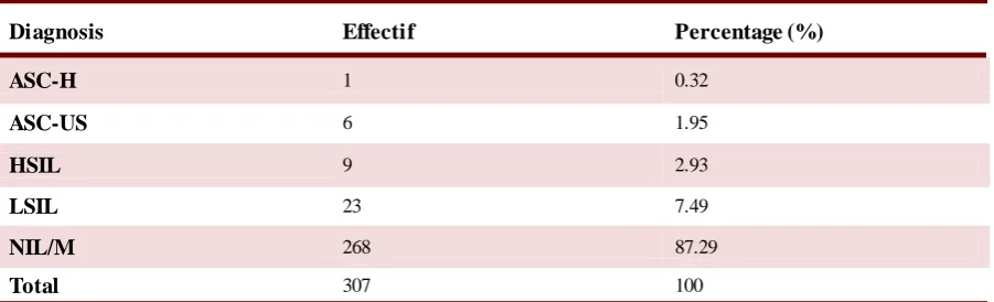
reported in the northern region of Cameroon. Table 1 shows the distribution of lesions according to
grade.
Performance of visual inspection after applica-tion of acetic acid (VIA)
According to VIA, 45 cases (14.65%) were
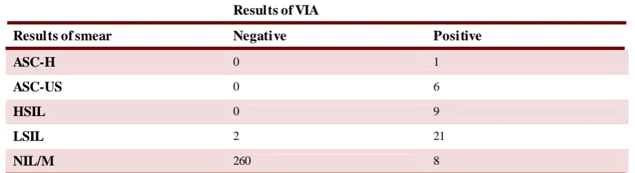
positive. Corresponding results are represented in Table 2 were 2 cases of false negative and 8 cases of
false positive were identified.
Performance of visual inspection after applica-tion of lugol’s iodine (VILI)
The visual inspection after application of lugol’s iodine revealed 44 (14.33%) positive cases. According to Table 3, 3 cases of false negative and
8 cases of false positive were identified.
The sensitivity, specificity, positive and
negative predictive values VIA and VILI tests are
presented in Table 4.
Relationship between socio-demographic charac-teristics and results of smear
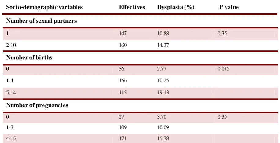
Table 5 represents the relationship between
socio-demographic characteristics and the onset of
cervical dysplasia.
Diagnosis Effectif Percentage (%)
ASC-H 1 0.32
ASC-US 6 1.95
HSIL 9 2.93
LSIL 23 7.49
NIL/M 268 87.29
Total 307 100
Table 1. Distribution of dysplastic lesions according to grade
ASC-H: atypical squamous cell cannot exclude high grade; ASC-US: atypical squamous cell of undefined signification ; HSIL: high squamous intra-epithelial lesion; LSIL: low squamous intra-epithelial lesion, NIL/M: no intra-epithelial lesion or malignancy.
Results of VIA
Results of smear Negative Positive
ASC-H 0 1
ASC-US 0 6
HSIL 0 9
LSIL 2 21
NIL/M 260 8
Table 2. Contingency of smear results with VIA results
ASC-H: atypical squamous cell cannot exclude high grade; ASC-US: atypical squamous cell of undefined signification; HSIL: high squamous intra-epithelial lesion; LSIL: low squamous intra-epithelial lesion, NIL/M: no intra-epithelial lesion or malignancy.
Results of VILI
Results of smear Negative Positive
ASC-H 0 1
ASC-US 0 6
HSIL 0 9
LSIL 3 20
NIL/M 260 8
Table 3. Contingency of smear results with VILI results
Se: sensitivity; Sp: specificity; PPV: positive predictive value; NPV: negative predictive value .
Se (%) Sp (%) PPV (%) NPV (%)
VIA
94.87
97.01
82.22
99.23
VILI 92.30 97.01 81.81 98.85
VIA/VILI
association 93.58 97.01 82.01 99.04
Table 4. Diagnostic values of VIA and VILI
Socio-demographic variables Effectives Dysplasia (%) P value
Age (years)
(20-29) 97 6.10 0.040
(30-39) 123 13.83
(40-62) 87 18.39
Marital statut
Married 236 11.44 0.027
Not married 71 16.90
Age of first sexual intercouse (years)
(13-18) 212 16.03 0.008
(19-30) 95 5.26
Table 5. Relationship between the socio-demographic characteristics and the results of the smear
Discussion
The prevalence of precancerous lesions of
cervix was 12.70% in the studied population. Significantly lower rates were reported in
Cameroon's rural and urban zones with 7.9% and
4.9% prevalence, respectively (5, 15). In the present study, low-grade lesions accounted for 74.43%
versus 25.64% for high-grade lesions. Doh and
Kouam found in 1999 a prevalence of 78.4% and
21.6% for low grade and high grade lesions,
respectively (16). These results show the real
necessity for screening of cervical cancer in the
Northern region and elsewhere in Cameroon.
Screening being the only mean of achieving
complete cure of the disease, after treatment.
We found that VIA was a good screening test
for precancerous lesions with a sensitivity of
94.87% and a specificity of 97.01%. The VIA
identified 100% of precancerous lesions of high
grade and mistook only 2 precancerous lesions of
low grade among the 29 cases confirmed by the Pap
smear. Therefore, an error of 6.89% on the lesions
of low grade occured. The sensitivity of VIA was
higher than that obtained in a study in Gabon (17).
The sensitivity and specificity of VILI were
almost similar to those of VIA. We found that VILI
had a sensitivity and specificity of 92.30% and
97.01%, respectively.
The association of the VIA with the VILI was beneficial. It reduced the false negatives left by one
or the other. This association gave a sensitivity,
specificity, positive and negative predictive values of 93.58%, 97.01%, 82.01%, and 99.04%,
respectively.
A higher proportion of dysplastic lesions
occurred in women over 30 years old. The average
age of beginning of precancerous lesions is
relatively low at 36.89 years. The study reports a
significant difference between age and appearance
of dysplasia (P< 0.04). This is consistent with data
reported by Mpiga et al. in Gabon in 2015 and
Nkegoum et al. in 2001 in Cameroon who found a
peak appearance of dysplasia between 36 and 40
years (15, 17).
Marital status was associated with the presence
of cervical dysplasia lesions. Unmarried women had
significantly (P< 0.02) more dysplasia than those
who were married. These results are in accordance
with the epidemiology of cervical cancer because
unmarried women are a priori more likely to have
multiple sexual partners and therefore a higher risk
of developing the disease. The present study
Table 5 Cont. Relationship between the socio-demographic characteristics and the results of the smear
Socio-demographic variables Effectives Dysplasia (%) P value Number of sexual partners
1 147 10.88 0.35
2-10 160 14.37
Number of births
0 36 2.77 0.015
1-4 156 10.25
5-14 115 19.13
Number of pregnancies
0 27 3.70 0.35
1-3 109 10.09
4-15 171 15.78
benefited from information on the number of sexual
partners for each participant. Although women with multiple sexual partners had a higher proportion of
dysplastic lesions, we did not find a significant
difference (P< 0.35) between the number of sexual partners and precancerous lesions.
The average age of first sexual intercourse was
17.52 years, and 75% of women had had sexual intercourse before the age of 19 years. We noticed a
statistically significant association (P< 0.008)
between age at first intercourse and dysplasia lesions. Those who had sexual intercourse between
13 and 18 years had significantly more dysplasia.
This result is in agreement with data reported by Duport in 2008 and the WHO report in 2007 which
evoke early first sexual intercourse as a determining
risk factor. As HPV is sexually transmitted, the early
sexual intercourse has an association with the risk of
infection (18).
Parity is also a cervical cancer risk factor. A
parity greater than or equal to 5 is considered as a
risk factor for cervical cancer (19). The present
study has considered at the same time the number of
pregnancies and parity. We noted an increasing
proportion of dysplasia in relation to the number of
pregnancies in the studied population. However, we
did not find a statistically significant association (P<
0.35) between the number of pregnancies and the
presence of precancerous lesions. On the other hand,
we found a significant association (P< 0.01)
between parity and dysplasia. Women with a
number of births greater than or equal to 5 had more
dysplasia than those with a lower number of births.
Taken together, reducing mortality and
morbidity caused by cervical cancer requires early
screening and the fight against the risk factors that
must be included in a national control program.
Acknowledgement
Authors thank the collaborators of their
respective institutions for the comments on the
manuscript.
Conflict of interest
The authors declared no conflict of interest.
References
1. Siegel R L, Miller K D, Jemal A. Cancer Statistics, 2017. CA
Cancer J Clin. 2017;67:7-30.
2. Parkin D M, Bray F, Ferlay J, et al. Global cancer statistics,
2002. CA Cancer J Clin. 2005;55:74-108.
3. Torre L A, Bray F, Siegel R L, et al. Global cancer statistics,
2012. CA Cancer J Clin. 2015;65:87-108.
4. Denny L. Cervical cancer: prevention and treatment. Discov
Med. 2012;14:125-31.
5. Tebeu P, Sandjong I, Nkele N, et al. Lesions pre cancereuses
du col uterin en zone rurale: Etude transversale. Medecin e
d’Afrique noire. 2005;52:27–31 [Article in French].
6. Arbyn M, Castellsague X, de Sanjose S, et al. Worldwide
burden of cervical cancer in 2008. Ann Oncol. 2011;22:2675- 8 6.
7. Maranga I O, Hampson L, Oliver A W, et al. Analysis of
factors contributing to the low survival of cervical cancer patients
undergoing radiotherapy in Kenya. PLoS One. 2013;8:e78411.
8. Janicek M F, Averette H E. Cervical cancer: prevention,
diagnosis, and therapeutics. CA Cancer J Clin. 2001;51:92-114.
9. Sherris J, Herdman C, Elias C. Cervical cancer in the
developing world. West J Med. 2001;175:231-3.
10. Monsonego J. Prevention of Cervical Cancer: Challenges and
Perspectives of HPV Prophylactic Vaccines: Karger Publishers;
2006.
11. Stoler M H, Schiffman M. Interobserver reproducibility of
cervical cytologic and histologic interpretations: realistic
estimates from the ASCUS-LSIL Triage Study. Jama .
2001;285:1500-5.
12. Sankaranarayanan R, Budukh A M, Rajkumar R. Effectiv e
screening programmes for cervical cancer in low and middle
-income developing countries. Bull World Health Organ.
2001;79:954-62.
13. Keshavarzi F, Nankali A, Fakheri T, et al. Cervical visual
inspection with acetic acid as an alternative screening test for
cervical cancer detection. Int J Collab Res Intern Med Public
Health. 2013;5:60-6
14. Ghosh P, Gandhi G, Kochhar P K, et al. Visual inspection of
cervix with Lugol's iodine for early detection of premalignant &
malignant lesions of cervix. Indian J Med Res. 2012;136:265- 7 1.
15. Nkegoum B, Belley Priso E, Mbakop A, et al. Precancerous
lesions of the uterine cervix in cameroonian women. Cytological
and epidemiological aspects of 946 cases. Gynecol Obstet Fertil.
2001;29:15-20 [Article in French].
16. Kouam A D, L. The management of pre-invasive cervical
lesions using cryotherapy in Yaounde, Cameroon. J Obstet
Gynaecol. 1999;19:640-2.
17. Mpiga É, Ivanga M, Koumakpayi I H, et al. Intérêt de
l’inspection visuelle à l’acide acétique et au soluté de Lugol avec
colposcope dans le dépistage des lésions du col utérin au Gabon.
Pan Afr Med J. 2015;22:165 [Article in French].
18. Duport N. Données épidémiologiques sur le cancer du col de
l’utérus. Etats des connaissances–Actualisation. 2008. [In
French].
19. Brinton L A, Reeves W C, Brenes M M, et al. Parity as a risk
factor for cervical cancer. Am J Epidemiol. 1989;130:486-96.