Witold Pilecki
1, Swietłana (Svetlana) Masgutova
2, Joanna Kowalewska
2,
Denys Masgutov
2, Nelly Akhmatova
3, Małgorzata
Poręba
1,
Małgorzata Sobieszczańska
1, Piotr Kolęda
1, Anna
Pilecka
1, Dariusz Kałka
1The Impact of Rehabilitation Carried out Using
the Masgutova Neurosensorimotor Reflex Integration
Method in Children with Cerebral Palsy on the Results
of Brain Stem Auditory Potential Examinations
Wpływ rehabilitacji metodą integracji odruchów neurosensorycznych
według Masgutowej u dzieci z porażeniem mózgowym na wyniki
badania wywołanych potencjałów słuchowych
1 Department of Pathophysiology, Wroclaw Medical University, Poland 2 Swietłana (Svetlana) Masgutova International Institute, Warszawa, Poland
3 Scientific-Research Institute I.I. (Élie) Metchnikoff Department of Sera and Vaccine Research, Laboratory of the
Immunoregulation Mechanisms, Moscow, Russia
Abstract
Background. Rehabilitation therapy in children with neuromotor development disorders can be carried out with the use of various methods.
Objectives. The aim of this study was to determine the efficiency of rehabilitation carried out with the use of the new therapeutic method MNRI® (Masgutova Neurosensorimotor Reflex Integration) in children with cerebral
palsy (CP) by objective measurements with a Brainstem Auditory Evoked Potentials (BAEP) examination.
Material and Methods. Besides the known parameters, Interpeak Latency I-V (IPL I-V) in BAEP, an original parameter proposed by Pilecki was introduced, called a relative IPL I-V value. The study involved a group of 17 children (9 girls and 8 boys) aged from 1.3 to 5.9 years (mean = 3.8 years, SD = 1.3) with cerebral palsy. Due to difficulty in co-operation, analysis of only 15 children could be finished.
Results. Analysis of the absolute IPL I-V values showed that after rehabilitation the percentage of the results with slowed transmission, i.e. those in which the IPL I-V value was prolonged, decreased from more than 88% to 60%. The assessment of the relative IPL I-V values showed that the results obtained after rehabilitation are more advan-tageous.
Conclusions. As a result of rehabilitation carried out by the MNRI® method in children with CP, a significant
improvement in the transmission in the brain stem section of the auditory pathway was observed based on the absolute and relative IPL I-V values. However, the change obtained in children was various (Adv Clin Exp Med 2012, 21, 3, 363–371).
Key words: cerebral palsy, rehabilitation, brainstem auditory evoked potentials.
Streszczenie
Wprowadzenie. Rehabilitacja dzieci z zaburzeniami neuromotorycznymi może być przeprowadzana z użyciem różnych metod.
Cel pracy. Ocena wpływu nowoczesnej metody rehabilitacji MNRI® (Masgutova Neurosensorimotor Reflex Integration) u dzieci z porażeniem mózgowym za pomocą obiektywnej metody słuchowych potencjałów wywoła-nych pnia mózgu (Brainstem Auditory Evoked Potentials – BAEP).
Materiał i metody. Oprócz znanych parametrów: Interpeak Latency I-V (IPL I-V) zastosowano nowy autorstwa Pileckiego, zwany względnym IPL I-V. Grupa badana obejmowała 17 dzieci (9 dziewczynek i 8 chłopców) w wieku 1,3–5,9 lat (średnia = 3,8 lat, SD = 1,3) z porażeniem mózgowym.
Adv Clin Exp Med 2012, 21, 3, 363–371 ISSN 1899–5276
ORIGINAL PAPERS
Wyniki. Analiza bezwzględnych wartości IPL I-V pokazała, że po rehabilitacji odsetek wyników ze zwolnioną transmisją, tj. tych których wartości były wydłużone, zmalał z ponad 88 do 60%. Ocena względnych IPL I-V wyka-zała, że wyniki uzyskane po rehabilitacji były lepsze.
Wnioski. W wyniku rehabilitacji przeprowadzonej metodą MNRI® u dzieci z porażeniem mózgowym stwierdzono
istotną poprawę przewodzenia w obszarze drogi słuchowej w pniu mózgu, co oparto na ocenie bezwzględnych oraz względnych wartości IPL I-V. Obserwowane zmiany u poszczególnych dzieci były jednak różne (Adv Clin Exp Med 2012, 21, 3, 363–371).
Słowa kluczowe: porażenie mózgowe, rehabilitacja, słuchowe potencjały wywołane pnia mózgu.
Neurological development deficits are condi-tioned to a significant extent by the disorders of the sensomotoric integration mechanisms of the primary reactions and reflex patterns. Rehabilita-tion therapy in children with neuromotoric de-velopment disorders can be carried out according to many methods and patterns, such as the Vojta, Doman-Delacato, Bobath, Wroclaw Improvement Model, Castillo Morales method and others [1–5].
In the presented studies the authors used the therapeutic program “Neurosensomotor integra-tion of the reflex patterns” – MNRI® (Masgutova
Neurosensorimotor Reflex Integration), which contains a diagnostic component and therapeutic procedures [6–11].
The basic point of the presented program is to support children’s neurostructural and physiologi-cal development while simultaneously securing the defense mechanisms and neurodevelopment. This study is based on the concept of the re-patterning and restoring of development and maturation of primary motor reflex patterns. The MNRI program develops the concept of reflex integration instead of the reflex inhibition and extinction strategy. It is based on the concept of L. Vygotsky (Wygotski) and J. Piaget about the role of the child’s primary motor function in the development of higher men-tal functions [12, 13].
The primary task of the therapeutic-rehabili-tation procedures of the MNRI® program is
neu-rosensomotor correction of the improper reflex patterns. The program includes the techniques and movement activities known by the name of “re-patterning” (imitating and pattern correction) and relies on the repetition of the dynamic and postural reflex pattern in order to stimulate the natural inborn mechanisms of brain neuroplastic-ity [6, 14].
To achieve the best possible outcome in chil-dren with diagnosed CNS damage or in chilchil-dren in danger of such damage, both early recognition of the damage and rapid introduction of the most effective form of rehabilitation are necessary.
Assessment of the clinical condition of pa-tients with neurodevelopmental deficits and other neurological disorders is usually carried out based on reflex examination. These methods, due to the large contribution of the human factor, are not
en-tirely objective, since their result is to a large extent dependent on the skills of the person performing the assessment. Furthermore, there is practically no method that is widely used, which makes it dif-ficult to compare results obtained by different re-searchers in different laboratories.
Moreover, the psychological attitude of the re-searcher, expecting to obtain a particular result, may increase the subjectivity of assessment [15, 16].
It would be very useful for the development of various programs and methods used in therapy of children with neurodevelopmental deficits and neurological disorders to develop a method which increases the objectivity of the assessment of the clinical condition of patients and of the effective-ness of the rehabilitation performed.
One of the courses of action aimed at increas-ing such objectivity could be to apply the method of evoked potentials.
This examination relies on the registration of an excitation wave appearing in the sensory path-ways of the central nervous system as a result of the activation of the appropriate receptor of ad-equate stimulus. The most commonly used forms are examinations of Brainstem Auditory Evoked Potentials (BAEP) and Visual Evoked Potentials (VEP) [17].
The result of the examination is independent of the patient’s will, of his state of consciousness, and is given as numerical values, which accounts for its repeatability and objectivity.
The evoked potentials method, including Brainstem Auditory Evoked Potentials, both for assessment of the clinical condition and for prog-nostic assessment has been used by many re-searchers. Such studies were presented by, among others, Majnemer, Jiang Ze Dong and Pilecki. De-scriptions concerned children with cerebral palsy (CP), Down syndrome, and those burdened with the risk of perinatal central nervous system dam-age [18–21].
investi-gate using the EP method can be analogous to the areas inaccessible for this examination [23, 24].
This method of description of the results and analysis of the patient condition was defined by Pilecki as indirect assessment [24].
One parameter which is particularly useful in the assessment of children with a variety of de-fects, disorders and neurodevelopmental deficits is the Interpeak Latency I-V (IPL I-V) described in the examination of the Brainstem Auditory Evoked Potentials (BAEP). The result obtained di-rectly points at the transmission efficiency in the brain stem part of the auditory pathway and speci-fies the time (given in milliseconds) that elapses between the excitation of structures related to the electrogenesis of peak I (i.e. the auditory nerve), and the inferior colliculi of the mesencephalon (electrogenesis of peak V). At the same time, this result, by way of an indirect assessment, allows us to predict with high probability the efficiency of transmission in other brain areas, including mo-tor pathways, and thus also on the range of momo-tor function [25].
Although the absolute (given in milliseconds) IPL I-V value is a widely accepted parameter, it has a significant disadvantage resulting from the fact that due to conditions of physiology in the first year of a child’s life, it is not constant and it is shortened from about 5.10 ms in the first week of life to approximately 4.05 ms after 12 months of age. For this reason, the statistical analysis of results obtained in this age group, especially when the age of respondents is various (which causes this parameter to be of high physiological changeability), has a very high standard deviation value. As a result, almost every result within the limits of two standard deviations (2 SD), result-ing from normal distribution of the feature, fits in the range of standard, even if it does not exist in practice. Such a high SD value also makes it dif-ficult to compare groups, (e.g. the control group with the study group or two study groups with each other) because the statistical analysis show no differences even when the mean values are very divergent.
To reduce this inconvenience in current analy-sis, besides the widely used parameter IPL I-V val-ues in milliseconds, the authors added an origi-nal parameter, proposed by Pilecki, that has been called the relative IPL I-V value, in contrast to the previous one, which is called the absolute IPL I-V value. This parameter was developed based on the analysis of the examination results of 411 chil-dren in adjusted age from –8 to +78 weeks to give the mean values for age groups in different weeks of life and standard deviation quantity, which was calculated at 0.1 ms. A new parameter was defined
as the quantity (a dimensionless number) that in-dicates by how many standard deviations the ex-amination result deviates from the mean value, predicted for each age group [25].
The benefit of this new parameter is impor-tant both in assessing individual children exam-ined several times, because it allows us to state the degree of change in subsequent studies, as well as in statistical assessment of the groups of examined children at different ages.
The aim of this study was to determine the efficacy of rehabilitation carried out with the use of the MNRI® method in children with cerebral
palsy using objective measurements taken using the BAEP examination. The analyzed parameters were the absolute and relative IPL I-V value.
Material and Methods
The study involved a group of 17 children (9 girls and 8 boys) aged from 1.3 to 5.9 years (mean = 3.8 years, SD = 1.3) with cerebral palsy (CP) caused by various factors. Due to the very di-verse nature of the experimental group, both when it comes to the etiology of disorders as well as to the clinical condition of the patients, and consid-ering that the study is to demonstrate the effective-ness of the impact of the rehabilitation carried out, the authors do not give a full clinical description of individual children, as each case would require a separate discussion.
The scheme of the studies performed consist-ed of three points, carriconsist-ed out in one continuous session, lasting about 1 hour:
1. Initial BAEP examination.
2. Neuromotor rehabilitation according to MNRI®.
3. Control BAEP examination.
The BAEP examination was performed by a stimulus in the form of a click at an intensity of 70 dB given with a frequency of 10 Hz. The right- and left-sided responses were analyzed separately.
The authors analyzed the IPL I-V parameter in the results of the brain stem auditory potentials examinations performed in children prior to and after the rehabilitation. Due to difficulty in co-op-eration, technically correct control examinations (without disruptions) could only be carried out in 15 children. Part of the statistical analysis was therefore carried out for a 17-person group, while the other part was for a group of 15 people.
IPL I-V value parameter (relative IPL I-V that is, rIPL I-V) [25].
Rehabilitation was a modification of the typi-cal therapeutic process and consisted of 6 con-secutive exercises that were chosen in such a way to affect the various body functions and mobilize individual motor functions. The exercises were re-peated six times. In the case of exercises performed for each body part separately (e.g. for the limbs), they were first done on the right side [7, 8, 10].
The duration of the exercises were modified to last about 35 minutes, while they usually last about 60 minutes. This change resulted from the need to finish the complete examination within 1 hour and was forced for technical reasons. Rehabilita-tion consisted of the following exercises from the MNRI® program:
1. Foot tendon guard reflex (automatic dorsal foot flexion reflex);
2. Hand supporting reflex (parachute reflex); 3. Leg cross flexion-extension reflex;
4. Galant reflex;
5. Asymmetric tonic neck reflex; 6. Reflex diaphragm mobilization.
The results were presented multilaterally using: Comparison of the incidence of disorders in the experimental group prior to rehabilitation and after rehabilitation with the results of the control group, which consisted of 30 healthy children at a corresponding age. These analyses were per-formed based on standards established for authors’ laboratory. It was assumed that the values falling within ± SD compared to the mean indicate a cor-rect result, the values lower than the mean of more than 2 SD indicating the accelerated transmissions were defined as shortened, and values higher by more than 2 SD indicated the slowed transmis-sions were defined as elongated.
1. The comparison of the results in the scope of the absolute IPL I-V values obtained prior to re-habilitation with the results of the control group.
2. The comparison of the results in the scope of the absolute IPL I-V values obtained prior to and after rehabilitation.
3. The comparison of the results in regard to the relative IPL I-V values obtained prior to and after rehabilitation.
The statistical analysis used the Student’s t-test for pairs and the Wilcoxon t-test.
Results
The present study has been focused on the as-sessment of one of the features described in the BAEP examinations, namely the IPL I-V value, which indicates the efficiency of transmissions in the brain stem segment of the auditory pathway.
Aside from the generally accepted parameter, i.e. the interpeak latency I-V assessed in millisec-onds, which in further analysis is defined as the absolute IPL I-V value, the assessment also includ-ed the original parameter, developinclud-ed by Pilecki, of the relative IPL I-V value.
Analysis of Absolute
IPL I-V Values
Assessment of the Frequency of Disorders
The basic assessment included the frequency with which the study results observed in the ex-perimental group deviated from the results of the control group and from the standard determined for authors’ laboratory. All results of the control group were in the range of the standard deter-mined in authors’ laboratory, which falls between the 3.85 ms and 4.25 ms.
In the experimental group, the number of the evaluated results in each rounds was different, be-cause in 2 children (i.e. in 4 responses, separately for each side), the technically correct results were registered only prior to the rehabilitation (the re-sults obtained after the rehabilitation were, due to the child’s anxiety, not possible to evaluate and were not included in the analysis). The results of these analyses are presented in Table 1.
Table 1. Assessment of the IPL I-V values in the analyzed groups
Tabela 1. Uzyskane wartości IPL I-V w analizowanych grupach
The assessment of the IPL I-V value
(Ocena wartość IPL I-V)
Experimental group (Grupa badana) Control group (Grupa kontrolna) 60 results (30 children) prior to rehabilitation
34 results (17 children)
after rehabilitation 30 results (15 children)
Proper (Prawidłowa) 4 (11.8%) 10 (33.3%) 60 (100%) Extended (Wydłużona) 30 (88.2%) 18 (60.0%) 0
The data presented in Table 1 indicates that, under the influence of rehabilitation, the percent-age of results with slowed transmission, i.e. those in which the IPL I-V value was elongated, de-creased from more than 88% to 60%. At the same time, the percentage of correct results increased from 12% to 33%. In 2 cases, after rehabilitation, the IPL I-V value proved to be even better than the laboratory standard.
Assessment of Absolute IPL I-V Value
In studies published by other authors, when describing the IPL I-V parameter, the result is shown in milliseconds and determines the time required by the excitation wave to pass the route from the auditory nerve (regarded as a structure related to the 1st segment) to the inferior colliculi of the mesencephalon (responsible for the electro-genesis of the V segment).
In an earlier study, Pilecki proposed a new way of describing this parameter, defined as the rela-tive value of IPL I-V, and to clarify which method of description is presented, he identified the cur-rent method used for the description as the abso-lute IPL I-V value (absoabso-lute IPL I-V).
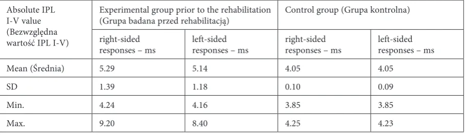
The comparison of the examination results in the scope of the absolute IPL I-V values is presented in Table 2. The data presented in Table 2 indicates that in children from the experimental group, both
the mean IPL I-V values and in terms of standard deviation, deviate to a large extent from the results of the control group. In the experimental group, these values were significantly higher both for the right- and left-sided responses; these differences were re-spectively 1.24 ms and 1.09 ms. The differences were statistically significant at the level of p < 0.001.
The intensification of the disorder may be tes-tified by the fact that the average value of the IPL I-V in the experimental group, whose average age was 3.8 years, was higher (i.e. worse) than in full-term newborns. At the same time, the dispersion of the results was enormous – from normal values to the results exceeding the average value of the con-trol group and the standard of authors’ laboratory by about 50 standard deviations. The complete re-sults in this regard are presented in Table 2.
Comparison of the Results Obtained Prior and After Rehabilitation
In order to prove that the rehabilitation im-proves the transmission efficiency in the auditory path, the results of examinations prior to and after the cycle of therapeutic exercises were compared.
As can be seen in Table 3, the mean values decreased after the rehabilitation; prior to reha-bilitation they amounted to, for the right and left side respectively, 5.29 ms and 5.14 ms, and after rehabilitation, 4.84 ms and 4.82 ms. The statistical
Table 2. Comparison of the absolute IPL I-V values in the experimental group prior to the rehabilitation with the control group
Tabela 2. Porównanie wartości bezwzględnych IPL I-V w grupie badanej przed rehabilitacją z grupą kontrolną
Absolute IPL I-V value (Bezwzględna wartość IPL I-V)
Experimental group prior to the rehabilitation
(Grupa badana przed rehabilitacją) Control group (Grupa kontrolna) right-sided
responses – ms left-sided responses – ms right-sided responses – ms left-sided responses – ms
Mean (Średnia) 5.29 5.14 4.05 4.05
SD 1.39 1.18 0.10 0.09
Min. 4.24 4.16 3.85 3.85
Max. 9.20 8.40 4.25 4.23
Table 3. Comparison of the absolute IPL I-V value in children prior to and after the rehabilitation
Tabela 3. Porównanie wartości bezwzględnych IPL I-V u dzieci przed rehabilitacją oraz po rehabilitacji
Absolute IPL I-V value (Bezwzględna wartość IPL I-V)
Prior to rehabilitation (Przed rehabilitacją) After rehabilitation (Po rehabilitacji) right-sided
responses – ms left-sided responses – ms right-sided responses – ms left-sided responses – ms
Mean (Średnia) 5.29 5.14 4.84 4.82
SD 1.39 1.18 1.41 1.14
Min. 4.24 4.16 3.76 3.84
analysis has shown that the differences were sig-nificant because, using the Wilcoxon test for the right-sided responses, p = 0.001 and for left-sided responses, p = 0.004, and using the Student’s t-test for pairs, the p-value for the respective sides was 0.001 and 0.002.
The data presented in Table 3 also indicates that the variation of results was very high in stud-ies carried out both prior to and after the reha-bilitation. Prior to the rehabilitation, the values ranged from 4.24 ms to 9.20 ms for the right-sided responses and from 4.16 to 8.40 for the left-sid-ed responses. After the rehabilitation, the values ranged from 3.76 ms to 8.96 ms for the right-sided responses and from 3.84 to 8.24 for the left-sided responses. As the authors can see, the values de-creased, which should be understood as the im-provement of the transmission efficiency of the brain stem segment of the auditory pathway sec-tion, for each side, both with regard to the mini-mum and maximini-mum values.
It has been found that, for the responses ob-tained from each side of the brain, the mean ab-solute value after rehabilitation was performed decreased by 0.45 ms (for the right side) and by 0.32 ms (for the left side).
Assessment of Relative IPL I-V Values
Additional information, important due to the fact that it is clear for both every researcher and every physician (not only those dealing with the research of evoked potentials), is delivered by the data presented in the form of the relative IPL I-V value.
The results indicating the severity of devia-tions from the standard are most apparent when the authors present them as a relative IPL I-V val-ue. Since in most studies, one assumes for param-eters whose distribution is normal, within nor-mal standards the results deviate by ± 2 SD from the standard, that is the values above and below compared to the mean. (± 3 SD are recognized as a wide standard.)
In the analyzed group, the following re-sults were found in the range of the relative IPL I-V value. Also the average values were very high, as they were 12.4 for the right-sided responses and of 10.9 for the left-sided responses in the studies prior to the rehabilitation (Table 4). Although the results obtained after the rehabilitation are much more advantageous, since they are 7.9 for the right-sided and 7.7 for the left-right-sided responses, they still remain very high. At the same time, the observed improvement was also huge, because it amounted 4.5 for the right-sided, and 3.2 for the left-sided responses.
The above data relates to mean values for the entire group. The analyses have not presented concrete results obtained in individual children in subsequent studies, but it is worth pointing out that the outcome of the rehabilitation in individual children varied widely.
The biggest improvement in the relative value of IPL I-V after MNRI® rehabilitation was
15.2 units.In this child, prior to rehabilitation the relative value was 14.7 and after rehabilitation, –0.5, meaning that in the analyzed case, the pri-mary delay compared to the standard, which was 14.7 standard deviations was normalized, because the result was even better than the standard of 0.5 standard deviation.
At the same time, beside the spectacular im-provement, in some children the result was only slightly improved, and in 2 cases there was a slight deterioration of the results (by 0.2 and 0.8 SD).
Discussion
In the present study, the authors analyzed the impact of one diagnostic and rehabilitative method on the results of a BAEP examination, and more specifically on efficiency (speed) of the transmis-sion of excitation in the selected area of the brain stem. Demonstration of improvement shows that the MNRI® method is very effective in this range.
Table 4. Comparison of the relative IPL I-V value in children prior to and after the rehabilitation
Tabela 4. Porównanie wartości względnych IPL I-V u dzieci przed rehabilitacją oraz po rehabilitacji
Relative IPL I-V value (Względna wartość IPL I-V)
Prior to rehabilitation (Przed rehabilitacją) After rehabilitation (Po rehabilitacji) right-sided responses left-sided responses right-sided responses left-sided responses
Mean (Średnia) 12.4 10.9 7.9 7.7
SD 14.0 11.8 14.1 11.4
Min. 1.9 1.1 –2.9 –2.1
The question, if the improvement observed in the auditory pathway is reflected in the improve-ment of mobility, which is clinically essential, re-mains open. Such a possibility has been pointed out by many authors, who have emphasized that examination of brain stem auditory potentials is of great prognostic importance when it comes to children’s motor development [22, 24, 26–29].
Such a possibility was pointed out by Pilecki, who showed that there is a close relationship be-tween mobility and the IPL I-V parameter [24]. Pilecki described drawing conclusions concerning mobility based on the results of BAEP examina-tions of brain stem auditory potentials ”indirect diagnosis” [21]. Although the term is of his au-thorship, the rule which he applied is widely ac-cepted both in terms of mobility and prognostic range [26, 29].
Another argument for the use of so-called in-direct assessment is the research results presented by Pilecki, who showed that in healthy children, during development, obtaining the ability of inde-pendent walking is associated with obtaining a cer-tain transmission speed in the brain stem auditory pathway segment of the child. As demonstrated by this author, it is not a coincidental time conver-gence of two processes occurring at one time, but the relationship is more precise. In children with slowed brain stem transmission, which is mani-fested by elongation of interpeak latency I-V, the development of the ability of independent walking is also delayed, and the child starts walking when the IPL I-V value decreases to about 4.25 ms [24].
The above analysis shows that the outcome of the rehabilitation obtained in individual children was variable. These results should not be surpris-ing, as the study concerned a clinically diverse group with various severities of clinical disorders and of different etiology of cerebral palsy syn-drome (CP).
The great improvement in examination results obtained after rehabilitation using the MNRI®
method, depending on the chosen statistical test p-value, ranged from 0.001 to 0.004.
It can be assumed that the cause of the im-provement in the examination results was the rehabilitation using the MNRI® method, because
only this factor had changed and the other exami-nation conditions in both rounds of the study were constant.
Although the above results clearly show an im-provement in the efficiency of transmission in the brain stem segment of the auditory pathway di-rectly after rehabilitation was performed, the ques-tion of the sustainability of this process remains open. Authors’ own observations suggest that the improvement observed directly after
rehabilita-tion is not durable, because in one of the children, whose examination was interrupted and then re-sumed due to technical reasons, it was found that initially there was very significant improvement, but when the examination was finished after about half an hour, the result was again analogous to the one obtained prior to the rehabilitation. This observation in no way undermines the effective-ness of the rehabilitation and only indicates that improvement is possible and regularly performed exercises should cause sustainable improvement of the BAEP results and, more importantly, improve-ment of the clinical condition of the rehabilitated children.
For doctors who do not deal with evoked po-tential examinations every day, the understand-ing of the intensification of changes in the results can be difficult, when the authors give the results for which the standards are not widely known. By using the parameter of the relative IPL I-V value, which indicates by how many quantities of SD the obtained result deviates from the mean de-termined for the healthy population, the average physician is able to determine the intensification of abnormalities.
Using this approach, the results obtained in the experimental group are interesting because in both groups, rIPL I-V (IPL I-V relative) val-ues reach even the value of about 50. That is, they differ from the mean determined for healthy chil-dren by up to 50 SD. Analysis in this regard will be carried out, because the children presented in this study are under constant control and the further studies are being conducted. After collect-ing a numerically larger group, the results will be published.
Using the equipment capabilities and the pro-fessional knowledge of the employees of the Chair of Pathophysiology of the Wroclaw Medical Uni-versity, the authors can believe that in the future, attempts will be made for further analysis (e.g. based on wavelet analysis or a single responses), allowing doctors to obtain broader information about the results using sophisticated mathemati-cal algorithms [30].
The authors have concluded that, as a result of the rehabilitation carried out using the MNRI®
References
[1] Vojta V: Current methods of rehabilitation in the treatment of motor disorders in perinatal encephalopathy. Cesk Neurol 1964, 27, 81–86.
[2] The Doman-Delacato method. J Iowa Med Soc 1968, 58, 507–509.
[3] Bobath CK: Neurophysiological bases of authors’ method of treatment of cerebral palsy. Acta Neurol Psychiatr Belg 1958, 58, 469–474.
[4] Sadowska L: Kompleksowa diagnostyka i stymulacja rozwoju dzieci z wrodzonymi i nabytymi dysfunkcjami ośrodkowego układu nerwowego według wrocławskiego modelu usprawniania. In: Psychospołeczne problemy rozwoju dziecka: aspekty diagnostyczne i terapeutyczne. Eds.: Czapiga A, Wydawnictwo Adam Marszałek, Toruń 2003, 81–97.
[5] Masgutowa S, Masgutow D: Integracja odruchów twarzy metodą Swietłany Masgutowej. Techniki pracy wspierające rozwój motoryki i mowy. MINK, Warszawa 2005, 85.
[6] Masgutova S: Odruchy jako podstawa rozwoju układu nerwowego i kształtowania schematów ruchowych w okre-sie niemowlęcym. In: Nowoczesne metody stymulacji rozwoju ruchowego i mowy. Eds.: Masgutova S, MINK, Krynica Górska 2005, 14–36.
[7] Masgutova S: Integration of Infant Dynamic and Postural Reflexes: Neuro-sensory-motor Reflex Integration®
Method for Children and Adults. SMEI, Florida 2009, 257.
[8] Masgutowa S, Akhmatowa N: Integracja dynamicznych i posturalnych odruchów z całym układem ruchowym. MINK, Warszawa 2004, 207.
[9] Masgutowa S, Kowal J, Mazur G, Masgutow D: Neurokinezjologiczna Terapia Taktylna™ dr S. Masgutowej. MINK, Warszawa 2005, 133.
[10] Masgutova S: MNRI® Neurosensorimotor Development: Visual and Auditory Reflexes Integration. Facilitation
Program of Development and Learning for Children and Adults. Florida 2009, 86.
[11] Masgutova SK, Akhmatova NK, Kiselevsky MV: Clinic-Immunological Assessment of Effect of the Therapy Program of Neuro-sensory-motor Integration of Reflex Patterns at Chronic Inflammatory Diseases of Respiratory System. Russ Immun J 2008, 2, 454–463.
[12] Piaget J: The Grasp of Consciousness: Action and Concept in the Young Child. Cambridge, M.A. Harvard 1976.
[13] Vygotsky LS: The Child Psychology. The Problems of Child Development. In 6 Books. Book – 4. Pedagogika. Moscow 1986.
[14] Masgutova S, Regner A: Rozwój mowy dziecka w świetle integracji sensomotorycznej. Continuo, MISM, Wrocław 2008, 167.
[15] Oswald ME, Grosjean S: Confirmation Bias. In: A Handbook on Fallacies and Biases in Thinking, Judgement and Memory. Cognitive Illusions. Eds.: Pohl RF, Psychology Press, Hove 2004, 79–96.
[16] Lilienfeld SO, Ammirati R, Landfield K: Giving debiasing away: Can psychological research on correcting cogni-tive errors promote human welfare? Persp Psycholog Sci 2009, 4, 390–398.
[17] Chiappa KH: Evoked Potentials in Clinical Medicine. Third edition. Lippincott, Raven 1997.
[18] Majnemer A, Rosenblatt B, Riley P: Prognostic significance of multimodality evoked response testing in high-risk newborns. Pediatr Neurol 1990, 6, 367–374.
[19] Majnemer A, Rosenblatt B: Evoked potentials as predictors of outcome in neonatal intensive care unit survivors: review of the literature. Pediatr Neurol 1996, 14, 189–195.
[20] Jiang ZD, Wu YY, Liu XY: Early development of brain stem auditory evoked potentials in Down’s syndrome. Early Hum Dev 1990, 23, 41–51.
[21] Pilecki W: Prognoza rozwoju ruchowego u dzieci z poważnie zaburzoną transmisją w pniu mózgu obserwowaną w badaniu BAEP. Wydawnictwo WTN, Wrocław 2002, 125–130.
[22] Pilecki W: Multimodalne potencjały wywołane w prognozie rozwoju ruchowego dzieci. In: Komputerowe ws-pomaganie badań naukowych. Wydawnictwo WTN, Wrocław – Polanica Zdrój 2003, 75–80.
[23] Pilecki W: Ocena transmisji w obwodowym odcinku drogi słuchowej u dzieci z mózgowym porażeniem dziecięcym (MPDz). Wydawnictwo WTN, Wrocław 2002, 107–112.
[24] Pilecki W, Szawrowicz T, Jagielski J, Janocha A, Borodulin-Nadzieja L: Zależność między prędkością transmisji w pniowej części drogi słuchowej a rozwojem ruchowym dziecka. Wydawnictwo WTN, Wrocław 2002, 119–124.
[25] Pilecki W: Względna wartość IPL I-V – nowy parametr przydatny w badaniu słuchowych potencjałów pniowych u dzieci. In: Komputerowe wspomaganie badań naukowych. Wydawnictwo WTN, Wrocław – Polanica Zdrój 2004, 93–98.
[26] Yilmaz Y, Degirmenci S, Akdas F, Kulekci S, Ciprut A, Yuksel S, Yildiz F, Karadeniz L, Say A: Prognostic value of auditory brain stem response for neurologic outcome in patients with neonatal indirect hyperbilirubinemia. J Child Neurol 2001, 16, 772–775.
[27] Scalais E: Multimodality evoked potentials as a prognostic tool in term asphyxiated newborns. Electroencephalogr Clin Neurophysiol 1998, 3, 137–144.
[28] Pike AA, Marlow N: The role of cortical evoked responses in predicting neuromotor outcome in very preterm infants. Early Hum Dev 2000, 57, 123–135.
[29] Majnemer A, Rosenblatt B: Prediction of outcome at school entry in neonatal intensive care unit survivours, with use of clinical and electrophysiological techniques. J Pediatrics 1995, 127, 823–830.
Address for correspondence:
Witold Pilecki
Department of Pathophysiology Wroclaw Medical University Marcinkowskiego 1
50-368 Wrocław Poland
E-mail: [email protected]
Conflict of interest: none declared