Arun Kumar Sharma
Research Scholar, Department of Pharmacology Amity Institute of Pharmacy, Amity University Sector-125, Noida - 201303 (U.P.), India
Landline: 0120-2445252| Mobile: +91 989 637 7853 Email address: [email protected]
Address for correspondence
Access this article online www.japer.in
Apoptosis: A Potential target site for natural bioactive agents
during myocardial infarction
INTRODUCTION
Myocardial infarction (MI) is a major end result of morbidity and mortality in developed and developing countries. Myocardial ischemia is a leading cause of MI. Although, reperfusion of blood after prolonged ischemia to heart cells is necessary for cardio protection, but it additionally causes the myocardial cell damage [1]. In-addition, myocardial ischemia-reperfusion injury significantly generates ROS due to anaerobic oxidation followed by oxidative stress. ROS generated oxidative stress doubles the risk of ischemia and reperfusion injury [2]. A number of imperative studies demonstrate both ischemia/reperfusion and oxidative stress are the primary factor for generation of apoptosis which consequently leads to MI. Clinical and preclinical
studies revealed the high level of ROS significantly induced MI through apoptosis [2-3]. Furthermore, I/R injury is an individual platform for apoptosis with or with generation of ROS, followed by MI [3-4]. Apoptosis is a genetically operated process of self-destruction of cells through the crumbling of DNA. It significantly differs from cellular necrosis which is premature cell death resulting from an exposure to external factors such as infection, toxins or trauma. Necrosis always leads to detrimental effects on surrounding cells. Whereas, apoptosis is a normal physiological process that eliminates DNA-damaged, or unwanted cells. Both necrosis and apoptosis cause cell death; they differ from each other in various morphological and cellular dictatorial characters. Necrosis is a process of rapid cellular swelling due to accumulation of fluid and electrolytes, premature burst of plasma membrane and distraction of cellular organelles resulting in to brisk loss of cellular homeostasis. Necrosis associated rupture and consequent leakage of broad array of cellular material through membrane culminates into the inflammation [5-6].
Myocardial infarction (MI) is an insidious disease, gently spreading in developed and developing countries. MI is the consequence of hypoxia to myocardial tissue which may leads to apoptosis, narcosis and followed by cardiac cell death. Activation of apoptotic pathways during myocardial infarction (MI) is frequently reported in clinical, preclinical and post-mortem studies. Several apoptosis inducing factors and signalling cascades leading to death of cardiomyocytes culminate into MI. Such involvement of ischemia induced apoptosis in MI is widely accepted. Apoptosis is a natural process for regulating the homeostasis in cellular organelles. Unlike the necrosis, it is a synchronize energy dependent process which is carried out by shrinkage of the cell. This contraction of cell leads to squeezing of nucleus and nuclear chromatin into brusquely demarcated masses. However, such programmed cell death in several tissues including myocardium becomes pathogenic under certain conditions. In-addition, reactive oxygen species (ROS) and generated oxidative stress is also play a key role in production of apoptosis and several associated signalling alteration which ultimately leads to MI. Recently, certain natural products especially from the plant kingdom have been evaluated for their anti-apoptotic potential. There is an up rise in the investigations delineating the exact mechanisms through which natural phytochemicals target MI associated apoptosis. This review explores novel signalling pathways and target sites for anti-apoptotic phytochemicals having potential to check the cellular apoptosis consequent to MI. A new vista may explore in the prospective treatment of MI by using apoptosis-modulating natural products.
Keywords: Reactive oxidative stress, Caspase, Apoptosis, Myocardial infarction, Phytochemicals, Herbal plants
ABSTRACT Arun Kumar Sharma#1
Shaikh Haidarali#2 Upendra Nagaich3
1Department of Pharmacology, Amity Institute of Pharmacy, Amity University, Sector-125, Noida - 201303 (U.P.), India 2Department of Pharmacology, R.C.Patel Institute of Pharmaceutical Education and Research, Shirpur- Dist-Dhule, Maharashtra, India 1Department of Pharmaceutics, Amity Institute of Pharmacy, Amity University, Sector-125, Noida- 201303 (U.P.), India
In contrast to necrosis, programmed cell death or apoptosis is an energy dependent and abundantly synchronized process. Apoptosis is carried out through the breaks up of cell after shrinkage of and formation of brusquely demarcated masses of the nucleus and nuclear chromatin squeezed together. Following this, cells become isolated from the surrounding tissues. The extensions buds come out from membrane and that ultimately close off to form membrane enclosed vesicles, known apoptotic bodies. These enclosed vesicles are moreover phagocytosed by adjacent cells or endure deprivation, which seems to be the process of necrosis, entitled as secondary necrosis. Usually apoptosis does not prompt an inflammatory response as compared to necrosis [7]. Apoptosis may be activated by a stimulus or deletion of a suppressing stimulus.
Impaired supply of oxygen and blood flow to the heart initiates a pathologic condition called ‘ischemic state’ which initiates cascades of destructive events. Earlier, it was postulated that myocardial cells in the region of obstructed coronary artery undergo necrosis and can’t regenerate. However, recent findings indicate apoptosis as another form of myocardial cell death playing an important role in the tissue destruction consequent to myocardial infarction (MI) [8]. Evidence suggests that a compartment of myocytes undergoes apoptosis during ischemia-reperfusion injury in addition to overt necrosis which was considered as a root cause of cell death in MI [4]. Thus, apoptosis may permanently damage the myocardial cells and lead to potential life-threatening arrhythmias consequently creating ventricular aneurysm. In addition to such damage, apoptosis in MI may trigger inflammation induced atherosclerotic plaque formation [9-10]. Hence, an increase in the plasma levels of C-reactive protein (CRP), a specific biomarker for inflammation, predicts the risk of MI [10-11].
Calcium deposition is another root cause of atherosclerotic plaque generation. Calcium depositions can be certainly detected by modern
devices, therefore, may be an alternative predictive source in preference to classical risk factors [12-14]. Another presentation allied with untimely atherosclerosis is high level of homocysteine (non-protein amino acid) in blood and resulting homocysteinuria [15]. However, predictive significance of homocysteinemia/ homecysteinuria is debated [16]. Data suggest that oxidative stress and disproportionate nitric oxide generation play extensive roles in ischemia/reperfusion injury that derange the cardiac functions [17]. It has been well described that medicinal plants have potent anti-apoptotic property against experimentally induced MI in animal models both in vivo and ex-vivo. Numerous studies on natural products and herbal compounds delineate the molecular pathways involved in apoptosis associated MI [18]. This review lays emphasis on some potential signalling pathways and target sites for anti-apoptotic herbal compounds showing efficacy against experimentally induced MI in preclinical studies. Moreover, review also encapsulates the catalogue of herbal compounds having prospective anti-apoptotic action.
Apoptosis vs. Myocardial Infarction
stimulated by both external and internal initiating the ‘extrinsic’ and ‘intrinsic’ pathways respectively. A number of accumulated evidences suggest that apoptosis contributes to the pathogenic alterations in cardiomyocytes [21]. Numerous clinical, subclinical and post-mortem studies have reported activation of apoptotic pathway in myocardial infarction [22-24]. Molecular magnetic resonance imaging technique demonstrates a huge apoptotic zone in the myocardium and viable myocardium in the central-myocardium early after an ischemic injury [25]. Therefore, myocardial rescue strategies after ischemic damage are the core interest in clinical approach. Several recent studies reported that I/R induce myocardial apoptosis in the ischemic myocardium in vivo [10-11, 21]. However, whether apoptosis is triggered during ischemia or during reperfusion is still controversial. Gottlieb et al. (1994) found that the hallmark of apoptosis, nucleosomal ladders of DNA fragments, was detected in ischemic myocardium after 30 min of ischemia and 4 h of reperfusion in the rabbit, but not in ischemic-only myocardium, suggesting that apoptosis may be expressed only during reperfusion [22].
Cardiac specific overexpression of α-1 adrenoceptors in mice induces cardiac hypertrophy, apoptosis, fibrosis, and heart failure, further emphasizing the role of β-adrenergic signaling in the pathogenesis of heart failure [17]. It has showed that apoptosis in adult cardiomyocytes caused by specific β-1 stimulation is mediated by a PKA-dependent pathway, and that β-1 and β-2 adrenoceptors deliver opposing signals with respect to cell-survival. Neurohormonal factors such as angiotensin II acting via the AT-1 R have been shown to produce cardiomyocyte apoptosis. Elevated circulating levels of antiotensin II and likely in situ cardiac antiotensin II production, may play an important role in this respect. It remains however, to be shown that drugs like ACE inhibitors or ARBs reduce cardiac apoptosis while improving heart failure condition in human [10-11, 17, 21-24].
Since I/R results in the reproducible induction of cardiomyocyte apoptosis, this is the model most often used to test the effect of antiapoptotic agents on cardiomyocyte survival. The following approaches were used to test the importance of different players in the pathways mediating apopotosis: (1) if reactive oxygen species (ROS) played a major role in causing apoptosis, radical scavengers or enzymes degrading ROS should reduce apoptosis. (2) Substrate deprivation as encountered in the course of ischemia and substrate withdrawal causes endoplasmic reticulam stress; if this was important in I/R or hypoxia, overexpression of molecular chaperones should diminish apoptosis. (3) Apoptosis occurs if the balance between pro-survival and pro-apoptotic pathway is tilted toward pro apoptotic pathways. If this concept applied to cardiac disease, activation of pro-survival pathways should reduce the extent of apoptosis. (4) If apoptosis contributed importantly to the cell damage inflicted during I/R and myocardial infarction, specific inhibitors of caspases should be expected to reduce apoptosis and infarct size [22-27].
anti-apoptotic effect in ischemia-reperfusion of cardiomyocytes [28].
Role of mitochondria in apoptosis
Mitochondria are the primary site for generation of ROS which may lead to the production of apoptosis. Mitochondria have four complexes of enzymes and at least eight distinct ROS- generating-complexes. One of these eight complexes releases ROS into an inter-membrane space from where it enters into cytosol and participates in ROS associated signalling cascade. Apart from this, complex I and complex II also possess a little potential to release ROS in inter-membrane space. The reactive species generated from ROS-generating-complexes contribute to apoptosis mediated MI [29]. Therefore, ROS maintain the homeostasis of oxidative stress associated pathophysiological events. Interestingly, ROS play a versatile role in apoptosis induction. On one hand mitochondrial ROS activate cytochrome-c that triggers caspase activation and leads to apoptosis and MI [30]. However, on the other hand ROS also possess anti-apoptotic effects. Major path for the induction of apoptosis and MI depend on Fas/Fas-ligand system and mitochondrial redox state. Mitochondrial superoxide is enhanced in apoptotic cells which were generated from reduction of oxygen, specifically during cytochrome-c release from the inter-membranous space to cytosol. This electron transfer starts activation of caspases [30-31].
Another important endogenous metabolite is nitric oxide (NO), which may increase the generation of superoxide related metabolites and oxidative stress [32]. This also enhances the intracellular free calcium ion [Ca+2] and activate Ca+2 dependent phospholipase A2 pathway, thus increasing the gateway of membrane permeability transition (MPT) pores [33]. Indeed, Ca+2 induces ROS production thereby increasing oxidation of mitochondrial proteins and lipids [33-34]. However, NO depolarizes the membrane potential, promotes the cytosolic Ca+2, and liberates cytochrome-c, which may activate the
arrangement of cellular incidents resulting in to apoptosis and MI [35-36]. Moreover, NO gets converted into peroxynitrite by reacting with ROS. Brown and Brown [37] revealed about NO reaction with mitochondrial ROS that may cause reversible inhibition of respiration, generatng super oxide and peroxynitrite, and irreversible inhibition which further leads to mitochondrial membrane permeability transition. This mitochondrial respiration inhibition may activate necrosis or apoptosis through mitochondrial membrane permeability transition activation and ultimately leads to MI [38].
Ischemia-reperfusion (I/R) induced MI via apoptosis
increased. Accordingly, reverse activation of 2Na+/Ca2+ ion exchange is increased which leads to intracellular Ca2+ overloading [40-42]. Intracellular Ca2+ overloading plays a consequent role in generation of apoptosis which is further a leading cause of MI [43]. Moreover, ROS itself results apoptosis by inducing numerous factors [2, 44]. Several evidences provide supportive data which indicates ROS play an important pathological role in cardiac injury through the apoptosis in cardiomyocytes cell death [44-45]. In-addition, I/R injury in myocardial cells significantly activate c-Jun N-terminal kinase (JNK) pathway which ultimately causes apoptosis followed by MI. JNK, or stress-activated protein kinase, is an imperative member of the mitogen-activated protein kinase (MAPK) superfamily [3]. JNKs play a key role in the activation of death receptor through both extrinsic and intrinsic apoptotic pathways. JNKs can upregulate pro-apoptotic genes through transactivation of specific transcription factors which significantly leads to activation of apoptotic signalling [46]. Prevalent evidences suggest the activation of JNK pathway during I/R injury in myocardial I/R resulting in apoptosis [47-48]. Abundance scientific literature revealed that oxidative stress can also influences the MAPK signaling pathways and associated ERK, JNK pathways [49]. Recent study also revealed apoptosis associated myocardial infarction. Additionally, author suggested tissue damage activity by apoptosis also seen after myocardial infarction [50].
Intrinsic (mitochondrial associated) pathway induced MI via apoptosis
Other than extrinsic pathways, several intracellular stimuli also participate in mitochondrial induced apoptosis and associated MI. Internal stimuli, such as irreversible genetic injury, hypoxia, extreme concentrations of cytosolic Ca+2, viral infection, or severe oxidative stress or destructive free radicals stimulate apoptosis by the intrinsic pathway. For instance physical stress, oxidative stress and DNA
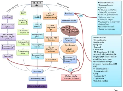
damage are the commonly known intrinsic factors for generating apoptosis and MI [51]. Apoptosis induced MI is the result of imbalance between pro-apoptotic i.e. Bid, Bax and, Bak and apoptotic Bcl-2and, Bcl-xL proteins of Bcl-2 family which critically regulate this signalling cascade [52]. After activation, Bax migrates to the outer mitochondrial membrane and forms a new complex by attaching with Bak. Formation of this complex makes outer mitochondrial membrane more permeable which insist cytochrome-c with other pro-apoptotic proteins [53]. If cytochrome-c intermingles with apoptotic protease activating factor-1 (APAF-1) in cytosol, this mixture stimulates caspase cascade. Cascade starts from caspase-9 activation leading to caspases-3 and caspases-7 which ultimately cause apoptotic cell death [54]. In spite of that some other apoptogens are also released by outer mitochondrial membrane on permeable involving second mitochondria-derived activator of caspase and apoptosis-inducing factor (AIF) [55], which can also initiate cleavage of BH3-interacting domain death agonist followed by mitochondrial pathway and leads to translocation of Bax/Bak proteins and apoptogens release [56]. As apoptosis plays a significant role in MI, all intrinsic and extrinsic cofactors, apoptotic and pro-apoptotic proteins may prove to be targetable for in the quest of new approaches to prevent MI and its consequences. Figure 1.
Extrinsic (TNF associated) pathway induced MI via apoptosis
family which leads to the apoptotic process [63]. The existing evidence intimates that the TNF receptor is present in the plasma membrane with accessible trimer. The cytoplasmic domain of all TNF receptor subunits include a subdivision of about 70 amino acids known as “death domain” which brings about protein–protein interactions [64]. TNF binding to trimeric receptor generates a conformation change in death receptor domain and that initiates the conscription of numerous proteins. The last proteins, procaspase-8 molecules, assemble at the inner surface of plasma membrane. Thus, the synthesis of caspases as proenzymes defend cells against unintended proteolytic injures. In-contrast, several proenzymes and procaspases reveal a low level of proteolytic movement. Therefore, consistent with one model, when two or more procaspases are aligned in close connection with each other, they are competent of splitting one another’s polypeptide chain and turn other molecule into the fully active caspases. At last matured enzyme caspase-8 contains four polypeptide chains, derived from two procaspase precursors. Caspase-8 activation is comparable in tenet to the initiation of effectors by hormone or growth factor [65]. Therefore, it may be concluded that all the signalling pathways involve the binding of extracellular ligand which leads to alteration in receptor conformation ultimately causing the downstream binding and activation pathway of proteins. Caspase-8 is considered as an originator caspase since it kicks off the apoptosis [66]. Mitochondria are able to regulate functional integrity of the cardiomyocytes. ATP production and calcium ion homeostasis are the necessary parameter to normalize mitochondria associated myocardium event. TNF-α and Fas-ligand participate in inducing death of myocardial cells by formation of Death Inducing Signaling Complex (DISC). DISC, once activated, leads to activation of procaspases-8 and -10 which further downstream other cascades of different caspases like as procaspases-3 and -7 and apoptosis followed by MI [36, 67-69]. Figure 1.
Targetable protein substrates
Potential caspases targeting during the apoptosis can be carried out on either regulatory proteins or structural and “housekeeping” intensive protein substrate groups. Regulatory proteins breakage strengthens apoptotic activation, whereas cleavage of structural proteins leads to cellular breakdown. First group involves a number of kinases, like as MEK kinase 1, P21-activated kinase 2, and Mst-1. Cleavage of caspases triggers these kinases which further leads to activation of c-Jun kinase and stress-activated protein kinase in response to manage apoptotic pathways [70-71]. In-addition, caspase –associated hydrolysis of proteins may also initiate the termination of negative regulators of apoptosis. For instance, focal adhesion kinase (FAK), phosphatidylinositol-3 kinase (PI-3 kinase), and the associated protein akt, raf-1, endogenous caspase inhibitors (or IAPs), anti-apoptotic members of Bcl-2 family, and inhibitors of caspase-activated DNAse (ICAD) are such kind of proteins. On the other hand, second class of targets, concerned in apoptotic structural changes, contain nuclear lamins, actin regulatory proteins like as fodrin, spectrin, and gelsolin, transcription factors, and DNA producer and renovator proteins for instance, PARP, MCM-3 [72-73].
Protective role of anti-apoptotic herbal compounds in MI
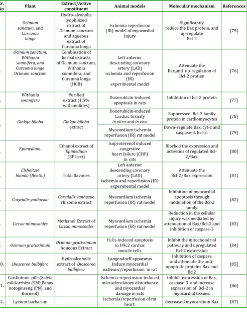
Table 1: Anti-apoptotic plant investigated for cardioprotective effects in experimental models
Sr.
No Plant Extract/Active constituent Animal models Molecular mechanism References
1.
Ocimum sanctum, and
Curcuma longa Hydro-alcoholic lyophilized extract of Ocimum sanctum and aqueous extract of Curcuma longa Ischemia reperfusion (IR) model of myocardial
Injury
Significantly reduce the Bax protein, and
up-regulate Bcl-2 [75] 2. Ocimum sanctum, Withania somnifera, and Curcuma longa Ocimum sanctum Combination of herbal extracts of Ocimum sanctum,
Withania somnifera, and Curcuma longa (HCB) Left anterior descending coronary artery (LAD) ischemia and reperfusion
(IR) experimental model
Attenuate the Bax,and up-regulation of
Bcl-2 protien [76]
3. somnifera Withania extract (1.5% Purified withanolides)
Doxorubicin-induced
apoptosis in rats Inhibition of bcl-2 protein [77]
4. Ginkgo biloba Ginkgo biloba extract
Doxorubicin-induced Cardiac toxicity
in vitro and in vivo
Suppressed Bcl-2 family
protein in cardiomyocytes [78]
Myocardium ischemia reperfusion (IR) rat model
Down-regulate Bax, cyt-c and
caspase-3. Bcl-2. [79]
5. Epimedium, Ethanol extract of Epimedium
(EPI-ext)
Isoproterenol induced congestive heart failure (CHF)
in rats
Blocked the expression and activities of regulated
Bcl-2/Bax. [80]
6. blanda (Benth.) Elsholtzia Total flavones
Left anterior descending coronary
artery (LAD) ischemia and reperfusion (IR)
experimental model
Attenuate the
Bcl-2/Bax expression [81]
7. Corydalis yanhusuo Corydalis yanhusuo rhizome extract reperfusion (IR) rat model Myocardium ischemia
Inhibition of myocardial apoptosis through modulation of the Bcl-2
family.
[82]
8. Cassia mimosoides Methanol Extract ofCassia mimosoides reperfusion (IR) rat model Myocardium ischemia
Reduction in the cellular injury was mediated by attenuation of Bax/Bcl-2 and
inhibition of caspase-3 [83]
9. Ocimum gratissimum Ocimum gratissimum Aqueous Extract H2O2-induced apoptosis in H9c2 cardiac muscle cells
Inhibit the mitochondrial pathway and upregulated
Bcl-2 expression [84]
10. Dioscorea bulbifera extract ofHydroalcoholic Dioscorea bulbifera
Langendorff apparatus Induce myocardial ischemic/reperfusion in rat
Inhibition of caspase and attenuate the anti-apoptotic proteins Bax and
Bcl2
[85]
11.
Cardiotonic pills(Salvia miltiorrhiza (SM),Panax notoginseng (PN), and
Borneol)
Ischemia-reperfusion-induced microcirculatory disturbance
and myocardial damage in rats
Inhibit expression of Bax, caspase-3 and increase
expression of Bcl-2 in myocardial tissues.
[86]
12. Lycium barbarum Ischemia/reperfusion of rat heart. decreased myocardium Bax [87]
Tulsi (Osscimum sanctum), is a herb cultivated throughout India for religious as well as medicinal purposes. Ocimum sanctum L. is suggested to possess hypolipidemic [88], hepatoprotective [89],and neuroprotective [90] properties. Its anticoagulant
longa) is a rhizomatous perennial plant belonging to zingiberaceae family. Curcumin is an active curcuminoid in turmeric which exhibits precise medical potential. The turmeric oil, which mostly contains curcumin is widely used for its antioxidant, antimutagenic [96], carcinogenic [97], anti-bacterial [98], and anti-fungal [99,100] activities. In addition, the extracts of osscimum sanctum and Curcuma longa show synergistic action in ischemia and reperfusion (I-R) model of MI. Interestingly, this combination attenuates the apoptotic proteins [75]. Moreover, in-addition Withania somnifera with above combination has been projected to attenuate Bax and up regulate the Bcl-2 protein in cardiovascular injury [76].
Withania somnifera (WS) (Ashwagandha), is an annual herb enriched with alkaloids and steroidal lactone. The Ashwagandha roots contain diverse withanolides, essential as well as non-essential fatty acids, amino acids, sterols, catecholamines, aromatic alcohols, gamma amino butyric acid, and glycerol [101,102]. The bioactive constituents of Withania somnifera possess anti-inflammatory [103], anti-oxidant [104], antitumor [105], antistress [106], immunomodulatory [107] and hematopoietic properties [108]. Furthermore, it possesses anti-apoptoic potential in myocardial infarction. WS attenuates the progression of Bcl-2 protein in doxorubicin-induced cardiac toxicity in rats [77].
Ginkgo biloba (GB) (Maidenhair tree), is an exceptional member which is unique of its species with no other similar members. The leaves of Ginko biloba contain the ginkgolides A, B and C which possess various pharmacological activities. In addition, GB has an antiplatelet action, along with neuroprotective [109] and cardioprotective [110] potentials. An investigation including western blot and immunohistochemical analysis has revealed that GB effectively down-regulates the Bax, cyt-c, Bcl-2 and caspase-3 in myocardium ischemia- reperfusion (IR) model in rat [78, 79].
Epimedium is a genus of flowering plants in the family Berberidaceae. In addition, the major active substances of epimedium include icariin, epimedoside, icariside, breviflavone and ikarisoside, most of which possess hepatotoxic, immunomodulatory, anti-tumor, antidepressant and anti-oxidant activities [111-112]. It has been documented that the use of epimedium extract attenuates myocardial apoptosis through remarkably attenuating Bcl-2/Bax protein [80]. Likewise, Elsholtzia blanda is a Chinese medicinal herb is reported to significantly inhibit the Bcl-2 and attenuat Bax expression in coronary occlusion induced myocardial infarction in rat [81]. Corydalis yanhusuo is a Chinese herbal medicine which contain the active constituent is protopine.Likewise, Protopine has posses the protective effects on cardiac myocytes electrophysiological [113], cerebral ischemic injury [114] ,inhibit platelet aggregation [115], depression [116], antithrombotic, anti-inflammatory [117], antioxidant, and neuroprotection effects [118] action. In addition study have revealed that the Corydalis yanhusuo attenuate the myocardial injury is closely associated with the inhibition of myocardial apoptosis through inhibition of the Bcl-2 family [82]. However, Cassia mimosoides is a species of legume which belongs to fabaceae family. Studies have revealed the cardioprotection action of Cassia mimosoides by inhibiting apoptosis in myocardial injury. In detail, Cassia mimosoides reduce the cellular injury by attenuation of Bax/Bcl-2, inhibition of caspase-3 activation from procas-pase-3 and subsequent reduction in the number of apoptotic cells [83].
H9c2 cells from H2O2-induced cell death [84]. Dioscorea bulbifera, (air potato) belongs to family Dioscoreaceae. It is mainly found in Africa and Asia. Abundantly, it content steroidal saponins i.e. dioscoreanosides A to K which show various pharmacological action [124]. In one study they revealed that the Dioscorea bulbifera inhibit anti-apoptotic proteins i.e. Bax and Bcl2 by Western blot analysis followed by TUNEL assay [85].
Cardiotonic pills (Salvia miltiorrhiza (SM),Panax notoginseng (PN), and Borneol) is a chinease traditional medicine. Therefore, the combination of three plant produce a synergistic action. Likewise, one study demonstrated that this cardiotonic pill prevent MI by inhibition of apoptosis.Interestingly,Cardiotonic pill attenuate the Bax,caspase-3 and increase the expression of bcl-2 in myocardial cell [86]. The Lycium barbarum is a chienese herb which belong to Solanaceae family. Likewise, Lycium barbarum has a
large variety of pharmaceutical activites, such as anti-aging, antitumor and immunomodulation [125-127].In addition one study Lycium barbarum possesses the antiapoptotic potential by effectively attenuate the myocardium Bax in Ischemia/reperfusion of rat heart [87].
The cardioprotective potential of medicinal plants through antiapototic pathway of myocardial preconditioning has been well evidenced in pre-clinical conditions. However, more studies are therefore needed to demonstrate a molecular mechanism of apoptosis which could certainly be a core procedure pertained to cardiovascular defensive potentials of herb. Therefore, the anti-apoptotic role of herbal plant is an imperative issue and offers new perspectives for the better exploitation of apoptosis induce cardiovascular disorders and improving cardiovascular outcomes.
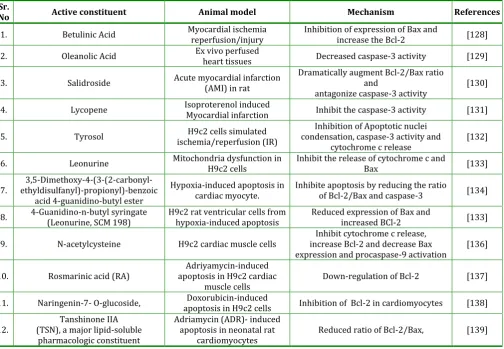
Table 2: Anti-apoptotic active constituent of plants investigated for cardioprotective effects in experimental models
Sr.
No Active constituent Animal model Mechanism References
1. Betulinic Acid Myocardial ischemia reperfusion/injury Inhibition of expression of Bax and increase the Bcl-2 [128]
2. Oleanolic Acid Ex vivo perfused heart tissues Decreased caspase-3 activity [129]
3. Salidroside Acute myocardial infarction (AMI) in rat Dramatically augment Bcl-2/Bax ratio and
antagonize caspase-3 activity [130] 4. Lycopene Isoproterenol induced Myocardial infarction Inhibit the caspase-3 activity [131]
5. Tyrosol ischemia/reperfusion (IR) H9c2 cells simulated condensation, caspase-3 activity and Inhibition of Apoptotic nuclei
cytochrome c release [132] 6. Leonurine Mitochondria dysfunction in H9c2 cells Inhibit the release of cytochrome c and Bax [133]
7. ethyldisulfanyl)-propionyl)-benzoic 3,5-Dimethoxy-4-(3-(2-carbonyl-acid 4-guanidino-butyl ester
Hypoxia-induced apoptosis in
cardiac myocyte. Inhibite apoptosis by reducing the ratio of Bcl-2/Bax and caspase-3 [134]
8. 4-Guanidino-n-butyl syringate (Leonurine, SCM 198) H9c2 rat ventricular cells from hypoxia-induced apoptosis Reduced expression of Bax and increased BCl-2 [133]
9. N-acetylcysteine H9c2 cardiac muscle cells increase Bcl-2 and decrease Bax Inhibit cytochrome c release,
expression and procaspase-9 activation [136]
10. Rosmarinic acid (RA) apoptosis in H9c2 cardiac Adriyamycin-induced
muscle cells Down-regulation of Bcl-2 [137] 11. Naringenin-7- O-glucoside, apoptosis in H9c2 cells Doxorubicin-induced Inhibition of Bcl-2 in cardiomyocytes [138]
12. (TSN), a major lipid-soluble Tanshinone IIA pharmacologic constituent
Adriamycin (ADR)- induced apoptosis in neonatal rat
Betulinic Acid is a triterpenoid obtained from various plant sources. It has posses antitumor [140], anti-HIV [141], antimalarial [142], anti-inflammatory [143] activity. In one study they demonstrate that betulinic acid attenuate the BAX protein and increase the expression of bcl-2 in the myocardial ischemia/reperfusion injury with help of western blot and TUNEL assay [128].
Oleanolic Acid is a naturally occurring triterpenoid, mostly distributed in food and medicinal plants. It has a variety of biological effects, such as anti-oxidants [144], antifungal, anti-inflammatory, anti-hyperlipdemia, hepatoprotective, tumor prevention, immunomodulatory, [145], HIV [147], anti-arrhythmic and cardiotonic [148]. In addition, Oleanolic acid posses antiapoptotic potential by significantly increased cardiac p-BAD/BAD and decreased caspase-3 peptide levels under high glucose perfusion condition [129].
Salidroside [2-(4-hydroxyphenyl) ethyl beta-D-glucopyranoside], active constituent of Rhodiola rosea. Salidroside possesses anti-aging, anti-cancer, anti-viral [149], anti-inflammatory [150], anti-hypoxia [151-152] and anti-oxidative [153-154] properties. The Salidroside, has been shown to exhibit protective effects in IR- and hypoxia-induced cardiomyocyte cell death [155].In one study the salidroside possesses the antiapoptotic potential by dramatically augment Bcl-2/Bax ratio and antagonize caspase-3 activity in acute myocardial infarction (AMI) in rat [130].
Lycopene is a red caratenoid found in various vegetables. Lycopene has the greatest ability of all the common carotenoids to quench singlet oxygen. It possesess the anti-cancer [156], antioxidant , low-density lipoprotein cholesterol reduction [157,158],
reduces proinflammatory cytokine and
chemokines [159,160].In addition,lycopene attenuate the caspase-3 protein in isoproterenol induced Myocardial infarction [131].
Tyrosol (2-(4-hydroxyphenyl) ethanol) is a natural phenolic antioxidant present in a various plants. Its mainly present in olive oil and white wine. Studies
suggested that Tyrosol possesses the free radical scavenging activity [161], anti-inflammatory [162] and neuroprotective [163] effects. Addition studies support that tyrosol attenuate the Apoptotic nuclei condensation, caspase-3 activity and cytochrome c release in H9c2 cells simulated ischemia/reperfusion (IR) [132]
Leonurine (LEO), an alkaloid isolated from Leonurus cardiaca. It possesses the beneficial effect against cardiovascular diseases [164-166], ischemic stroke [167], atherosclerosis [168, 169],vasodilating [170], antioxidative [164,171], antiapoptotic [172,173], and anti-inflammatory [168] activities. Interestingly,leonurine used to treat the myocardial problem in various way. In addition, LEO inhibit the release of cytochrome c and Bax in H9c2 cells simulated ischemia/reperfusion (IR) in rat [133]. Additional studies demonstrate that the formation of new synthetic compound i.e. 3,5-Dimethoxy-(3-(2-carbonyl-ethyldisulfanyl)-propionyl)-benzoic acid 4-guanidino-butyl ester which possesses the antiapototic active with inhibition of Bcl-2/Bax and caspase-3 in Hypoxia-induced apoptosis in cardiac myocytes [134].Likewise, another compound synthesized form Leonurine that is 4-Guanidino-n-butyl syringate (Leonurine, SCM 198) which possesses the antiapoptotic activity by inhibiting Bax and increased expression of BCl-2 in H9c2 rat ventricular cells [135].
N-acetylcysteine(NAC) is a well known pharmaceutical agent for paracetamol poisoning. Acetylcysteine is a derivative of cysteine in which an acetyl group is attached to the nitrogen atom.NAC which contain a thiol group that responsible for scavenging the oxygen free radical [174].Additional studies revealed that NAC Inhibit the cytochrome c release and increase expression of Bcl-2 and decrease Bax expression and procaspase-9 activation in H9c2 cardiac muscle cells [136].
Lamiaceae. Likewise, Rosmarinic acid has a number of interesting biological activities such as antiviral, antibacterial, antioxidant and anti-inflammatory activity [175]. A study reveals RA markedly inhibits the apoptotic characteristics by reducing intracellular ROS generation and by recovering the mitochondrial membrane potential. In addition, RA reverses the down regulations of GSH, SOD and Bcl-2 in adriyamycin induced apoptosis in H9c2 cardiac muscle cells [176].
Dracocephalum rupestre (Lamiaceae), is a rhizomatous herb mainly found in China. It contains abundant amount of flavanoids. The Eriodictyol-7-O-glucoside and Naringenin-7-O-Eriodictyol-7-O-glucoside is a major flavanoid obtained from Dracocephalum rupestre [177]. Experimental investigations suggest that Naringenin-7-O-glucoside reduces mRNA expression of caspase-3 and caspase-9 [178]. Salvia miltiorrhiza
(danshen), also belongs to Chinese traditional herbs and is officially noted in the Chinese Pharmacopeia [179]. Active constituents of Salvia miltiorrhiza possess various pharmacological activities including hepatoprotective, fibrotic, oxidative, anti-inflammatory and anti-apoptotic activities [180-186]. Interestingly, one of these studies reveals that Salvia miltiorrhizia markedly reduces the ratio of Bcl-2/Bax protein in adriamycin-induced apoptosis in cardiomyocyte [139]. Taken together, it may be suggested that the multiple herbal compounds and extracts have been systematically proved to prevent myocardial infarction through apoptotic pathways. Further investigations are warranted to elucidate molecular mechanisms involved in such specific apoptosis modulating effects of these potential substances.
CONCLUSION
Apoptosis or programmed cell death is a series of genetically controlled events that result in killing the cells. Growing body of evidence suggests that herbal medicine is increasingly gaining greater acceptance from the public and medical profession due to understanding the mechanisms by which herbs positively influence health and quality of life. Numerous pharmacological intervention apparent that experimental evaluation of herbal drugs for the treatment of cardiovascular diseases is rather impressive, but very few have reached clinical trials and still fewer have been marketed. In fact, elaborate studies with such compounds with respect to their abilities to inhibit apoptosis and understanding their mechanism of action may provide valuable information for their possible application in myocardial infarction treatment and prevention. Hence, pharmacologists need to take more active interest in the evaluation of herbal drugs for potential antiapoptotic activity and their standardization to allow them to be clinically effective and globally competitive.
ACKNOWLEDGMENT
We express our gratefulness to Dr. Pitchai Balakumar, Associate Professor and Head, Pharmacology Unit, Faculty of Pharmacy, Asian Institute of Medicine, Science and Technology (AIMST) University, Semeling, 08100 Bedong, Kedah Darul Aman, Malaysia for the expert suggestion, inspiration and review.
CONFLICT OF INTEREST No conflict of interest.
REFERENCE
1. Raedschelders, K.; Ansley, D.M.; Chen, D.D. The cellular and molecular origin of reactive oxygen species generation during myocardial ischemia and reperfusion. Pharmacol Ther, 2012, 133(2), 230-55. 2. Braunersreuther, V.; Jaquet, V. Reactive oxygen
species in myocardial reperfusion injury: from
physiopathology to therapeutic approaches. Curr Pharm Biotechnol. 2012, 13(1), 97-114.
3. Shen, H.M.; Liu, Z.G. JNK signaling pathway is a key modulator in cell death mediated by reactive oxygen and nitrogen species. Free Radic Biol Med. 2006,
40(6), 928-39.
4. Saraste, A.; Pulkki, K.; Kallajoki, M.; Henriksen, K.; Parvinen, M.; Voipio-Pulkki, L.M. Apoptosis in human acute myocardial infarction. Circulation, 1997, 95(2), 320-323.
5. Kerr, J.F.; Winterford, C.M.; Harmon, B.V. Apoptosis. Its significance in cancer and cancer therapy. Cancer, 1994, 73(8), 2013-2026.
6. Kerr, J.F.; Wyllie, A.H.; Currie, A.R. Apoptosis: a basic biological phenomenon with wide-ranging implications in tissue kinetics. Br J Cancer, 1972,
26(4), 239-257.
7. Saraste, A.; Pulkki, K. Morphologic and biochemical hallmarks of apoptosis. Cardiovasc Res, 2000, 45(3), 528-537.
8. Krijnen, P.A.; Nijmeijer, R.; Meijer, C.J.; Visser, C.A.; Hack, C.E.; Niessen, H.W. Apoptosis in myocardial ischaemia and infarction. J ClinPathol, 2002, 55(11), 801-811.
9. Van de Werf, F.; Bax, J.; Betriu, A.;Blomstrom-Lundqvist, C.; Crea, F.; Falk, V.;Filippatos, G.; Fox, K.; Huber, K.; Kastrati, A.; Rosengren, A.; Steg, P.G.; Tubaro, M.; Verheugt, F.; Weidinger, F.; Weis, M. ESC Committee for Practice Guidelines (CPG).Management of acute myocardial infarction in patients presenting with persistent ST-segment elevation: the Task Force on the Management of ST-Segment Elevation Acute Myocardial Infarction of the European Society of Cardiology. Eur Heart J, 2008, 29(23), 2909-2945.
10. Wilson, A.M.; Ryan, M.C.; Boyle, A.J. The novel role of C-reactive protein in cardiovascular disease: risk marker or pathogen. Int J Cardiol, 2006, 106(3), 291-297.
professionals from the Centers for Disease Control and Prevention and the American Heart Association.
Circulation, 2003, 107(3), 499-511.
12. Greenland, P.;LaBree, L.;Azen, S.P.; Doherty, T.M.;Detrano, R.C. Coronary artery calcium score combined with Framingham score for risk prediction in asymptomatic individuals. JAMA, 2004, 291(2), 210-215.
13. Detrano, R.; Guerci, A.D.; Carr, J.J.; Bild, D.E.; Burke, G.; Folsom, A.R.; Liu, K.; Shea, S.; Szklo, M.; Bluemke, D.A.; O'Leary, D.H.; Tracy, R.; Watson, K.; Wong, N.D.; Kronmal, R.A. Coronary calcium as a predictor of coronary events in four racial or ethnic groups. N Engl J Med, 2008, 358(13), 1336-1345.
14. Arad, Y.; Goodman, K.J.; Roth, M.; Newstein, D.; Guerci, A.D. Coronary calcification, coronary disease risk factors, C-reactive protein, and atherosclerotic cardiovascular disease events: the St. Francis Heart Study. J Am CollCardiol, 2005, 46(1), 158-165. 15. Clarke, R.; Halsey, J.; Bennett, D.; Lewington, S.
Homocysteine and vascular disease: review of published results of the homocysteine-lowering trials. J Inherit Metab Dis, 2011, 34(1), 83-91. 16. Lonn, E. Homocysteine in the prevention of ischemic
heart disease, stroke and venous thromboembolism: therapeutic target or just another distraction?
CurrOpinHematol, 2007, 14(5), 481-487.
17. Annapurna, A.; Reddy, C.S.; Akondi, R.B.; Rao, S.R. Cardioprotective actions of two bioflavonoids, quercetin and rutin, in experimental myocardial infarction in both normal and streptozotocin-induced type I diabetic rats. J Pharm Pharmacol, 2009, 61(10), 1365-1374.
18. Zhang, R.; Fang, W.; Han, D.; Sha, L.; Wei, J.; Liu, L.; Li, Y. Clematichinenoside attenuates myocardial infarction in ischemia/reperfusion injury both in vivo and in vitro. Planta Med, 2013, 79(14), 1289-1297.
19. Saikumar, P.; Dong, Z.; Mikhailov, V.; Denton, M.; Weinberg, J.M.; Venkatachalam, M.A. Apoptosis: definition, mechanisms, and relevance to disease. Am J Med, 1999, 107(5), 489-506.
20. Elmore, S. Apoptosis: a review of programmed cell death. Toxicol Pathol, 2007, 35(4), 495-516.
21. Haunstetter, A.; Izumo, S. Apoptosis: basic mechanisms and implications for cardiovascular disease. Circ Res, 1998, 82, 1111–1129.
22. Gottlieb, R.A.; Burleson, K.O.; Kloner, R.A.; Babior, B.M.; Engler, R.L. Reperfusion injury induces apoptosis in rabbit cardiomyocytes. J Clin Invest, 1994, 94, 1621–1628.
23. Itoh, G.; Tamura, J.; Suzuki, M.; Suzuki, Y.; Ikeda, H.; Koike, M.; et al. DNA fragmentation of human infarcted myocardial cells demonstrated by the nick end labeling method and DNA agarose gel electrophoresis. Am J Pathol, 1995, 146, 1325–1331. 24. Saraste, A.; Pulkki, K.; Kallajoki, M.; Henriksen, K.;
Parvinen, M.; Voipio-Pulkki, L.M. Apoptosis in human acute myocardial infarction. Circulation, 1997, 95, 320–323.
25. Sosnovik, D.E.; Garanger, E.; Aikawa, E.; Nahrendorf, M.; Figuiredo, JL.; Dai, G.; et al. Molecular MRI of cardiomyocyte apoptosis with simultaneous delayed-enhancement MRI distinguishes apoptotic and necrotic myocytes in vivo: potential for midmyocardial salvage in acute ischemia.
CircCardiovasc Imaging, 2009, 2, 460–467.
26. Christia, P.; Frangogiannis, N.G. Targeting inflammatory pathways in myocardial infarction. Eur J Clin Invest., 2013, 43(9), 986-95.
27. Chiong, M.; Wang, Z.V.; Pedrozo, Z.; Cao, D.J.; Troncoso, R.; Ibacache, M.; Criollo, A.; Nemchenko, A.; Hill, J.A.; Lavandero, S. Cardiomyocyte death: mechanisms and translational implications. Cell Death Dis, 2011, 22;2:e244.
28. Gross, A.; McDonnell, J.M.; Korsmeyer, S.J. BCL-2 family members and the mitochondria in apoptosis.
Genes Dev, 1999, 13, 1899–1911.
29. Baines, C.P. How and when do myocytes die during ischemia and reperfusion: the late phase. J CardiovascPharmacolTher, 2011, 16(3-4), 239-43. 30. Oerlemans, M.I.; Koudstaal, S.; Chamuleau, S.A.; de
Kleijn D.P.; Doevendans, P.A.; Sluijter, J.;P. Targeting cell death in the reperfused heart: pharmacological approaches for cardioprotection. Int J Cardiol, 2013,
165(3), 410-422.
31. Nagata, S. Fas ligand-induced apoptosis. Annu Rev Genet, 1999, 33, 29-55.
32. Brown, G.C. Regulation of mitochondrial respiration by nitric oxide inhibition of cytochrome c oxidase.
BiochimBiophysActa, 2001, 1504(1), 46-57.
34. Kowaltowski, A.J.; Castilho, R.F.; Vercesi, A.E. Ca(2+)-induced mitochondrial membrane permeabilization: role of coenzyme Q redox state. Am J Physiol, 1995,
269(1 Pt 1), C141-7.
35. Kowaltowski, A.J.; Castilho, R.F.; Vercesi, A.E. Mitochondrial permeability transition and oxidative stress. FEBS Lett, 2001, 495(1-2), 12-5.
36. Konstantinidis, K.; Whelan, RS, Kitsis, R.N. Mechanisms of cell death in heart disease.
ArteriosclerThrombVascBiol, 2012, 32(7), 1552-1562. 37. Brown, G.C.; Borutaite, V. Nitric oxide, mitochondria,
and cell death. IUBMB Life, 2001, 52 (3-5), 189-95. 38. Huang, C.; Cui, Y.; Ji, L.; Zhang, W.; Li, R.; Ma, L.; Xing,
W.; Zhou, H.; Chen, B.; Yu, J.; Zhang, H. Catalpol decreases peroxynitrite formation and consequently exerts cardioprotective effects against ischemia/reperfusion insult. Pharm Biol, 2013,
51(4), 463-473.
39. Hausenloy, D.J.; Yellon, D.M. Myocardial ischemia-reperfusion injury: a neglected therapeutic target. J Clin Invest. 2013, 123(1), 92-100.
40. Avkiran, M.; Marber, M.S. Na(+)/H(+) exchange inhibitors for cardioprotective therapy: progress, problems and prospects. J Am Coll Cardiol. 2002,
39(5), 747-53.
41. Herzog, W.R.; Vogel, R.A.; Schlossberg, M.L.; Edenbaum, L.R.; Scott, H.J.; Serebruany, V.L. Short-term low dose intracoronary diltiazem administered at the onset of reperfusion reduces myocardial infarct size. Int J Cardiol. 1997, 59(1), 21-7.
42. De Stefani, D.; Raffaello, A.; Teardo, E.; Szabò, I.; Rizzuto, R. A forty-kilodalton protein of the inner membrane is the mitochondrial calcium uniporter.
Nature. 2011, 476(7360), 336-40.
43. Gao, F.; Gong, B.; Christopher, T.A.; Lopez, B.L.; Karasawa, A.; Ma, X.L. Anti-apoptotic effect of benidipine, a long-lasting vasodilating calcium antagonist, in ischaemic/reperfused myocardial cells. Br J Pharmacol. 2001, 132(4), 869-78.
44. [44] von Harsdorf, R.; Li, P.F.; Dietz, R. Signaling pathways in reactive oxygen species-induced cardiomyocyte apoptosis. Circulation. 1999, 99(22), 2934-41.
45. Liou, S.F.; Hsu, J.H.; Liang, J.C.; Ke, H.J.; Chen, I.J.; Wu, J.R.; Yeh, J.L. San-Huang-Xie-Xin-Tang protects cardiomyocytes against hypoxia/reoxygenation
injury via inhibition of oxidative stress-induced apoptosis. J Nat Med. 2012, 66(2), 311-20.
46. Dhanasekaran, D.N.; Reddy, E.P. JNK signaling in apoptosis. Oncogene. 2008, 27(48), 6245-51. 47. Ferrandi, C.; Ballerio, R.; Gaillard, P.; Giachetti, C.;
Carboni, S.; Vitte, P.A.; Gotteland, J.P.; Cirillo, R. Inhibition of c-Jun N-terminal kinase decreases cardiomyocyte apoptosis and infarct size after myocardial ischemia and reperfusion in anaesthetized rats. Br J Pharmacol. 2004, 142(6), 953-60.
48. Armstrong, S.C. Protein kinase activation and myocardial ischemia/reperfusion injury. Cardiovasc Res. 2004, 61(3), 427-36.
49. McCubrey, J.A.; Lahair, M.M.; Franklin, R.A. Reactive oxygen species-induced activation of the MAP kinase signaling pathways. Antioxid Redox Signal. 2006, 8 (9-10), 1775-89.
50. Webster, K.A. Mitochondrial membrane permeabilization and cell death during myocardial infarction: roles of calcium and reactive oxygen species. Future Cardiol. 2012, 8(6), 863-84.
51. Lenihan, C.R.; Taylor, C.T. The impact of hypoxia on cell death pathways. BiochemSoc Trans, 2013, 41(2), 657-663.
52. Youle, R.J.; Strasser, A. The BCL-2 protein family: opposing activities that mediatecell death. Nat Rev Mol Cell Biol, 2008, 9, 47–59.
53. Zou, H.; Li, Y.; Liu, X.; Wang, X. An APAF-1 cytochrome c multimeric complex is a functional apoptosome that activates procaspase-9. J Biol Chem, 1999, 274,11549–11556.
54. Boatright, K.M.; Salvesen, G.S. Mechanisms of caspase activation. CurrOpin Cell Biol, 2003, 15, 725–731. 55. Zamzam, N.; Kroemer, G. The mitochondrion in
apoptosis: how Pandora's boxopens. Nat Rev Mol Cell Biol. 2001, 2, 67–71.
56. Li, H.; Zhu, H.; Xu, C.J.; Yuan, J. Cleavage of BID by caspase 8 mediates the mitochondrial damage in the Fas pathway of apoptosis. Cell, 1998, 94, 491–501. 57. Hopkins, P.N. Molecular biology of atherosclerosis.
Physiol Rev, 2013, 93(3), 1317-542.
58. Fulda, S.; Gorman, M.; Hori, O.; and Samali, A. “Cellular Stress Responses: Cell Survival and Cell Death,” International Journal of Cell Biology, 2010,
2010: 214074.
59. Haimovitz-Friedman, A.; Kan, C.C.; Ehleiter, D.; Persaud, R.S.; McLoughlin, M.;Fuks, Z.; Kolesnick, R.N. Ionizing radiation acts on cellular membranes to generate ceramide and initiate apoptosis, J Exp Med. 1994, 180(2), 525-535.
60. Jiang, Q.; DeTolla, L.; van Rooijen, N.; Singh, I.S.; Fitzgerald, B.; Lipsky, M.M.; Kane, A.S.; Cross, A.S.; Hasday, J.D. Febrile-range temperature modifies early systemic tumor necrosis factor alpha expression in mice challenged with bacterial endotoxin. Infect Immun, 1999, 67(4), 1539-1546. 61. Thio, C.L.; Goedert, J.J.; Mosbruger, T.; Vlahov, D.;
Strathdee, S.A.; O'Brien, S.J.; Astemborski, J.; Thomas, D.L. An analysis of tumor necrosis factor alpha gene polymorphisms and haplotypes with natural clearance of hepatitis C virus infection. Genes Immun, 2004, 5(4), 294-300.
62. Wang, X.; Lin, Y.; Tumor necrosis factor and cancer, buddies or foes? ActaPharmacol Sin, 2008, 29(11), 1275-1288.
63. Pinckard, J.K.; Sheehan, K.C.; Schreiber, R.D. Ligand-induced formation of p55 and p75 tumor necrosis factor receptor heterocomplexes on intact cells, J Biol Chem, 1997, 272(16), 10784-10789.
64. Miki, K.; Eddy, E.M. Tumor necrosis factor receptor 1 is an ATPase regulated by silencer of death domain,
Mol Cell Biol, 2002, 22(8), 2536-2543.
65. McIlwain D.R.; Berger, T.; Mak, T.W. Caspase functions in cell death and disease. Cold Spring HarbPerspect Biol, 2013, 5(4), a008656.
66. Salvesen, G.S.; Dixit, V.M. Caspase activation: the induced-proximity model. ProcNatlAcadSci U S A, 1999, 96(20), 10964-10967.
67. Orogo, A.M.; Gustafsson, Å.B. Cell death in the myocardium: my heart won't go on. IUBMB Life, 2013, 65(8), 651-656.
68. Tian, X.F.; Cui, M.X.; Yang, S.W.; Zhou, Y.J.; Hu, D.Y. Cell death, dysglycemia and myocardial infarction.
Biomed Rep, 2013, 1(3), 341-346.
69. Oerlemans, M.I.; Koudstaal, S.; Chamuleau, S.A.; de Kleijn, D.P.; Doevendans, P.A.; Sluijter, J.P. Targeting cell death in the reperfused heart: pharmacological approaches for cardioprotection. Int J Cardiol, 2013,
165(3), 410-22.
70. Widmann, C.; Gibson, S.; Johnson, G.L. Caspase-dependent cleavage of signaling proteins during
apoptosis. A turn-off mechanism foranti-apoptotic signals. J Biol Chem, 1998, 273, 7141–7147.
71. Saikumar, P.; Dong, Z.; Mikhailov, V.; Denton, M.; Weinberg, J.M.; Venkatachalam, M.A. Apoptosis: definition, mechanisms, and relevance to disease. Am J Med, 1999, 107,489-506.
72. Sanghavi, D.M.; Thelen, M.; Thornberry, N.A.; et al. Caspase-mediatedproteolysis during apoptosis: insights from apoptotic neutrophils. FEBS Lett, 1998,
422, 179–184.
73. Schwab, B.L.; Leist, M.; Knippers, R.; Nicotera, P. Selective proteolysisof the nuclear replication factor MCM3 in apoptosis. Exp Cell Res, 1998, 238, 415– 421.
74. P A J,Krijnen.; R, Nijmeijer.; C J L M, Meijer.; C A, Visser.; C E, Hack.; H W M, Niessen. Apoptosis in myocardial ischaemia and infarction. J Clin Pathol., 2002, 55, 801–811.
75. Mohanty, I.; Arya, DS.; Gupta, SK. Effect of Curcuma longa and Ocimum sanctum on myocardial apoptosis in experimentally induced myocardial ischemic-reperfusion injury. BMC Complement Altern Med., 2006, 6 , 1–12.
76. Mohanty, I.; Gupta, SK.; Arya, DS .Antiapoptotic and cardioprotective effects of a herbal combination in rats with experimental myocardial infarction. Int J Integr Biol.,2007,1, 178–188.
77. Hamza, A.; Amin, A.; Daoud, S. The protective effect of a purified extract of Withania somnifera against doxorubicin-induced cardiac toxicity in rats. Cell Biol Toxicol., 2008,24, 63–73.
78. Liu, TJ.; Yeh, YC.; Ting, CT.; Lee, WL.; Wang, LC.; Lee, HW.; Wang, KY.;Lai, HC.; Lai, HC. Ginkgo biloba extract 761 reduces doxorubicininduced apoptotic damage in rat hearts and neonatal cardiomyocytes.
Cardiovasc Res., 2008,80, 227–235.
79. Qiao, ZY.; Huang, JH.; Ma, JW.; Xu, YW.; Xie, J.; Liu, HJ.; Xiong, SJ.; Ge, GH. Ginkgo biloba extract reducing myocardium cells apoptosis by regulating apoptotic related proteins expression in myocardium tissues.
Mol Biol Rep., 2014,41, 347-53.
with congestive heart failure. Int J Mol Med., 2008,
21,117–124.
81. Ling, HY.; Lou, YJ. Total flavones from Elsholtzia blanda reduce infarct size during acute myocardial ischemia by inhibiting myocardial apoptosis in rats. J Ethnopharmacol.,2005,101, 169–175.
82. Ling, H.; Wu, L.; Li, L. Corydalis yanhusuo rhizoma extract reduces infarct size and improves heart function during myocardial ischemia/reperfusion by inhibiting apoptosis in rats. Phytother Res., 2006 ,20,448-53.
83. Jongwon, Lee.; Sun, Ha Lim. Methanol Extract of Cassia mimosoides var. nomame Attenuates Myocardial Injury by Inhibition of Apoptosis in a Rat Model of Ischemia-Reperfusion .Prev Nutr Food Sci., 2012,17,177-183.
84. Mu-Jang Lee, Han-Min Chen,Bor-Show Tzang, Chiu-Wen Lin,Chau-Jong Wang,Jer-Yuh Liu, and Shao-Hsuan Kao. Ocimum gratissimum Aqueous Extract Protects H9c2 Myocardiac Cells from H2O2-Induced Cell Apoptosis through Akt Signalling. Evid-Base Complemen Alter Med., 2011,2011,8.
85. D K, Das.;HR, Vasanthi.; S, Mukherjee.; D, Ray.; KSP, Jayachandran.; I, Leklib. Protective role of air potato (Dioscorea bulbifera) of yam family in myocardial ischemic reperfusion injury. Food Funct., 2010,1, 278-283
86. Na, Zhao.; Yu-Ying, Liu.; Fang, Wang.; Bai-He, Hu.; Kai, Sun.; Xin, Chang.; Chun-Shui, Pan.; Jing-Yu, Fan.; Xiao-Hong, Wei.; Xiang, Li.; Chuan-She,Wang.; Zhi-Xin, Guo.; Jing-Yan, Han. Cardiotonic pills, a compound Chinese medicine, protects ischemia-reperfusion-induced microcirculatory disturbance and myocardial damage in rats., Am J Physiol Heart Circ Physiol., 2010,298,1166–1176,.
87. Lu, SP.; Zhao, PT. Chemical characterization of Lycium barbarum polysaccharides and their reducing myocardial injury in ischemia/reperfusion of rat heart. Int J Biol Macromol., 2010, 47(5),681-4. 88. Hussain, E.H.; Jamil, K.; Rao, M. Hypoglycaemic,
hypolipedemic and antioxidant properties of Tulsi (Ocimum sanctum Linn) on streptozotocin induced diabetes in rats. Ind JClin Bioche., 2001, 16,190–194. 89. Akilavalli,N.;Radhika,J.; Brindha, P. Hepatoprotective
activity of Ocimum sanctum Linn. Against lead induced toxicity in albino rats. Asn J Phrmctcl Clncl Resrh., 2014, 84–89.
90. Yanpallewar,S.U.;Rai,S.,Kumar,M.;Acharya,S.B. Evaluation of antioxidant and neuroprotective effect of Ocimum sanctum on transient cerebral ischemia and long-term cerebral hypoperfusion. Pharmacol Bioch Beha., 2004,79,155–164.
91. Khan,
I.Newaz.;Habib,Md.R.;Rahman,Md.M.;Mannan,A.;Sark er,M.I.;Hawlader, S. Thrombolytic potential of Ocimum sanctum L., Curcuma longa L., Azadirachta indica L. and Anacardium occidentale L. J Basic Clin Pharm., 2011,2,125–127.
92. Kalabharathi,H.L.;Suresh,R.N.;Pragathi,B.;Pushpa,V.H .;Satish,A.M. Anti inflammatory activity of fresh tulasi leaves (Ocimum sanctum) in Albino rats. Internat J Pharma and BioScien., 2011, 2,45–50.
93. Kath, RK.; Gupta, RK.. Antioxidant activity of hydro alcoholic leaf extract of Ocimum sanctum in animal models of peptic ulcer. Ind J Physi Pharmaco., 2006,50, 391–396.
94. Mediratta, PK.; Sharma,K.K.; Singh,S. Evaluation of immune modulatory Potential of Ocimum sanctum seed oil and its possible mechanism of action. J Ethnopharmacol., 2002,80, 15–20.
95. Joseph, B.; Nair, V. M. Ethanopharmacological and phytochemical aspects of Ocimum sanctum Linn— The Elixir of Life. Brit J Pharma Res., 2013,3, 273– 292.
96. Jayaprakasha, GK.; Jena, BS.; Negi, PS.; Sakariah, KK. Evaluation of antioxidant activities and antimutagenicity of turmeric oil: A byproduct from curcumin production. Zeitschrift für Naturforschung C.,2002,57, 828–835.
97. Aratanechemuge, Y.; Komiya, T.; Moteki, H.; Katsuzaki, H.; Imai, K.;Hibasami, H. Selective induction of apoptosis by Ar-turmerone isolated from turmeric (Curcuma longa L.) in two human leukemia cell lines, but not in human stomach cancer cell line. Inter J Mol Med.,2002,9, 481–484.
98. Negi, P. S.; Jayaprakasha, G. K.; Jagan Mohan Rao, L.; Sakariah, K. K. Antibacterial activity of turmeric oil: a byproduct from curcumin manufacture. J Agri Food Chemist., 1999, 47, 4297–4300.
99. [86] Apisariyakul, A.; Vanittanakom, N.; Buddhasukh, D. Antifungal activity of turmeric oil extracted from Curcuma longa (Zingiberaceae).
100. Gowda, NKS.; Malathi, V.; Suganthi, R. U. Effect of some chemical and herbal compounds on growth of Aspergillus parasiticus and aflatoxin production.
Animal Feed Sci Techy., 2004,116, 281–291.
101. Chatterjee, S.; Srivastava ,S.; Khalid, A.; Singh, N.; Sangwan, R.S.; Sidhu,O.P.; Roy, R.;. Khetrapal, C.L,; Tuli, R. Comprehensive metabolic fingerprinting of Withania somnifera leaf and root extracts.
Phytochemistry., 2010, 71,1085–1094
102. Sankar, S.R.; Manivasagam, T.; Sankar V.; Prakash, S.; R. Muthusamy, A. Krishnamurti, S. Surendran. Withania somnifera root extract improves catecholamines and physiological abnormalities seen in a Parkinson's disease model mouse. J Ethnopharco., 2009,125, 369–373
103. Anbalagan K, Sadique J. Influence of an Indian medicine (Ashwagandha) on acutephase reactants in inflammation. Indian J ExpBiol .,1981,19,245-249. 104. Panda S, Kar A. Evidence for free radical scavenging
activity of ashwagandha root powder in mice. Indian J Physiol Pharmacol .,1997; 41:424-426.
105. Devi PU, Sharada AC, Solomon FE, Kamath MS. In vivo
growth inhibitory effect of Withania somnifera (Ashwagandha) on a transplantable mouse tumor, Sarcoma 180. Indian J Exp Biol., 1992, 30,169-172. 106. Singh, N.; Nath, R.; Lata, A. Withania somnifera
(ashwagandha), a rejuvenating herbal drug which enhances survival during stress (an adaptogen). Int J Crude Drug Res.,1982,20,29-35.
107. Ghosal, S.; Lal, J. Srivastava R. Immunomodulatory and CNS effects of sitoindosides IX and X, two new glycowithanolides from Withania somnifera.
Phytotherapy Res.,1989,3,201-206.
108. Davis ,L.; Kuttan, G. Suppressive effect of cyclophosphamide-induced toxicity by Withania somnifera extract in mice. J Ethnopharmacol.,
1998,62,209-214.
109. Smith, P.; MacLennan, K.; Darlington, CL.The neuroprotective properties of the Ginkgo biloba leaf: a review of the possible relationship to platelet-activating factor (PAF). J Ethnopharmacology., 1996,50 , 131–9.
110. Curtis-Prior, P.; Vere, D.; Fray, P. Therapeutic value of Ginkgo biloba in reducing symptoms of decline in mental function. J Pharm Pharmacol., 1999;1:535– 541.
111. Lee, MK.; Choi, YJ.; Sung, SH.; Shin, DI.; Kim, JW.; Kim, YC. Antihepatotoxic activity of icariin, a major constituent of Epimedium koreanum. Planta Med., 1995,61, 523-526.
112. Yap, SP.; Shen, P.; Butler, MS.; Gong, Y.; Loy, CJ.; Yong, EL. New estrogenic prenylflavone from Epimedium brevicornum inhibits the growth of breast cancer cells. Planta Med., 2005, 71,114-119.
113. L S, Song.; G J, Ren.; Z L, Chen.; Z H, Chen.; Z N, Zhou.; H P, Cheng. Electrophysiological effects of protopine in cardiac myocytes: inhibition of multiple cation channel currents. Bri J Pharmacol., 2000,129, 893– 900.
114. XH, Xiao.; JT, Liu.; JW, Hu.; TX, Li.; YH, Zhang. Protective effect of protopine on the focal cerebral ischaemic injury in rats. Basic Clin Pharmacol and Toxico., 2007,101 , 85–89.
115. FN, Ko.; TS, Wu.; YC, Lu.; TF, Huang.; C.M, Teng. Antiplatelet effects of protopine isolated from Corydalis tubers. Thrombo Res., 1989,56, 289–298 116. LF, Xu.; WJ, Chu.; XY, Qing.; S, Li.; XS, Wang.; GW,
Qing.; J, Fei.; LH, Guo. Protopine inhibits serotonin transporter and noradrenalinensporter and has the antidepressant-like effect in mice models.
Neuropharmacology., 2006,50 ,934–940.
117. SA, Saeed.; AH, Gilani.; RU, Majoo.; BH, Shah. Anti-thrombotic and anti-inflammatory activities of protopine. Pharmacological Research., 1997,36 ,1–7. 118. XH, Xiao.; JT, Liu.; JW, Hu.; XP, Zhu.; H, Yang.; CY,
Wang.; YH, Zhang. Protective effects of protopine on hydrogen peroxide-induced oxidative injury of PC12 cells via Ca2+ antagonism and antioxidant mechanisms. Euro J Pharmacol., 2008,591,21–27 119. LM, Pessoa.; SM, Morais.; CM, Bevilaqua.
Anthelmintic activity of essential oil of Ocimum gratissimum Linn. and eugenol against Haemonchus contortus. Vet Parasitol., 2002,109, 59–63.
120. KW, Martin.; E, Ernst. Herbal medicines for treatment of bacterial infections: a review of controlled clinical trials. J Antimicrob Chemother., 2003, 51,241–246
121. MR, Silva.; JG, Oliveira Jr.; OF, Fernandes. Antifungal activity of Ocimum gratissimum towards dermatophytes. Mycoses., 2005,48, 172–175
Elaeophorbia drupifera against HIV-1 and HIV-2 infections. Antiviral Res., 2003, 58, 25–33.
123. SK, Mahapatra.; SP, Chakraborty.; S, Roy. Immunomodulatory role of Ocimum gratissimum and ascorbic acid against nicotine-induced murine peritoneal macrophages in vitro. Oxid Med Cell Longev., 2011, 734319.
124. Tapondjou, LA.; Jenett-Siems, K.; Böttger, S.; Melzig, MF. Steroidal saponins from the flowers of Dioscorea bulbifera var. sativa. Phytochemistry. 2013 ,95 ,341-50.
125. Wang, JH.; Wang, HZ.; Zhang, M.; Zhang, SH. Effect of anti-aging Lycium barbarum polysaccharide. Acta Nutrimenta Sinica 2002,24, 189–191.
126. L,Gan.; SH, Zhang. Effect of Lycium barbarum polysaccharides on anti-tumor activity and immune function. Acta Nutrimenta Sinica., 2003,25, 200–202 127. XM, Peng.; LJ, Huang.; CH, Qi.; YX, Zhang.; GY, Tian.
Studies on chemistry and immuno-modulating mechanism of a glycoconjuate from Lycium barbarum L. Chinese J Chemistry., 2001, 19, 1190– 1197.
128. Anzhou, Xia.; Zhi, Xue.; Yong, Li.; Wei, Wang.; Jieyun, Xia.; Tiantian, Wei.; Jing, Cao.; Weidong, Zhou.; Cardioprotective Effect of Betulinic Acid on Myocardial Ischemia Reperfusion Injury in Rats.
Evid-Based Comple Alter Med., 2014, 2014,1-10 129. Rudo F, Mapanga.; Uthra, Rajamani.; Nonkululeko,
Dlamini.; Makhosazane, Zungu-Edmondson.; Roisin, Kelly-Laubscher.; Mohammed, Shafiullah.; Athiq, Wahab.; Mohamed Y, Hasan.; Mohamed A, Fahim.; Philippe, Rondeau.; Emmanuel, Bourdon.; M,Faadiel Essop. Oleanolic Acid: A Novel Cardioprotective Agent That Blunts Hyperglycemia-Induced Contractile Dysfunction. PLoS One., 2012, 7, 47322 130. Zhong, H.; Xin, H.; Wu, LX.; Zhu, YZ. Salidroside
attenuates apoptosis in ischemic cardiomyocytes: a mechanism through a mitochondria-dependent pathway. J Pharmacol Sci., 2010, 114(4), 399-408. 131. Aman, Upaganlawar.; Vaibhav, Patel.; Balaraman, R.
Tomato lycopene attenuates myocardial infarction induced by isoproterenol: Electrocardiographic, biochemical and anti-apoptotic study. Asian Pac J Trop Biomed., 2012; 2(5): 345-351.
132. Sun, L.; Isaak, CK.; Zhou, Y.; Petkau, JC.; O,K.; Liu, Y.; Siow, YL. Salidroside and tyrosol from Rhodiola
protect H9c2 cells from ischemia/reperfusion-induced apoptosis. Life Sci., 2012, 91, 151–158. 133. Liu, XH.; Pan, LL.; Gong, QH.; Zhu, YZ. Antiapoptotic
effect of novel compound from Herba leonuri - leonurine (SCM-198): a mechanism through inhibition of mitochondria dysfunction in H9c2 cells.
Curr Pharm Biotechnol., 2010, 11(8), 895-905. 134. C, Liu.; W, Guo.; S, Maerz.; X, Gu.; Y, Zhu. 3,
5-Dimethoxy- 4-(3-(2-carbonyl-ethyldisulfanyl)-propionyl)-benzoic acid 4- guanidino-butyl ester: a novel twin drug that prevents primary cardiac myocytes from hypoxia-induced apoptosis. Euro J Pharmacology., 2013, 700, 118–126.
135. Liu, XH.; Chen, PF.; Pan, LL.; Silva, RD.; Zhu, YZ. 4-Guanidino-n-butyl syringate (Leonurine, SCM 198) protects H9c2 rat ventricular cells from hypoxia-induced apoptosis. J Cardiovasc Pharmacol., 2009,
54(5), 437-44.
136. Kumar, S.; Sitasawad, SL. N-acetylcysteine prevents glucose/glucose oxidase-induced oxidative stress, mitochondrial damage and apoptosis in H9c2 cells.
Life Sci. 2009, 13, 328-36.
137. Kim, DS.; Kim, HR.; Woo, ER.; Hong, ST.; Chae, HJ.; Chae, SW. Inhibitory effects of rosmarinic acid on adriamycin-induced apoptosis in H9c2 cardiac muscle cells by inhibiting reactive oxygen species and the activations of c-Jun N-terminal kinase and extracellular signal-regulated kinase. Biochem Pharmacol., 2005, 70, 1066–1078.
138. Han, X.; Ren, D.; Fan, P.; Shen, T.; Lou, H .Protective effects of naringenin- 7-O-glucoside on doxorubicin-induced apoptosis in H9C2 cells. Eur J Pharmacol., 2008, 581, 47–53.
139. Gao, J.; Yang, G.; Pi, R.; Li, R.; Wang, P.; Zhang, H.; Le, K.; Chen, S.; Liu, P. Tanshinone IIA protects neonatal rat cardiomyocytes from adriamycin- induced apoptosis. Transl Res., 2008, 151, 79–87.
140. Pisha, E.; Chai, H.; Lee, IS. Discovery of betulinic acid as a selective inhibitor of human melanoma that functions by induction of apoptosis., Nat med., 1995,
1, 1046-1051.